

Mitochondrial bioenergetic alterations after focal traumatic brain injury in the immature brain
- PMID: 26028309
- PMCID: PMC4586357
- DOI: 10.1016/j.expneurol.2015.05.009
Mitochondrial bioenergetic alterations after focal traumatic brain injury in the immature brain
Abstract
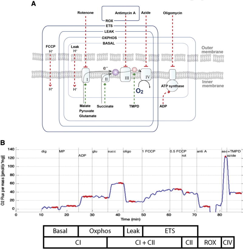
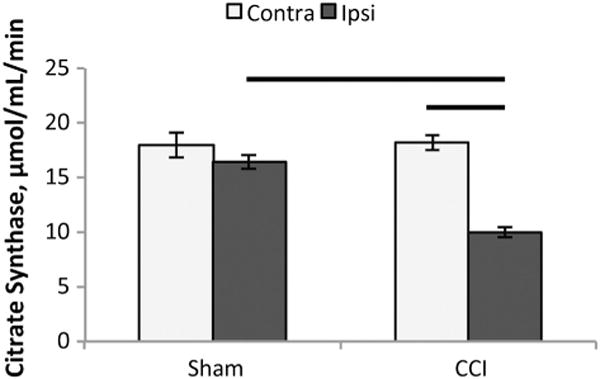
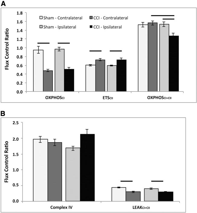
Traumatic brain injury (TBI) is one of the leading causes of death in children worldwide. Emerging evidence suggests that alterations in mitochondrial function are critical components of secondary injury cascade initiated by TBI that propogates neurodegeneration and limits neuroregeneration. Unfortunately, there is very little known about the cerebral mitochondrial bioenergetic response from the immature brain triggered by traumatic biomechanical forces. Therefore, the objective of this study was to perform a detailed evaluation of mitochondrial bioenergetics using high-resolution respirometry in a high-fidelity large animal model of focal controlled cortical impact injury (CCI) 24h post-injury. This novel approach is directed at analyzing dysfunction in electron transport, ADP phosphorylation and leak respiration to provide insight into potential mechanisms and possible interventions for mitochondrial dysfunction in the immature brain in focal TBI by delineating targets within the electron transport system (ETS). Development and application of these methodologies have several advantages, and adds to the interpretation of previously reported techniques, by having the added benefit that any toxins or neurometabolites present in the ex-vivo samples are not removed during the mitochondrial isolation process, and simulates the in situ tricarboxylic acid (TCA) cycle by maximizing key substrates for convergent flow of electrons through both complexes I and II. To investigate alterations in mitochondrial function after CCI, ipsilateral tissue near the focal impact site and tissue from the corresponding contralateral side were examined. Respiration per mg of tissue was also related to citrate synthase activity (CS) and calculated flux control ratios (FCR), as an attempt to control for variability in mitochondrial content. Our biochemical analysis of complex interdependent pathways of electron flow through the electron transport system, by most measures, reveals a bilateral decrease in complex I-driven respiration and an increase in complex II-driven respiration 24h after focal TBI. These alterations in convergent electron flow though both complex I and II-driven respiration resulted in significantly lower maximal coupled and uncoupled respiration in the ipsilateral tissue compared to the contralateral side, for all measures. Surprisingly, increases in complex II and complex IV activities were most pronounced in the contralateral side of the brain from the focal injury, and where oxidative phosphorylation was increased significantly compared to sham values. We conclude that 24h after focal TBI in the immature brain, there are significant alterations in cerebral mitochondrial bioenergetics, with pronounced increases in complex II and complex IV respiration in the contralateral hemisphere. These alterations in mitochondrial bioenergetics present multiple targets for therapeutic intervention to limit secondary brain injury and support recovery.
Keywords: Axonal injury; Bioenergetics; Contusion; Mitochondria; Pediatric traumatic brain injury; Swine.
Copyright © 2015 Elsevier Inc. All rights reserved.
Figures
References
-
- Adelson PD, Pineda J, Bell MJ, Abend NS, Berger RP, Giza CC, Hotz G, Wainwright MS, Pediatric TBID, Clinical AssessmentWorking G Common data elements for pediatric traumatic brain injury: recommendations from the working group on demographics and clinical assessment. J Neurotrauma. 2012;29:639–653. - PMC - PubMed
-
- Almeida A, Brooks KJ, Sammut I, Keelan J, Davey GP, Clark JB, Bates TE. Postnatal development of the complexes of the electron transport chain in synaptic mitochondria from rat brain. Dev Neurosci. 1995;17:212–218. - PubMed
-
- Armstead WM. Age and cerebral circulation. Pathophysiology. 2005;12:5–15. - PubMed
-
- Armstead W, Kurth C. Different cerebral hemodynamic responses following fluid percussion brain injury in the newborn and juvenile pig. J Neurotrauma. 1994;11:487–497. - PubMed
Publication types
MeSH terms
Substances
Grants and funding
LinkOut - more resources
Full Text Sources
Other Literature Sources
