. 2015 Sep;149(3):728-40.e15.
doi: 10.1053/j.gastro.2015.05.043. Epub 2015 May 29.

Fibroblast Growth Factor Signaling Controls Liver Size in Mice With Humanized Livers

Affiliations

Fibroblast Growth Factor Signaling Controls Liver Size in Mice With Humanized Livers

Willscott E Naugler et al. Gastroenterology. 2015 Sep.

Abstract

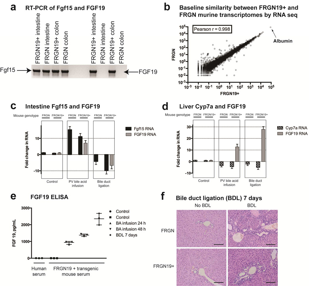
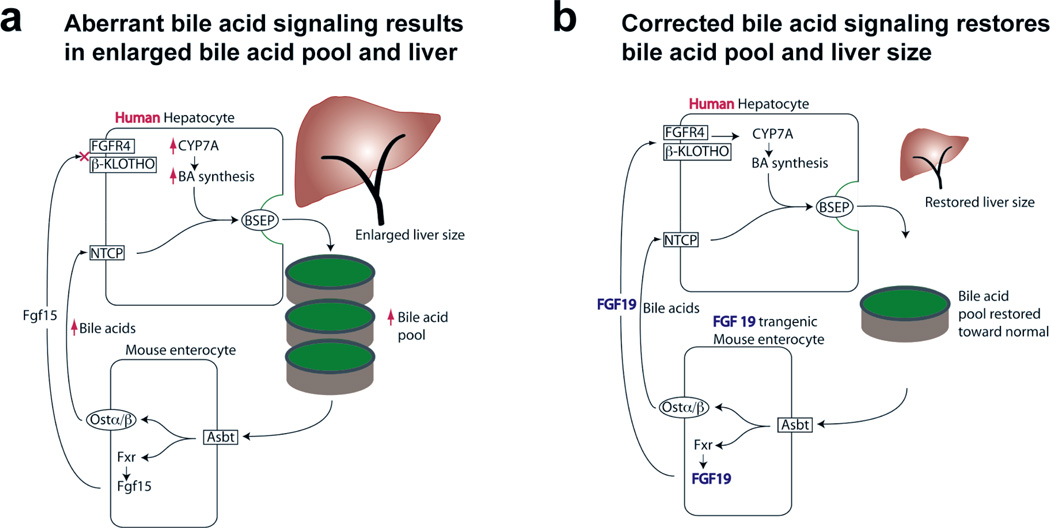
Background & aims: The ratio of liver size to body weight (hepatostat) is tightly controlled, but little is known about how the physiologic functions of the liver help determine its size. Livers of mice repopulated with human hepatocytes (humanized livers) grow to larger than normal; the human hepatocytes do not recognize the fibroblast growth factor (FGF)-15 produced by mouse intestine. This results in up-regulation of bile acid synthesis in the human hepatocytes and enlargement of the bile acid pool. We investigated whether abnormal bile acid signaling affects the hepatostat in mice.

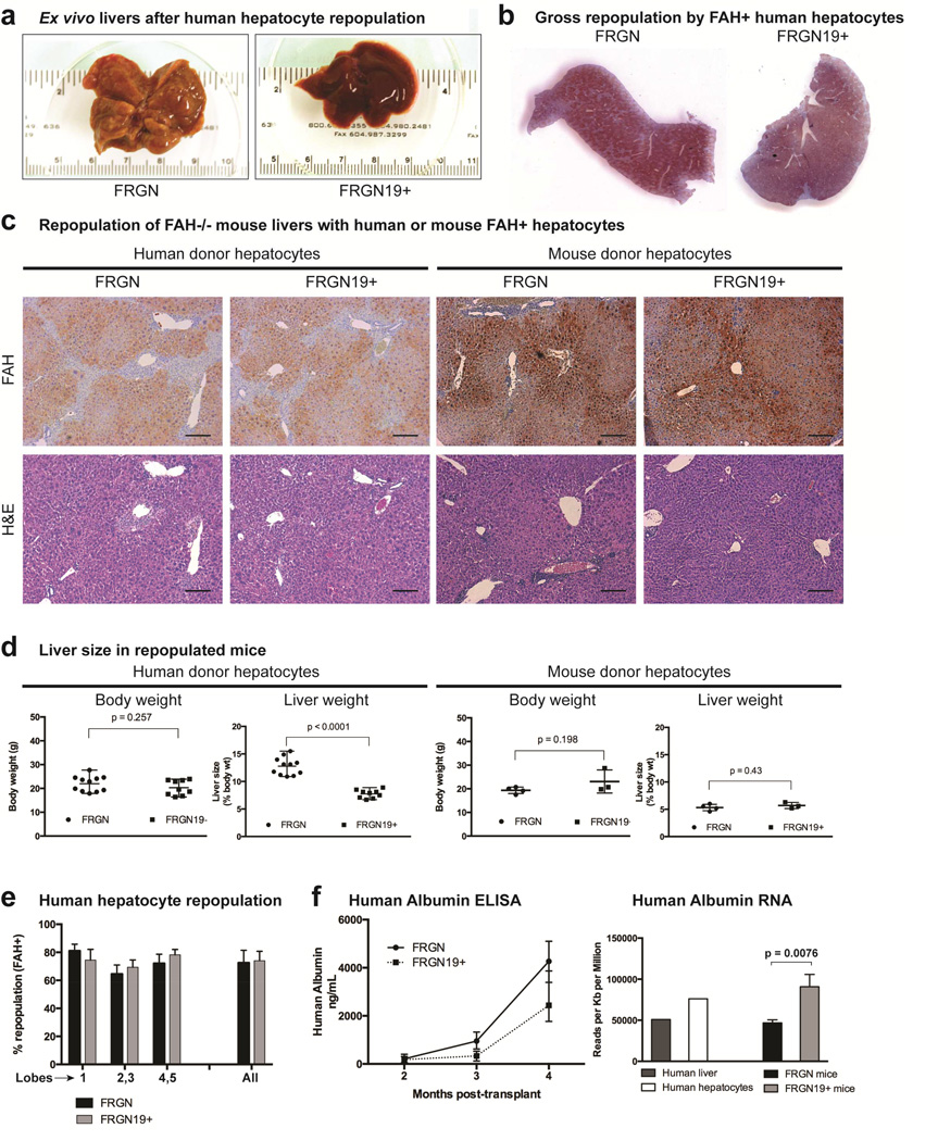
Methods: We crossed Fah(-/-), Rag2(-/-), Il2r(-/-) mice with nonobese diabetic mice to create FRGN mice, whose livers can be fully repopulated with human hepatocytes. We inserted the gene for human FGF19 (ortholog to mouse Fgf15), including regulatory sequences, into the FRGN mice to create FRGN19(+) mice. Livers of FRGN19(+) mice and their FRGN littermates were fully repopulated with human hepatocytes. Liver tissues were collected and bile acid pool sizes and RNA sequences were analyzed and compared with those of mice without humanized livers (controls).

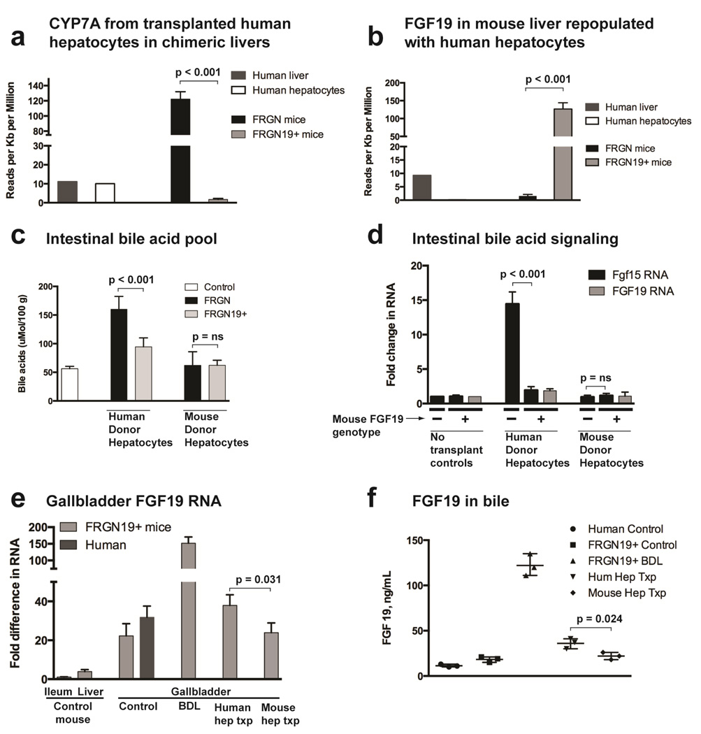
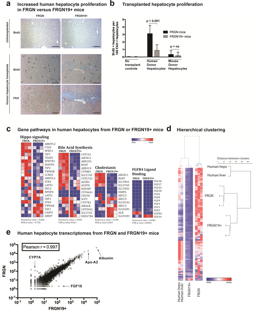
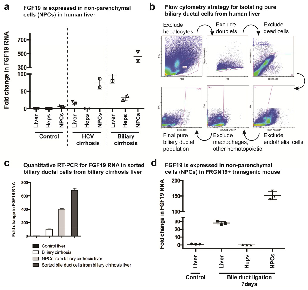
Results: Livers were larger in FRGN mice with humanized livers (13% of body weight), compared with control FRGN mice; they also had much larger bile acid pools and aberrant bile acid signaling. Livers from FRGN19(+) normalized to 7.8% of body weight, and their bile acid pool and signaling more closely resembled that of control FRGN19(+) mice. RNA sequence analysis showed activation of the Hippo pathway, and immunohistochemical and transcription analyses revealed increased hepatocyte proliferation, but not apoptosis, in the enlarged humanized livers of FRGN mice. Cell sorting experiments showed that although healthy human liver does not produce FGF19, nonparenchymal cells from cholestatic livers produce FGF19.

Conclusions: In mice with humanized livers, expression of an FGF19 transgene corrects bile acid signaling defects, resulting in normalization of bile acid synthesis, the bile acid pool, and liver size. These findings indicate that liver size is, in part, regulated by the size of the bile acid pool that the liver must circulate.

Keywords: CYP7A; Mouse Model; Regeneration; Signal Transduction.


Conflict of interest statement

Conflicts of interest

The authors declare no conflicts of interest related to this manuscript.

Figures

Figure 1
Figure 1. FRGN19+ transgenic mice demonstrate physiological regulation of FGF19 and bile acid signaling
(a) RT-PCR shows presence of RNA for Fgf15 in the small intestine and colon of both FRGN19+ transgenic mice and their FRGN littermates, while only showing FGF 19 RNA in the transgenic mice. (b) Scatterplot of RNA sequencing of mouse transcriptomes from FRGN19+ transgenic mice and their FRGN littermates are near indistinguishable (Pearson r = 1 signifies identical datasets). (c) Intestinal regulation of Fgf 15 and FGF 19 under 3 conditions: control, portal vein (PV) bile acid infusion and bile duct ligation (BDL), n = 3 mice of each genotype for each condition. (d) Liver regulation of Cyp 7a and FGF 19 under 3 conditions: control, portal vein (PV) bile acid infusion and bile duct ligation (BDL), n = 3 mice of each genotype for each condition. (e) ELISA for serum FGF19 on human and mouse serum. (f) H&E histology of liver (10×) with BDL for 7 days in FRGN19+ and FRGN mice, showing typical biliary ductal proliferation seen in BDL. Scale bar = 100 µm.
Figure 2
Figure 2. Human hepatocyte repopulated mouse livers are larger in FGF 19 – mice compared to FRGN19+ mice
(a) Representative gross pictures of human hepatocyte repopulated mouse livers 4 months after hepatocyte transplantation. (b) Low powered view of representative lobes of human hepatocyte repopulated livers stained for FAH (brown) in FRGN and FRGN19+ mice. (c) FAH staining (brown) identifies transplanted human or mouse hepatocytes in FRGN and FRGN19+ transgenic mice. H&E staining of repopulated livers shows normal parenchymal architecture. Scale bar = 100 µm. (d) Body and liver weights of mice with livers fully repopulated with either human or mouse hepatocytes in FRGN and FRGN19+ mice. (e) Percent of liver lobes positive for FAH (indicating level of repopulation) by morphometric analysis. (f) ELISA for human albumin in serum of FRGN and FRGN19+ mice transplanted with human hepatocytes, n = 10 FRGN mice and n = 9 FRGN19+ mice. Human albumin RNA quantified by RNA sequencing from human hepatocytes 4 months after transplant in FRGN and FRGN19+, n = 6 FRGN and n = 6 FRGN19+ mice.
Figure 3
Figure 3. FRGN19+ mice with humanized livers show normalized bile acid signaling compared to FRGN mice
(a) Complete RNA sequencing of chimeric human hepatocyte repopulated mouse livers showed a marked decrease in human CYP7A in FRGN19+ compared to FRGN mice, indicating restoration of intestine-liver signaling of bile acid homeostasis (n = 3 each genotype; n = 1 each for human liver and human hepatocytes). (b) RNA sequencing of chimeric livers revealed FGF 19 in liver tissue from FRGN19+ mice, but no FGF 19 in chimeric livers from FRGN mice, indicating that human hepatoctyes do not produce FGF 19 in this model (n = 3 each genotype). (c) Intestinal contents were collected from control C57/B6 (n = 3), human hepatocyte-transplanted FRGN (n = 5) and FRGN19+ mice (n = 5), and mouse hepatocyte-transplanted FRGN (n = 4) and FRGN19+ (n = 3) mice, and the bile acid pool quantitated after ethanol extraction of bile acids. (d) Fgf 15 and FGF 19 RNA was quantitated by qPCR in small intestine tissue, fold change normalized to un-transplanted controls; human hepatocyte-transplanted FRGN (n = 10), FRGN19+ (n = 9), and mouse hepatocyte-transplanted FRGN (n = 4) and FRGN19+ (n = 3) mice. (e) FGF 19 RNA was quantitated by qPCR in gallbladder tissue, normalized to FGF 19 RNA in the ileum of an FRGN19+ mouse; FRGN19+ controls (n = 4), human GB controls (n = 3), FRGN19+ mice after 7 days of BDL (n = 3), FRGN19+ mice repopulated with human hepatocytes (n = 4) and FRGN19+ mice repopulated with mouse hepatocytes (n = 3). (f) FGF 19 ELISA of bile from human controls (n = 3, obtained during resection of hepatic hemangioma n = 1 or adenoma n = 2), and FRGN19+ mice (controls, 7 days after BDL, liver repopulated with human hepatocytes or liver repopulated with mouse hepatocytes).
Figure 4
Figure 4. Transcriptional and proliferative differences in human hepatocytes when transplanted into FRGN and FRGN19+ mice
(a) Immunohistochemistry showing BrdU or FAH staining in untransplanted or human-hepatocyte repopulated livers 4 months after transplant. Serial sections used for BrdU and FAH staining in repopulated livers, and arrows indicate BrdU positive hepatocyte nuclei in FAH + hepatocytes. Scale bar = 100 µm. (b) Quantitation of BrdU positive nuclei in FAH + hepatocytes in FRGN and FRGN19+ mice, controls (n = 3 each), human hepatocyte-transplanted FRGN (n = 4) and FRGN19+ (n = 4) mice, and mouse hepatocyte-transplanted FRGN (n = 3) and FRGN19+ (n = 3) mice. (c) Gene pathways indicated from RNA sequencing data (human transcriptome only) from liver tissue of human hepatocyte repopulated mice, either FRGN (n = 3) or FRGN19+ (n = 3) mice; statistics generated through GSEA, heatmap generated with GENEE. (d) Clustering heatmap of human transcriptomes from liver tissue of human hepatocyte repopulated FGF 19 – or + mice (n = 3 each); Ward’s unsupervised hierarchical clustering shows two distinct transcriptome clusters for single-donor human hepatocytes when transplanted into FRGN versus FRGN19+ mice. (e) Scatterplot of RNA sequencing of human hepatoctye transcriptomes (single donor) from FRGN19+ transgenic mice and their FRGN littermates are similar; arrows indicate specific genes.
Figure 5
Figure 5. Location of FGF 19 expression in human liver
(a) Quantitative RT-PCR (qPCR) for FGF 19 was performed on whole liver tissue or specific populations of cells (hepatocytes or non-parenchymal cells, NPCs) from the same liver tissue; control humam liver, n = 3, obtained during resection of hepatic hemangioma n = 1 or adenoma n = 2, HCV cirrhotic liver and biliary cirrhosis liver (n = 2 each) obtained from explants prior to liver transplantation. (b) Flow cytometry isolated pure biliary ductal cells from an explanted human liver with biliary cirrhosis from biliary atresia. (c) Quantitative RT-PCR for FGF19 RNA done on human biliary cirrhosis liver, NPC cell fraction, and sorted bile duct cells (all from 1 liver, 3 qPCR replicates). (d) Similar FGF 19 qPCR was done in FRGN19+ transgenic mice 7 days after bile duct ligation (BDL) had been performed (n = 3 mice).
Figure 6
Figure 6. Relationship between bile acid pool size and liver size
(a) Aberrant signaling perturbs bile acid homeostasis when transplanted human hepatocytes fail to recognize Fgf15, the mouse intestinal signal for inhibiting bile acid synthesis in the liver. This leads to an enlarged bile acid pool and consequent increase in hepatocyte mass needed to circulate the pool. (b) Introduction of the human inhibitory signal, FGF19, allows transplanted human hepatocytes to properly regulate bile acid synthesis, and the bile acid pool is normalized. Hepatocyte mass is decreased, proportional to the bile acid pool which is circulated.

Comment in

References

    1. Taub R. Liver regeneration: from myth to mechanism. Nat Rev Mol Cell Biol. 2004;5:836–847. - PubMed
    1. Michalopoulos GK, DeFrances MC. Liver regeneration. Science. 1997;276:60–66. - PubMed
    1. Michalopoulos GK. Liver regeneration. J Cell Physiol. 2007;213:286–300. - PMC - PubMed
    1. Camargo FD, Gokhale S, Johnnidis JB, et al. YAP1 increases organ size and expands undifferentiated progenitor cells. Curr Biol. 2007;17:2054–2060. - PubMed
    1. Anakk S, Watanabe M, Ochsner SA, et al. Combined deletion of Fxr and Shp in mice induces Cyp17a1 and results in juvenile onset cholestasis. J Clin Invest. 2011;121:86–95. - PMC - PubMed

Publication types

MeSH terms