. 2015 Apr;28(2):109-20.
doi: 10.1293/tox.2014-0061. Epub 2015 Mar 1.

Time course of the incidence/multiplicity and histopathological features of murine colonic dysplasia, adenoma and adenocarcinoma induced by benzo[a]pyrene and dextran sulfate sodium

Affiliations

Time course of the incidence/multiplicity and histopathological features of murine colonic dysplasia, adenoma and adenocarcinoma induced by benzo[a]pyrene and dextran sulfate sodium

Jiro Sonoda et al. J Toxicol Pathol. 2015 Apr.

Erratum in

  • Errata (Printer's correction).
    [No authors listed] [No authors listed] J Toxicol Pathol. 2016 Jan;29(1):74. Epub 2016 Feb 17. J Toxicol Pathol. 2016. PMID: 26989306 Free PMC article.

Abstract

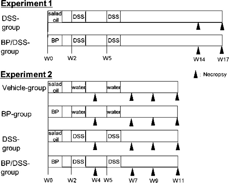
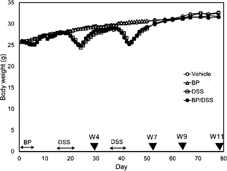
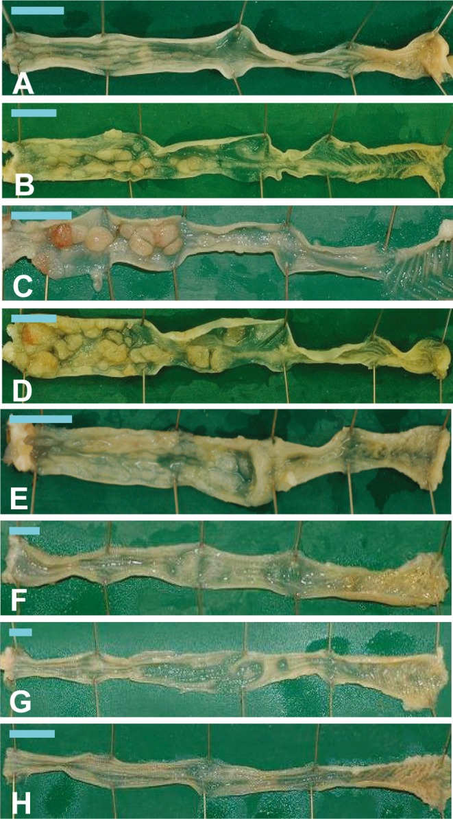
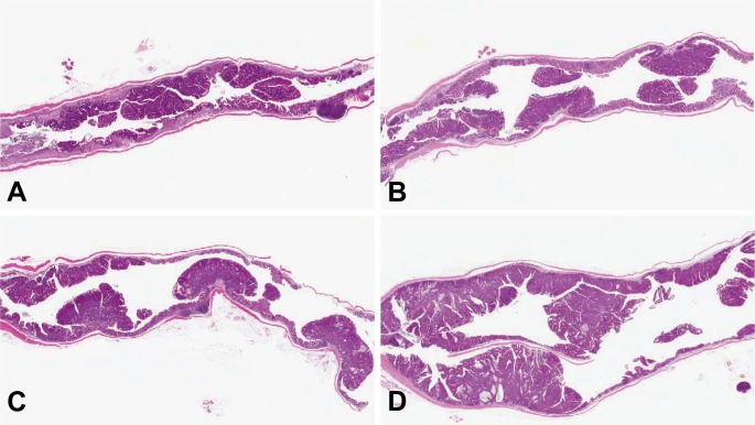
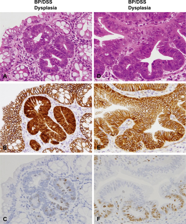
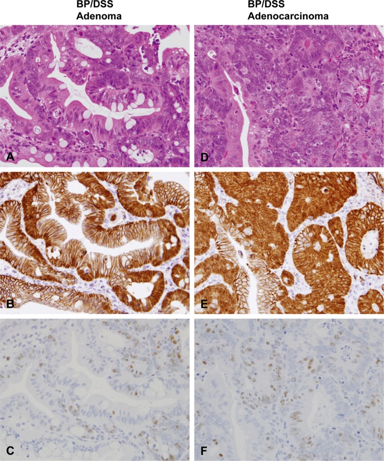
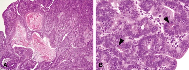
Benzo[a]pyrene (BP) is mutagenic but noncarcinogenic in the murine colon. Recently, we reported rapid induction of colonic tumors by treatment of CD2F1 mice with BP (125 mg/kg for 5 days) followed by a colitis inducer, dextran sulfate sodium (DSS) (4% in drinking water for 1 or 2 weeks). However, there are no reports on detailed time course and histopathological features of colonic proliferative lesions in this model. Here, we show the detailed time course of colonic dysplasia, adenoma and adenocarcinoma induced by treatment with BP, DSS, and a combination of the two (BP/DSS). In the colon of mice exposed to BP/DSS, 14.6 dysplastic foci per mouse were present one week after DSS treatment (week 4). The number of dysplastic foci decreased with time to 3.1 at week 9 and thereafter remained almost constant. At week 4, 1.5 adenocarcinomas were also observed, with a marked increase in numbers with time, reaching 29.3 at week 14. In contrast, the number of dysplastic foci induced by DSS alone showed a time course similar to that following BP/DSS treatment; however, only a few tumors appeared. Neither dysplastic foci nor neoplastic lesions were induced by BP only. In mice exposed to BP/DSS, β-catenin was demonstrated immunohistochemically in the nucleus and/or cytoplasm of the tumor cells, and this translocation from the cell membrane was evident in subsets of dysplastic foci. In dysplastic foci induced by DSS alone, β-catenin was absent in the nucleus/cytoplasm. These finding suggest that aberrant β-catenin accumulation in dysplastic foci is associated with tumor progression in this BP/DSS model.

Keywords: benzo[a]pyrene; colon; dextran sulfate sodium; dysplastic foci; tumor; β-catenin.


Figures

Fig. 1.
Fig. 1.
Outline of the protocols for the experiments for histopathology of the colon. Experiment 1 consisted of DSS and BP/DSS groups, and the mice in these groups (6 to 12 mice per group) were necropsied at weeks 14 and 17. In this report, the first week of the experiment is defined as week 0 (indicated as W0 in these figures). Experiment 2 consisted of vehicle, BP, DSS, and BP/DSS groups, and all mice were necropsied at weeks 4, 7, 9 and 11 (6 or 8 mice/group). Male CD2F1 mice were treated with BP at an oral dose of 125 mg/kg/day for 5 consecutive days, and starting 10 days after the last dose, mice were given 4% DSS in drinking water for 1 week once (at week 2) or twice (intermittently, at weeks 2 and 5 separated by a 2-week DSS treatment free interval period). Salad oil and distilled water were used as vehicle controls for BP and DSS solutions, respectively.
Fig. 2.
Fig. 2.
Body weights of mice. The symbols (▼) indicate the time of tissue collection (necropsy) for histopathology.
Fig. 3.
Fig. 3.
Macroscopic view of the colons of mice from the BP/DSS group at weeks 4 (A), 7 (B), 9 (C) and 11 (D), DSS group at weeks 7 (E) and 9 (F), vehicle group at week 7 (G) and BP group at week 7 (H). In the BP/DSS group, a rough surface was seen in the distal colon at week 4 (A), and nodular or polypoid masses were found in the distal and/or middle colons at weeks 7 (B), 9 (C) and 11 (D). No obvious changes were found in the vehicle (G) and BP groups (H), while masses were sporadically observed in the DSS group (E and F). Bar: 1 cm.
Fig. 4.
Fig. 4.
Histopathology of the colons of mice from the BP/DSS group at weeks 4, 7, 9 and 11. Neoplastic lesions including tubular adenoma and polypoid tubular adenocarcinoma can be seen in the distal part of the colonic mucosa at week 4 (A). Thereafter, the size of the neoplastic lesions gradually increases with time at weeks 7 (B), 9 (C) and 11 (D). H& E stain.
Fig. 5.
Fig. 5.
Histopathology with H & E stain (A and D) and immunohistochemistry of β-catenin (B and E) and Ki-67 (C and F) in dysplasia of mice from the BP/DSS group. Figs. (A) to (C) and Figs. (D) to (F) are serial sections of different dysplasias, respectively. Dysplasia was characterized by irregular crypt branching and distortion of the crypt architecture. β-catenin was localized on the cell membrane of most dysplastic foci (E) and distributed in the nucleus and/or cytoplasm of a small number of dysplastic foci (B). Ki-67-positive cells were diffusely distributed in dysplastic foci (C and F).
Fig. 6.
Fig. 6.
Histopathology with H & E stain (A and D) and immunohistochemistry of β-catenin (B and E) and Ki-67 (C and F) in adenomas and adenocarcinomas of mice from the BP/DSS group. Adenomas consisted of variably sized glands lined by single/multiple layers of epithelial cells (A). Adenocarcinomas were characterized by variable sized, irregularly branched and distorted glands lined by marked stratified epithelial cells (D). β-catenin was distributed in the nucleus and cytoplasm of adenoma (B) and adenocarcinoma cells (E). Ki-67-positive cells were most numerous in colonic adenoma (C) and adenocarcinoma cells (F).
Fig. 7.
Fig. 7.
Histopathology with H & E stain and immunohistochemistry of β-catenin and Ki-67 in the colonic mucosa of mice from the vehicle group (A–C) and BP group (D–F) and in dysplasias (G–I) and adenomas (J–L) of mice from the DSS group. No obvious histological changes were found in the vehicle (A) and BP group (D). Histological characteristics of dysplastic foci (G) and adenomas (J) found in the DSS group were similar to those observed in the BP/DSS group. β-catenin was localized on the cell membrane of the normal mucosal cells in the vehicle (B) and BP groups (E) and dysplastic foci (H) in the DSS group. The β-catenin was distributed in the nucleus and cytoplasm of adenomas (K) in the DSS group. Ki-67-positive cells were mainly localized in the lower zone of normal colonic crypts (C and F) and in diffusely distributed dysplastic foci (I) and adenomas (L) in the DSS group.
Fig. 8.
Fig. 8.
Histopathology of the colons of mice from the BP/DSS group. Adenocarcinomas were occasionally accompanied by squamous metaplasia (A) or differentiation into Paneth cells (B, arrowhead). H& E stain.

References

    1. Diggs DL, Huderson AC, Harris KL, Myers JN, Banks LD, Rekhadevi PV, Niaz MS, and Ramesh A. Polycyclic aromatic hydrocarbons and digestive tract cancers: a perspective. J Environ Sci Health C Environ Carcinog Ecotoxicol Rev. 29: 324–357. 2011. - PMC - PubMed
    1. Rosenberg DW, Giardina C, and Tanaka T. Mouse models for the study of colon carcinogenesis. Carcinogenesis. 30: 183–196. 2009. - PMC - PubMed
    1. Westbrook AM, Szakmary A, and Schiestl RH. Mechanisms of intestinal inflammation and development of associated cancers: lessons learned from mouse models. Mutat Res. 705: 40–59. 2010. - PMC - PubMed
    1. Clapper ML, Cooper HS, and Chang W-CL. Dextran sulfate sodium-induced colitis-associated neoplasia: a promising model for the development of chemopreventive interventions. Acta Pharmacol Sin. 28: 1450–1459. 2007. - PubMed
    1. Tanaka T. Colorectal carcinogenesis: Review of human and experimental animal studies. J Carcinog. 8: 5 2009. - PMC - PubMed

LinkOut - more resources