Case Reports
. 2015 Apr;28(2):121-4.
doi: 10.1293/tox.2014-0060. Epub 2015 Mar 1.

Spontaneous and bilateral necrosis of the femoral head in a young experimental beagle dog

Affiliations
Case Reports

Spontaneous and bilateral necrosis of the femoral head in a young experimental beagle dog

Ryosuke Kobayashi et al. J Toxicol Pathol. 2015 Apr.

Erratum in

  • Errata (Printer's correction).
    [No authors listed] [No authors listed] J Toxicol Pathol. 2016 Jan;29(1):74. Epub 2016 Feb 17. J Toxicol Pathol. 2016. PMID: 26989306 Free PMC article.

Abstract

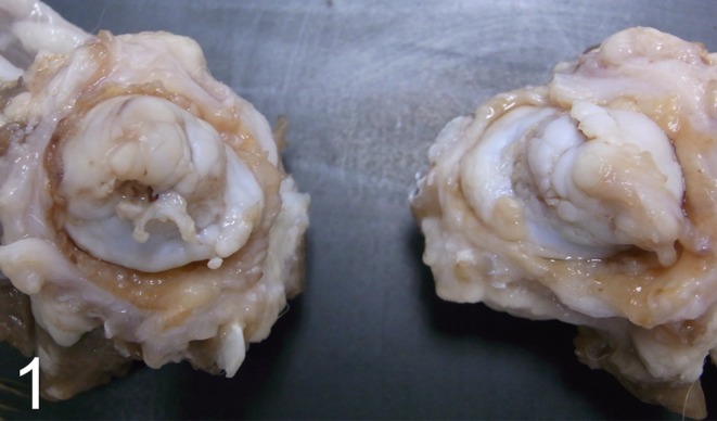
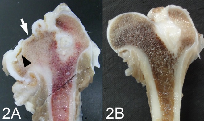
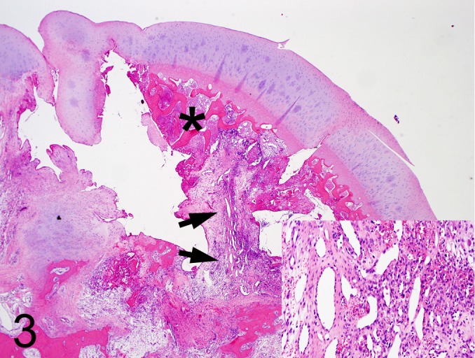
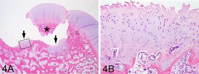
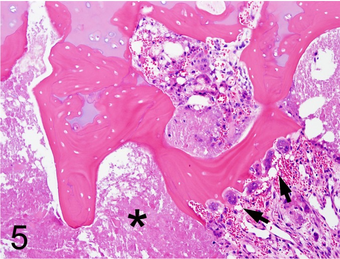
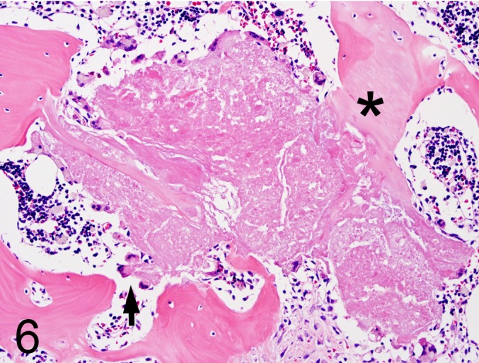
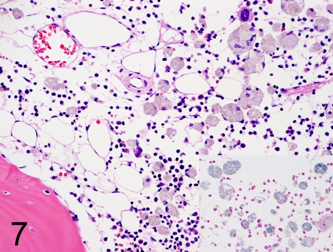
This report describes the pathological characterizations of a rare case of necrosis of the femoral head that was spontaneous, bilateral, avascular and nontraumatic. A 14-month-old beagle dog was presented with pain in the hind limbs. At necropsy, the articular surface in the bilateral femoral head was markedly irregular. There were no gross abnormalities other than in the hip joints. Microscopically, a wide range of trabecular bone necrosis localized in the subchondral area was observed in both femoral heads. In the right femoral head, fibrosis and proliferative vessels were noted in the subchondral area. The articular cartilage was thickened irregularly, but there was no evidence of cartilage necrosis. The bone marrow adjacent to the affected area showed severe depression. In the metaphysis, atrophic bone marrow, but not bone necrosis, was observed. This was a rare case of spontaneous necrosis of the femoral head in an experimental beagle dog.

Keywords: Legg-Calvé-Perthes disease; avascular osteonecrosis; dog; necrosis of the femoral head; spontaneous lesion.


Figures

Fig. 1.
Fig. 1.
Macroscopic changes in the femoral heads. The spherical shape of the bilateral femoral head is completely collapsed, and the articular surface shows marked irregularity.
Fig. 2.
Fig. 2.
The longitudinal cut surface of the left femoral head (A). B: age-matched control at the same magnification. The articular cartilage thickened irregularly. Cracks in the subchondral area (black arrow head) or ulceration (white arrow) form.
Fig. 3.
Fig. 3.
The lesions in the right femoral head. Subchondral bone necrosis (asterisk) and formation of the clefts are marked, and articular cartilage is thickened. Proliferation of the vessels is noted (arrows and inset). H&E, (20×).
Fig. 4.
Fig. 4.
The lesions in the left femoral head. The rectangle in A indicates the area magnified in B. A: In the left femoral head, similar subchondral bone necrosis (asterisk) and thickened articular cartilage can be seen on the right side. Trabecular bone is exposed to the articular cavity due to loss of articular cartilage. Chondroid metaplasia of the trabecular bone can be seen in the basal part of the ulceration (arrows). H&E, (20×). B: In the metaplastic cartilage, abnormal arrangements of the chondrocytes are observed. H&E, (200×).
Fig. 5.
Fig. 5.
Bone necrosis in the subchondral area. Loss of osteocytes and empty lacunae of the trabecular bone are noted. Some osteoclasts come into contact with necrotic bones (arrows). Eosinophilic amorphous matrices can be seen surrounding necrotic bone (asterisk). H&E, (200×).
Fig. 6.
Fig. 6.
The eosinophilic amorphous matrix surrounding necrotic bone. The matrix is partially connected to necrotic bone (asterisk). Some multinucleated giant cells can be seen at the periphery of the matrix (arrow). H&E, (200×).
Fig. 7.
Fig. 7.
The lesions in the bone marrow. The hematopoietic tissues show depression. Atrophic adipose tissues and lipofuscin-laden macrophages positive for Schmorl’s method (inset) can be seen. H&E, inset = Schmorl’s method, (400×).

References

    1. Hottendorf GH, and Hirth RS. Lesions of spontaneous subclinical disease in Beagle dogs. Vet Pathol. 11: 240–258. 1974. - PubMed
    1. Kobayashi K, Hirouchi Y, Iwata H, Yamakawa S, Mikami S, Yamamoto S, Hashiguchi J, and Enomoto M. Historical control data of spontaneous lesions in beagle dogs. J Toxicol Pathol. 7: 329–343. 1994.
    1. Sato J, Doi T, Wako Y, Hamamura M, Kanno T, Tsuchitani M, and Narama I. Histopathology of incidental findings in beagles used in toxicity studies. J Toxicol Pathol. 25: 103–134. 2012. - PMC - PubMed
    1. Thompson K. Osteonecrosis. In: Pathology of Domestic Animals 5th ed. MG Maxie (ed). Elsevier, Philadelphia. Vol. 1. 88-92. 2007.
    1. Carlson CS, and Weisbrode SE. Aseptic necrosis of bone. In: Pathologic Basis of Veterinary Disease, 5th ed. JF Zachary, and MD Mcgavin (eds). Elsevier, Missouri. 953-955. 2012.

Publication types

LinkOut - more resources