. 2015 Mar 25;49(2):141-6.
doi: 10.2478/raon-2014-0037. eCollection 2015 Jun.

Artery of Percheron infarction: review of literature with a case report

Affiliations

Artery of Percheron infarction: review of literature with a case report

Urska Lamot et al. Radiol Oncol. .

Abstract

Background: Clinical features indicating an ischemic infarction in the territory of posterior cerebral circulation require a comprehensive radiologic examination, which is best achieved by a multi-modality imaging approach (computed tomography [CT], CT-perfusion, computed tomography angiography [CTA], magnetic resonance imaging [MRI] and diffusion weighted imaging [DWI]). The diagnosis of an acute ischemic infarction, where the damage of brain tissue may still be reversible, enables selection of appropriate treatment and contributes to a more favourable outcome. For these reasons it is essential to recognize common neurovascular variants in the territory of the posterior cerebral circulation, one of which is the artery of Percheron.

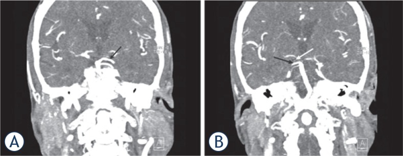
Case report: A 69 year-old woman, last seen awake 10 hours earlier, presented with two typical clinical features of the artery of Percheron infarction, which were vertical gaze palsy and coma. Brain CT and CTA of neck and intracranial arteries upon arrival were interpreted as normal. A new brain CT scan performed 24 hours later revealed hypodensity in the medial parts of thalami. Other imaging modalities were not performed, due to the presumption that the window for the application of effective therapy was over. The diagnosis of an artery of Percheron infarction was therefore made retrospectively with the re-examination of the CTA of neck and intracranial arteries.

Conclusions: A multi-modality imaging approach is necessary in every patient with suspicion of the posterior circulation infarction immediately after the onset of symptoms, especially in cases where primary imaging modalities are unremarkable and clinical features are severe, where follow-up examinations are indicated.

Keywords: Percheron; imaging; infarction.


Figures

FIGURE 1.
FIGURE 1.
The non-contrast head CT scan performed on the day of admission was normal, in particular without early signs of ischemia at the level of both thalami (A) the mesencephalon (B).
FIGURE 2.
FIGURE 2.
Another non-contrast head CT scan was performed 24 hours later and showed symmetrical ill-defined areas of hypodensity in the medial part of both thalami, corresponding to occlusion of the artery of Percheron (white arrows) (A). The hypodense area in the right thalamus extended into the anterior part of the mesencephalon and cerebral peduncle. The hypodense area in the left thalamus extended only into the anterior part of the mesencephalon (white arrow) (B).
FIGURE 3.
FIGURE 3.
Computed tomography angiography (CTA) of the neck and intracranial arteries. The basilar artery and the left posterior cerebral artery (PCA) (black arrow) were both transient (A). At first glance, the right PCA appears to be fully opacified. Detailed examination of the CTA images revealed a filling defect of the P1 segment of the right PCA (white arrow) and another rare anatomic variant: duplication of the right superior cerebellar artery (black arrow), which could have been mistaken for a transient right PCA (B).
FIGURE 4.
FIGURE 4.
Anatomic variations of the arterial supply to the paramedian thalamic-mesencephalic region as described by Percheron: Variant I (A), variant IIa (B), variant IIb (C) – the artery of Percheron, variant III (D). Vessels marked by initials: thalamic perforators (TP), midbrain perforators (MP), posterior cerebral artery (PCA), superior cerebellar artery (SCA), basilar artery (BA), anterior inferior cerebellar artery (AICA) and artery of Percheron (AOP).

References

    1. Krampla W, Schmidbauer B, Hruby W. Iscmaemic stroke of the artery of Percheron. Eur Radiol. 2008;18:192–4. - PubMed
    1. Matheus MG, Castillo M. Imaging of acute bilateral paramedian thalamic and mesencephalic infarcts. AJNR Am J Neuroradiol. 2003;24:2005–8. - PMC - PubMed
    1. Godani M, Auci A, Torri T, Jensen S, Sette DM. Coma with vertical gaze palsy: Relevance of angio-CT in acute percheron artery syndrome. Case Rep Neurol. 2010;2:74–9. - PMC - PubMed
    1. Cassourret G, Prunet B, Sbardella F, Bordes J, Maurin O, Boret H. Ishemic stroke of the artery of Percheron with normal initial MRI: a case report. Case Rep Med. 2010;2010:425734. doi: 10.1155/2010/425734. - DOI - PMC - PubMed
    1. Amin OSM, Shwani SS, Zangana HM, Hussein EMH, Ameen NA. Bilateral infarction of paramedian thalami: a report of two cases of artery of Percheron occlusion and review of the literature. BMJ Case Rep. 2011;2011 doi: 10.1136/bcr.09.2010.3304. bcr0920103304. - DOI - PMC - PubMed

LinkOut - more resources