Review
. 2015 May 13:6:478.
doi: 10.3389/fmicb.2015.00478. eCollection 2015.

The Min system and other nucleoid-independent regulators of Z ring positioning

Affiliations
Review

The Min system and other nucleoid-independent regulators of Z ring positioning

Veronica W Rowlett et al. Front Microbiol. .

Abstract

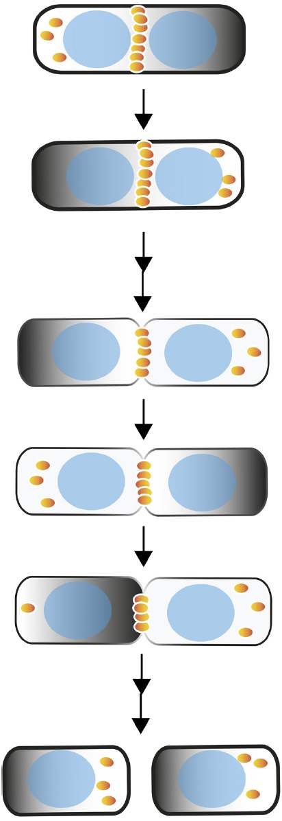
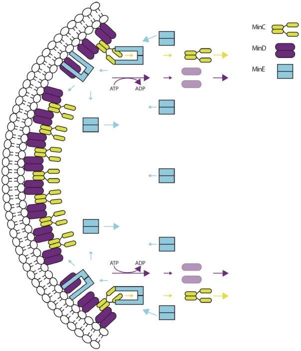
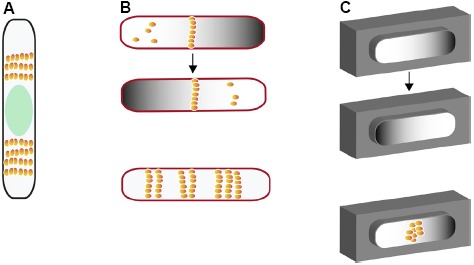
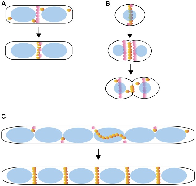
Rod-shaped bacteria such as E. coli have mechanisms to position their cell division plane at the precise center of the cell, to ensure that the daughter cells are equal in size. The two main mechanisms are the Min system and nucleoid occlusion (NO), both of which work by inhibiting assembly of FtsZ, the tubulin-like scaffold that forms the cytokinetic Z ring. Whereas NO prevents Z rings from constricting over unsegregated nucleoids, the Min system is nucleoid-independent and even functions in cells lacking nucleoids and thus NO. The Min proteins of E. coli and B. subtilis form bipolar gradients that inhibit Z ring formation most at the cell poles and least at the nascent division plane. This article will outline the molecular mechanisms behind Min function in E. coli and B. subtilis, and discuss distinct Z ring positioning systems in other bacterial species.

Keywords: FtsZ; Min system; Z-ring positioning; bacterial cell division; divisome.


Figures

FIGURE 1
FIGURE 1
Negative spatial regulation of Z ring positioning in E. coli by the Min system. Shown are Min oscillations (dark gradients), dynamics of FtsZ (orange spheres) and eventual assembly as a Z ring at the midpoint of a wild-type cell through the process of cell division and partitioning of the Min system to daughter cells. Nucleoids are depicted as blue ovals; passage of time in seconds is represented by an arrow, passage of time in minutes by a double arrow.
FIGURE 2
FIGURE 2
The molecular mechanism of Min oscillation. Membrane-bound complexes of MinC (chartreuse) and MinD (purple) are targeted by MinE (cyan). MinE dimers change conformation and bind MinD and the membrane, displacing MinC and stimulating the ATPase activity of MinD and its removal from the membrane. MinC and MinD-ADP move toward the opposite pole and begin another cycle of oscillation. MinE can stay membrane bound to remove more MinCD complexes, or change conformation and follow MinD to the opposite pole. Adapted from Rowlett and Margolin (2013).
FIGURE 3
FIGURE 3
Different model systems for investigating nucleoid-independent Z ring positioning in E. coli. (A) Multiple Z rings in cells lacking both Min and topoisomerase IV with large nucleoid-free regions on either side of unpartitioned nucleoid (green oval). (B) (top) Min oscillations and FtsZ positioning in nucleoid-free maxicells (outlined in red); (bottom) Z ring positioning in maxicells containing FtsZ that is unresponsive to MinC. (C) Cell-free oscillation of MinCDE in artificial cell-like compartments coated with a lipid bilayer before (top and middle) and after addition of purified FtsZ (bottom). FtsZ is depicted as orange spheres.
FIGURE 4
FIGURE 4
Positive control of Z ring positioning. Shown are models for positive spatial regulation of Z rings by PomZ in Myxococcus xanthus (A); MapZ/LocZ in Streptococcus pneumoniae (B); SsgB in Streptomyces aerial mycelia that are developing into spores (C). A sample cell cycle progression is shown for each. The above regulators are represented by pink spheres, FtsZ as orange spheres, and nucleoids as blue ovals.

References

    1. Adler H. I., Fisher W. D., Cohen A., Hardigree A. A. (1967). Miniature Escherichia coli cells deficient in DNA. Proc. Natl. Acad. Sci. U.S.A. 57, 321–326. - PMC - PubMed
    1. Åkerlund T., Bernander R., Nordström K. (1992). Cell division in Escherichia coli minB mutants. Mol. Microbiol. 6, 2073–2083. 10.1111/j.1365-2958.1992.tb01380.x - DOI - PubMed
    1. Åkerlund T., Gullbrand B., Nordström K. (2002). Effects of the Min system on nucleoid segregation in Escherichia coli. Microbiology 148, 3213–3222. - PubMed
    1. Arumugam S., Petrášek Z., Schwille P. (2014). MinCDE exploits the dynamic nature of FtsZ filaments for its spatial regulation. Proc. Natl. Acad. Sci. U.S.A. 111, E1192–E1200. 10.1073/pnas.1317764111 - DOI - PMC - PubMed
    1. Bailey M. W., Bisicchia P., Warren B. T., Sherratt D. J., Männik J. (2014). Evidence for divisome localization mechanisms independent of the Min system and SlmA in Escherichia coli. PLoS Genet. 10:e1004504. 10.1371/journal.pgen.1004504 - DOI - PMC - PubMed

LinkOut - more resources