. 2015 May 15:4:9.
doi: 10.1186/s40035-015-0030-4. eCollection 2015.

Posttranslational modifications of α-tubulin in alzheimer disease

Affiliations

Posttranslational modifications of α-tubulin in alzheimer disease

Fan Zhang et al. Transl Neurodegener. .

Abstract

Background: In Alzheimer disease (AD), hyperphosphorylation of tau proteins results in microtubule destabilization and cytoskeletal abnormalities. Our prior ultra-morphometric studies documented a clear reduction in microtubules in pyramidal neurons in AD compared to controls, however, this reduction did not coincide with the presence of paired helical filaments. The latter suggests the presence of compensatory mechanism(s) that stabilize microtubule dynamics despite the loss of tau binding and stabilization. Microtubules are composed of tubulin dimers which are subject to posttranslational modifications that affect the stability and function of microtubules.

Methods: In this study, we performed a detailed analysis on changes in the posttranslational modifications in tubulin in postmortem human brain tissues from AD patients and age-matched controls by immunoblot and immunocytochemistry.

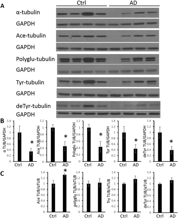
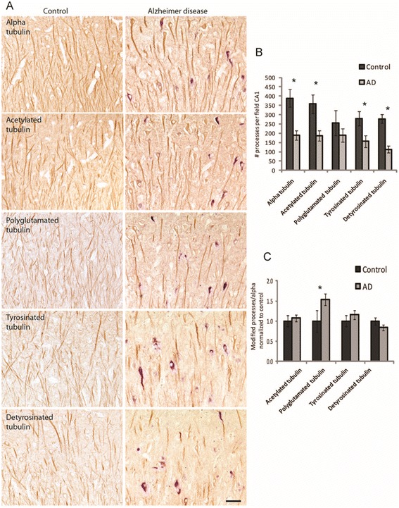
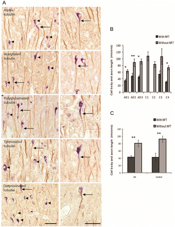
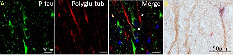
Results: Consistent with our previous study, we found decreased levels of α-tubulin in AD brain. Levels of tubulin with various posttranslational modifications such as polyglutamylation, tyrosination, and detyrosination were also proportionally reduced in AD brain, but, interestingly, there was an increase in the proportion of the acetylated α-tubulin in the remaining α-tubulin. Tubulin distribution was changed from predominantly in the processes to be more accumulated in the cell body. The number of processes containing polyglutamylated tubulin was well preserved in AD neurons. While there was a cell autonomous detrimental effect of NFTs on tubulin, this is likely a gradual and slow process, and there was no selective loss of acetylated or polyglutamylated tubulin in NFT-bearing neurons.

Conclusions: Overall, we suggest that the specific changes in tubulin modification in AD brain likely represent a compensatory response.

Keywords: Acetylation; Alzheimer disease; Polyglutamylation; Tau; Tubulin.


Figures

Fig. 1
Fig. 1
Immunoblot analysis of tubulins in AD brain. (a) Representative immunoblot analysis of tubulin expression and post-translational modifications in brain homogenates from hippocampal tissues from AD and age-matched control patients. GAPDH was used as the internal loading control. (b) The quantification results, normalized to GAPDH levels, confirmed a significant decrease in α-tubulin, acetylated tubulin, polyGlu-tubulin, tyrosinated tubulin, and detyr-tubulin levels. (c) The quantification results, normalized to α-tubulin levels, demonstrated an increase in acetylated tubulin (Ace TUB). Data are means ± SEM. * indicates significant difference between AD and control with p < 0.05
Fig. 2
Fig. 2
Immunocytochemical analysis of tubulins in AD brain. Representative images of the CA1 region demonstrate specific staining of the tubulin antibodies for neuron cell bodies and axonal processes in both control and AD cases (a). Tubulin antibodies are stained brown and NFT, using tau antibody, are stained blue. Qualitatively it appears that there are fewer processes stained in the AD cases and that only the thicker processes are retained (A). Quantification found there are significantly fewer processes stained for alpha, acetylated, tyrosinated, and detyrosinated tubulin in AD cases (b). When normalized to α-tubulin levels, an increase in stable glutamylated tubulin was found in AD cases (c). *p < 0.05. Scale bar = 50 μm
Fig. 3
Fig. 3
The relationship between neuronal tubulin levels and NFTs. Most neurons with NFT (mean 78 %) did not demonstrate axonal processes stained for tubulins (a, arrowheads left panels). However, some neurons with NFT (blue), had tubulin staining in the cell body and axonal process (A, arrows). Every tubulin modification was retained in the axonal process in some NFT-bearing neurons (A, right panels). Scale bars = 50 μm. Measuring the lengths of the cell bodies and axonal processes in the NFT and normal surrounding neurons in the CA1 region found the NFTs were indeed significantly shorter (b) in all AD cases and one control case. Taken together, the NFT in both AD and control cases, were significantly shorter when compared to the normal neuron population (c), yet no difference in length was found when comparing AD and control NFT, or AD and control normal neurons, suggesting the loss of axonal process length is a result of NFT formation and not disease. *p < 0.001, **p < 0.05
Fig. 4
Fig. 4
NFT-bearing neurons do not necessarily contain less acetylated tubulin. (a) Confocal microscopy demonstrated that in AD hippocampal tissues, those neurons containing neurofibrillary pathology (red, arrowheads) display levels of acetylated tubulin (green) comparable to those without NFT (arrows). (b) The same pattern was found using light level microscopy with acetylated tubulin stained brown, and phospho-tau stained blue
Fig. 5
Fig. 5
NFT-bearing neurons do not necessarily contain less polyglutamylated tubulin. (a) Levels of polyglutamylated tubulin (red) are similar in both normal and NFT-bearing neurons stained with phosphorylated tau (green). Blue: DAPI. (b) The same pattern was found using light level microscopy with glutamylated tubulin stained brown, and phospho-tau stained blue

References

    1. Wang JZ, Xia YY, Grundke-Iqbal I, Iqbal K. Abnormal hyperphosphorylation of tau: sites, regulation, and molecular mechanism of neurofibrillary degeneration. J Alzheimers Dis. 2013;33(Suppl 1):S123–39. - PubMed
    1. Iqbal K, Gong CX, Liu F. Microtubule-associated protein tau as a therapeutic target in Alzheimer's disease. Expert Opin Ther Targets. 2014;18(3):307–18. doi: 10.1517/14728222.2014.870156. - DOI - PubMed
    1. Iqbal K, Liu F, Gong CX, Alonso Adel C, Grundke-Iqbal I. Mechanisms of tau-induced neurodegeneration. Acta Neuropathol. 2009;118(1):53–69. doi: 10.1007/s00401-009-0486-3. - DOI - PMC - PubMed
    1. Cash AD, Aliev G, Siedlak SL, Nunomura A, Fujioka H, Zhu X, et al. Microtubule reduction in Alzheimer's disease and aging is independent of tau filament formation. Am J Pathol. 2003;162(5):1623–7. doi: 10.1016/S0002-9440(10)64296-4. - DOI - PMC - PubMed
    1. Scheff SW, DeKosky ST, Price DA. Quantitative assessment of cortical synaptic density in Alzheimer's disease. Neurobiol Aging. 1990;11(1):29–37. doi: 10.1016/0197-4580(90)90059-9. - DOI - PubMed

LinkOut - more resources