

Cerebral tuberculomas - A clinical challenge
- PMID: 26029627
- PMCID: PMC3949551
- DOI: 10.1016/j.rmcr.2013.04.003
Cerebral tuberculomas - A clinical challenge
Abstract
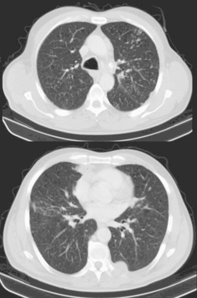
Cerebral tuberculomas are a rare and serious form of tuberculosis (TB) due to the haematogenous spread of Mycobacterium Tuberculosis (MT). Symptoms and radiologic features are nonspecific, leading sometimes to misdiagnosis. Anti-TB drugs are essential for the successful treatment of cerebral tuberculomas but there is no agreement regarding the duration of therapy. The authors present a case of a 55 years old male, presented to the emergency room with sudden onset of diplopia. Cerebral computerized tomography revealed multiple brain lesions, with contrast enhancement and peri-lesional oedema. The patient was HIV negative and because of previous malignancy the first suspicion was metastatic disease. Cultural exam of the bronchial wash showed MT sensitive to all first-line drugs. The patient started antituberculosis treatment with 4 drugs (HRZE) for 2 months, followed by maintenance therapy (HR). Treatment was prolonged for 24 months because at 12th and 18th months of treatment one of the brain lesions, although significantly smaller, still showed contrast enhancement. Even though it is not clear if contrast enhancement lesions represent active lesions or just inflammation, continuing treatment until total resolution of the tuberculomas is probably prudent.
Keywords: Cerebral tuberculomas; Disseminated tuberculosis.
Figures


References
-
- Pimentel M.L.V., Alves S.M.V., Novis S.A.P., Brandão R.Z., Neto E.B. Intracranial tuberculomas developing during treatment of pulmonary tuberculosis: case report. Arq Neuropsiquiatri. 2000;58(2-B):572–577. - PubMed
-
- Hejazi N., Hassler W. Multiple intracranial tuberculomas with atypical response to tuberculostatic chemotherapy: literature review and a case report. Infection. 1997;25(4):41–46. - PubMed
-
- Sahaiu-Srivastava S., Jones B. Brainstem tuberculoma in the immunocompetent: case report and literature review. Clinical Neurology and Neurosurgery. 2008;110:302–304. - PubMed
LinkOut - more resources
Full Text Sources
Other Literature Sources
