. 2015 Jun 1;10(6):e0128216.
doi: 10.1371/journal.pone.0128216. eCollection 2015.

The ACE2/Ang-(1-7)/Mas Axis Regulates the Development of Pancreatic Endocrine Cells in Mouse Embryos

Affiliations

The ACE2/Ang-(1-7)/Mas Axis Regulates the Development of Pancreatic Endocrine Cells in Mouse Embryos

Lin Wang et al. PLoS One. .

Abstract

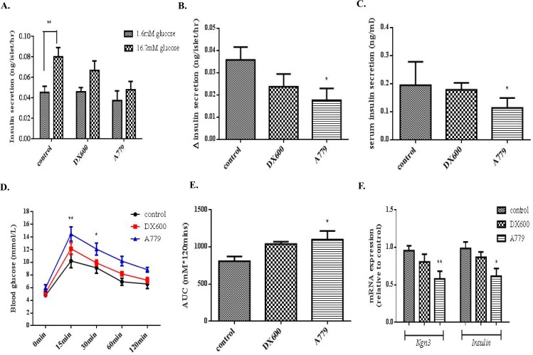
Angiotensin-converting enzyme 2 (ACE2), its product Angiotensin-(1-7) [Ang-(1-7)], and Ang-(1-7) receptor Mas, have been shown to regulate organogenesis during embryonic development in various species. However, it is not known whether a local ACE2/Ang-(1-7)/Mas axis is present in the fetal pancreas. It is hypothesized that there is a local ACE2/Ang-(1-7)/Mas axis in the embryonic pancreas in mice that is involved in regulating islet cell development. To address this issue, the endogenous expression profile of axis constituents in embryonic mouse pancreata was examined. Involvement of the ACE2 axis in the regulation of pancreatic development was also examined. The present experiments showed in an in vivo animal model that endogenous expression levels of ACE2 and the Mas receptor were upregulated in mouse pancreata in late embryogenesis, peaking on embryonic day E16.5, when it reached 3 folds compared to that seen at E12.5. Consistently, endogenous expression of Ang-(1-7) also peaked at E16.5. Treatment with the ACE2 inhibitor DX600 did not alter islet development. However, prenatal treatment with A779, a Mas receptor antagonist, reduced the β-cell to α-cell ratio in neonatal islets, impaired islet insulin secretory function, and impaired the pups' glucose tolerance. In ex vivo pancreas explant cultures, A779 again decreased the β-cell to α-cell ratio, apparently through its effects on β-cell proliferation (reduced proliferation shown with Ki67 staining), and also decreased Insulin and Ngn3 mRNA expression. Furthermore, treatment of explant cultures with Ang-(1-7) increased mRNA levels of Insulin and pancreatic progenitor marker Ngn3, as well as Nox4, the ROS generation enzyme; these stimulatory effects were attenuated by co-treatment with A779, suggesting that Ang-(1-7), via Mas receptor signaling, may promote differentiation of pancreatic progenitors into insulin-producing cells via modulation of reactive oxygen species. These data together suggest that a Mas receptor-mediated mechanism may stimulate pancreatic cell development.


Conflict of interest statement

Competing Interests: The authors declare no conflict of interest.

Figures

Fig 1
Fig 1. Expression of ACE2 and the Mas receptor in embryonic mouse pancreas from E12.5 to neonatal period.
(A-B) ACE2 and Mas receptor protein expression levels in relation to E12.5 levels. n = 6 per time point. (C) Relative mRNA expression of ACE2 and Mas receptor in the developing pancreas. n = 4 per time point. (D) Quantification of Ang-(1–7) levels in the developing pancreas. n = 4 per time point. One-way ANOVA followed by Tukey’s post hoc tests were used. All data are expressed as means ± SEM. *p < 0.05, **p < 0.01, ***p < 0.001 vs. E12.5, # p < 0.05 vs. E16.5.
Fig 2
Fig 2. Localization of ACE2 and the Mas receptor at E12.5, E14.5, E16.5, in embryonic mouse pancreata.
Florescent IHC for ACE2 and the Mas receptor are shown above and below, respectively (both are shown in red). Both series were nuclear counterstained with DAPI (blue). Scale bar = 50 μm.
Fig 3
Fig 3. Effects of ACE2 inhibition and Mas receptor antagonism on endocrine cell composition of neonatal mouse pancreas (day 4 postnatal).
(A) Ratio of pancreas weight to total body weight of neonates. n = 7 and each group included 3 neonatal mice each time. One-way ANOVA followed by Tukey’s post hoc tests were performed. (B) H&E sections showing altered structure (blue arrows) of pancreata from pups in the A779 (Mas receptor antagonist) group, but not the DX600 (ACE2 inhibitor) group, compared to controls. Scale bar = 50 μm. (C-D) Assessment of β-cell and α-cell areas based on fluorescent IHC through insulin and glucagon staining. At least six islets were examined from each animal and at least 5 mice were employed in each group. One-way ANOVA followed by Tukey’s post hoc tests were performed. All data are expressed as means ± SEM. *p < 0.05 vs. control.
Fig 4
Fig 4. Insulin secretory function and glucose tolerance in mouse neonates.
(A-B) Insulin release from neonatal islets (day 4 postnatal) in response to a high-glucose (16.7 mM) challenge. One-way ANOVA followed by Tukey’s post hoc tests were used. (C) Serum insulin concentration in serum collected from neonates (day 4) from each group. One-way ANOVA followed by Tukey’s post hoc tests were performed. (D) IPGTTs were performed on 4-weeks-old pups from each experimental group. Changes in blood glucose level were measured immediately before (t = 0 min), and at four time points after (t = 15, 30, 60 and 120 min), glucose loading. Two-way ANOVA followed by Tukey’s post hoc tests were used. (E) Comparison of areas under the curve (AUCs) across groups. (F) mRNA expression of Ngn3 and Insulin in neonatal islets isolated from DX600 or A779 group pups. One-way ANOVA followed by Tukey’s post hoc tests were performed. All data are expressed as means ± SEM, n = 6 per group. *p < 0.05, **p < 0.01 vs. control.
Fig 5
Fig 5. Effects of Ang-(1–7) and A779 on endocrine cell differentiation in cultured pancreatic rudiments.
(A-B) Expression of endocrine cell marker transcripts following administration of Ang-(1–7) at a range of doses. (C) Real-time assessment of the mRNA expression of Ngn3 and Insulin. n = 6. (D-E) Assessment of β-cell and α-cell areas as identified by immunofluorescent staining with anti-insulin and anti-glucagon antibodies. (F-G) Quantitation of proliferating β-cells, recognized as insulin+Ki67+cells, in pancreas explant cultures exposed to exogenous 1 μM Ang-(1–7) or 1 μM A779. Scale bar = 50 μm. One-way ANOVA followed by Tukey’s post hoc tests were used. All data are expressed as means ± SEMs. n = 6 from at least 7 pancreatic rudiments were used in each group. *p < 0.05, **p < 0.01, *** p <0.001 vs. control, # p < 0.05, ## p < 0.01, ### p <0.001 vs. Ang-(1–7).
Fig 6
Fig 6. Assessment of the interaction of Ang-(1–7) and ROS in cultured pancreatic rudiments.
(A-B) DHE-labelled sections. (C-D) Assessment of mRNA expression changes of NADPH oxidase subunits Nox1, Nox2 and Nox4. (E-F) IHC for Nox4. (G) Protein expression of p22phox in pancreatic rudiments treated with 1μM A779 (Mas receptor antagonist) in the presence and absence of 1 μM Ang-(1–7). (H) mRNA expression of Ngn3 and Insulin in pancreatic rudiments treated with 1 μM Ang-(1–7) in the presence and absence of 0.5 μM DPI (Nox4 inhibitor). Scale bar = 50 μm. One-way ANOVA followed by Tukey’s post hoc tests were used. All data are expressed as means ± SEM. n = 5 per group. *p < 0.05, **p < 0.01, *** p < 0.001 vs. control, # p < 0.05, ## p <0.01 vs. Ang-(1–7).

References

    1. Norwood VF, Craig MR, Harris JM, Gomez RA (1997) Differential expression of angiotensin II receptors during early renal morphogenesis. Am J Physiol 272: R662–668. - PubMed
    1. Perlegas D, Xie H, Sinha S, Somlyo AV, Owens GK (2005) ANG II type 2 receptor regulates smooth muscle growth and force generation in late fetal mouse development. Am J Physiol Heart Circ Physiol 288: H96–102. 10.1152/ajpheart.00620.2004 - DOI - PubMed
    1. Schutz S, Le Moullec JM, Corvol P, Gasc JM (1996) Early expression of all the components of the renin-angiotensin-system in human development. Am J Pathol 149: 2067–2079. - PMC - PubMed
    1. Leung PS (2007) The physiology of a local renin-angiotensin system in the pancreas. J Physiol 580: 31–37. 10.1113/jphysiol.2006.126193 - DOI - PMC - PubMed
    1. Donoghue M, Hsieh F, Baronas E, Godbout K, Gosselin M, Stagliano N, et al. (2000) A novel angiotensin-converting enzyme-related carboxypeptidase (ACE2) converts angiotensin I to angiotensin 1–9. Circ Res 87: E1–9. - PubMed

Publication types

MeSH terms

LinkOut - more resources