. 2015 Jun 1;10(6):e0128301.
doi: 10.1371/journal.pone.0128301. eCollection 2015.

Interferon-gamma and nitric oxide synthase 2 mediate the aggregation of resident adherent peritoneal exudate cells: implications for the host response to pathogens

Affiliations

Interferon-gamma and nitric oxide synthase 2 mediate the aggregation of resident adherent peritoneal exudate cells: implications for the host response to pathogens

Bhagawat S Chandrasekar et al. PLoS One. .

Abstract

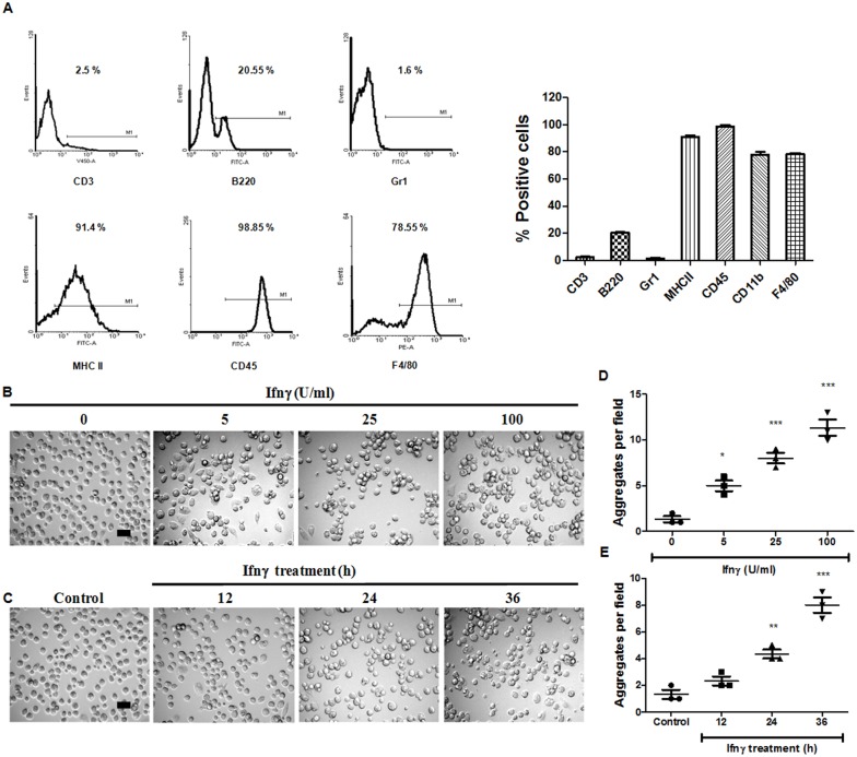
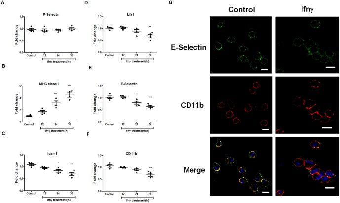
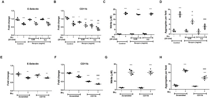
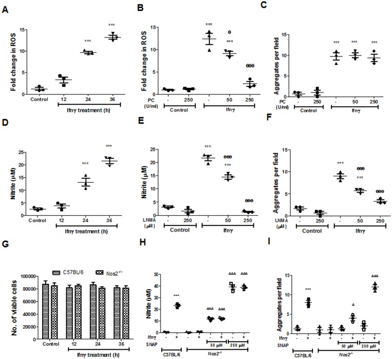
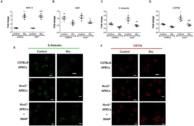
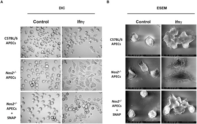
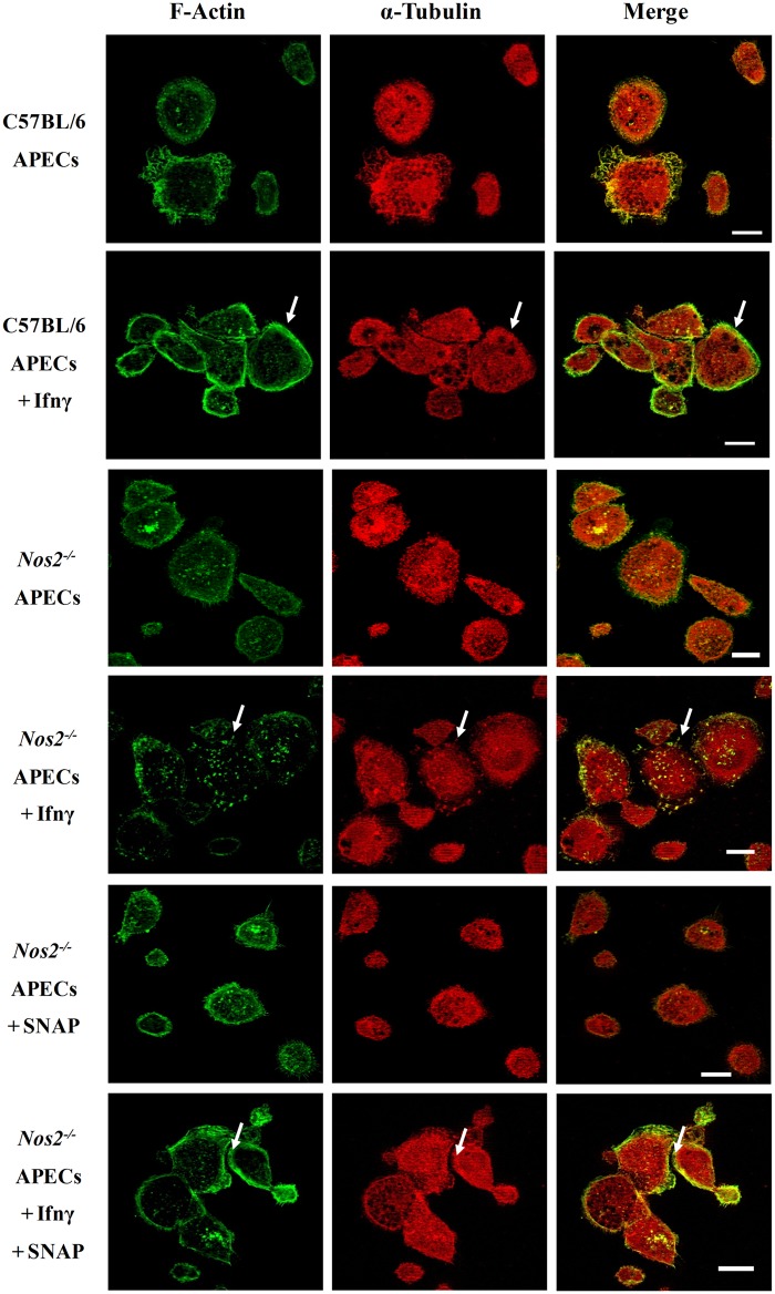
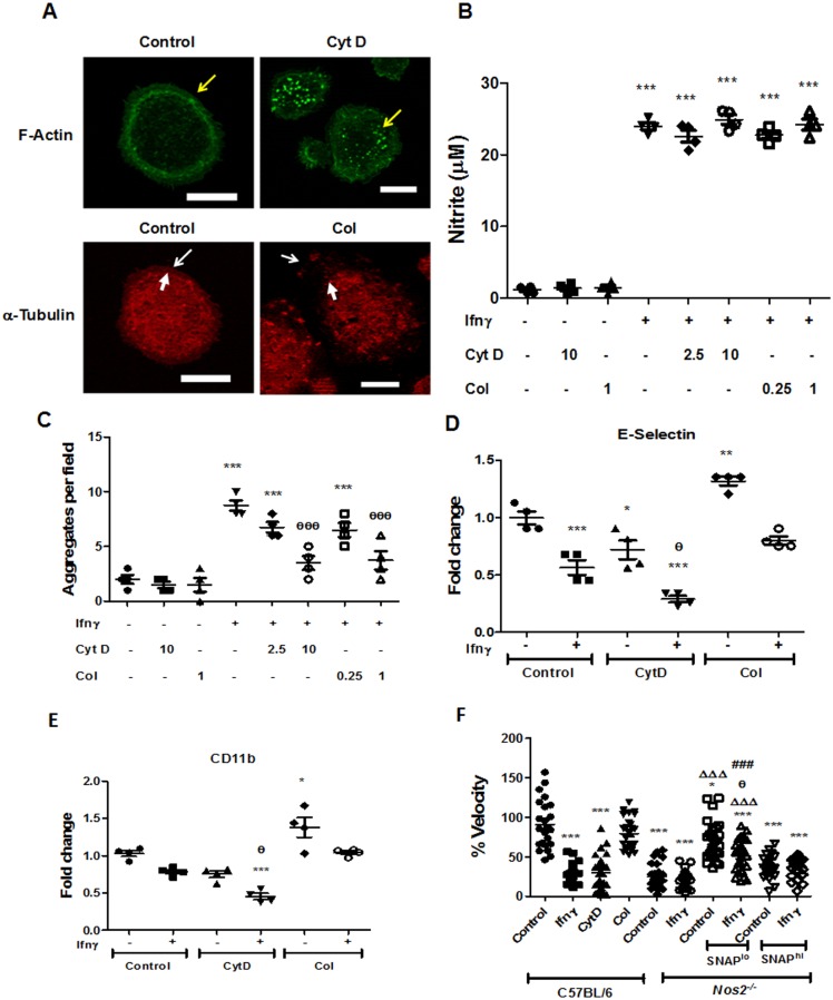
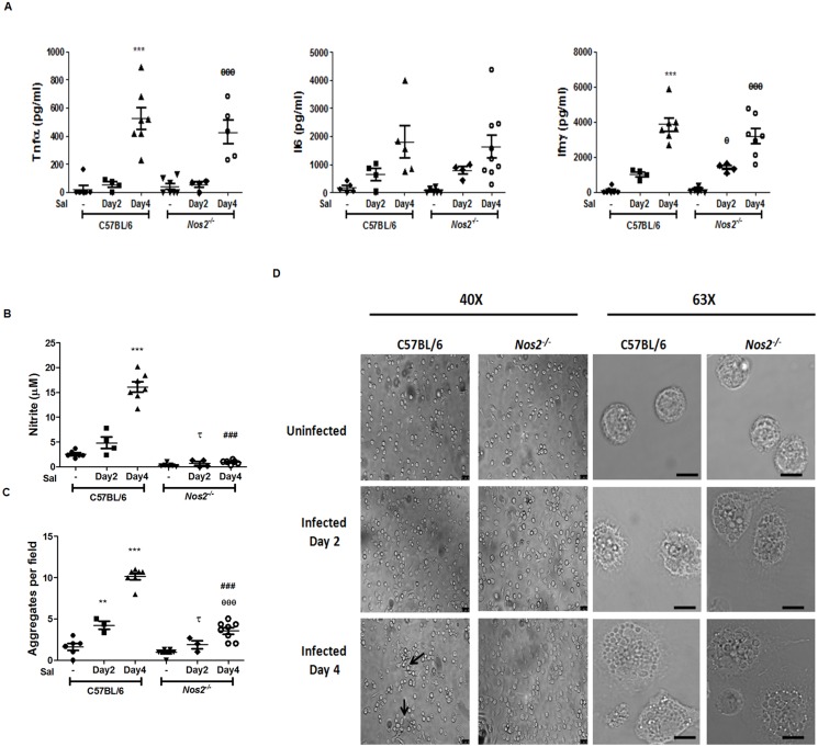
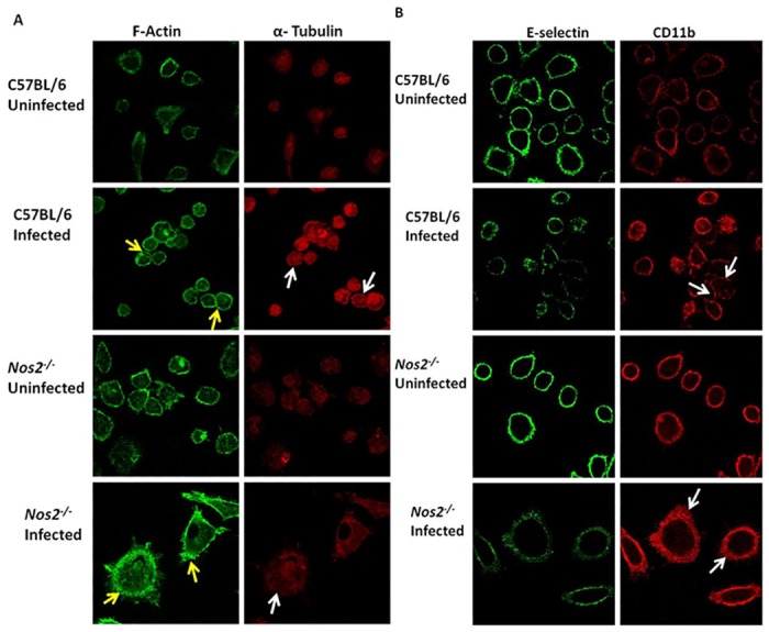
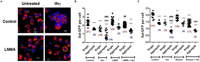
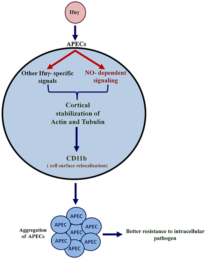
Interferon-gamma (Ifnγ), a key macrophage activating cytokine, plays pleiotropic roles in host immunity. In this study, the ability of Ifnγ to induce the aggregation of resident mouse adherent peritoneal exudate cells (APECs), consisting primarily of macrophages, was investigated. Cell-cell interactions involve adhesion molecules and, upon addition of Ifnγ, CD11b re-localizes preferentially to the sites of interaction on APECs. A functional role of CD11b in enhancing aggregation is demonstrated using Reopro, a blocking reagent, and siRNA to Cd11b. Studies with NG-methyl-L-arginine (LNMA), an inhibitor of Nitric oxide synthase (Nos), NO donors, e.g., S-nitroso-N-acetyl-DL-penicillamine (SNAP) or Diethylenetriamine/nitric oxide adduct (DETA/NO), and Nos2-/- mice identified Nitric oxide (NO) induced by Ifnγ as a key regulator of aggregation of APECs. Further studies with Nos2-/- APECs revealed that some Ifnγ responses are independent of NO: induction of MHC class II and CD80. On the other hand, Nos2 derived NO is important for other functions: motility, phagocytosis, morphology and aggregation. Studies with cytoskeleton depolymerizing agents revealed that Ifnγ and NO mediate the cortical stabilization of Actin and Tubulin which contribute to aggregation of APECs. The biological relevance of aggregation of APECs was delineated using infection experiments with Salmonella Typhimurium (S. Typhimurium). APECs from orally infected, but not uninfected, mice produce high amounts of NO and aggregate upon ex vivo culture in a Nos2-dependent manner. Importantly, aggregated APECs induced by Ifnγ contain fewer intracellular S. Typhimurium compared to their single counterparts post infection. Further experiments with LNMA or Reopro revealed that both NO and CD11b are important for aggregation; in addition, NO is bactericidal. Overall, this study elucidates novel roles for Ifnγ and Nos2 in regulating Actin, Tubulin, CD11b, motility and morphology during the aggregation response of APECs. The implications of aggregation or "group behavior" of APECs are discussed in the context of host resistance to infectious organisms.


Conflict of interest statement

Competing Interests: The authors have declared that no competing interests exist.

Figures

Fig 1
Fig 1. Ifnγ induces APECs to aggregate.
APECs were isolated from C57BL/6 mice, cultured in tissue culture media for ~ 24 h and were characterized by staining with a panel of antibodies to different cell surface proteins followed by FACS analysis. Representative plots and data (A) for different markers is shown which are representative of multiple experiments with APECs from 4 mice. Bright field images of APECs treated with different doses of Ifnγ for 36 h (B) and kinetics of APECs treated with 25 U/ml Ifnγ for indicated time points (C). The scale bar represents 20 μm. Quantification of the extent of APECs aggregates formed as a function of dose of Ifnγ (D) and incubation time (E). An aggregate consists of six or more interacting cells in any given field acquired at 20X magnification. The data is represented as mean ± S.E from three independent experiments.
Fig 2
Fig 2. Modulation of expression of different cell surface proteins on APECs in response to Ifnγ.
Kinetic analysis of cell surface amounts of P-Selectin (A), MHC class II (B), Icam1 (C), Lfa1 (D), E-Selectin (E) and CD11b (F) upon Ifnγ treatment (25 U/ml) of APECs. Fluorescence microscopic images acquired at 63X with a scale of 10 μm of E-Selectin and CD11b on the cell surface of APECs treated with 25 U/ml of Ifnγ for 36 h (G). The data is represented as mean ± S.E from two independent experiments.
Fig 3
Fig 3. CD11b is required for aggregation of APECs in response to Ifnγ.
The relative amounts of E-Selectin (A, E) and CD11b (B, F) on APECs upon treatment with Ifnγ (25 U/ml) in the absence or presence of indicated amounts of Reopro (A-D) or siRNA to Cd11b (E-H) was measured after 36 h. The amount of nitrite (C,G) and cell aggregates (D,H) upon 25 U/ml of Ifnγ treatment in the absence or presence of indicated doses of Reopro or 200 nM siRNA to Cd11b is shown. The data is represented as mean ± S.E from three independent experiments and control refers to untreated cells alone. The significance with respect to untreated controls and Ifnγ treated C57BL/6 APECs controls are represented as * and θ respectively.
Fig 4
Fig 4. Nos2 derived NO mediates the Ifnγ induced aggregation of APECs.
Kinetic analysis of amounts of ROS (A), nitrite (D) of APECs treated with 25 U/ml of Ifnγ. The amounts of ROS (B) and the number of cell aggregates (C) in APECs treated with Ifnγ in the presence or absence of indicated doses of PEG-Catalase (PC) for 36 h. The amount of nitrite (E) and the number of cell aggregates (F) of APECs treated with Ifnγ in the presence or absence of indicated doses of LNMA for 36 h. Kinetic analysis of the number of viable APECs from C57BL/6 and Nos2 -/- mice left untreated or upon treatment with 25 U/ml of Ifnγ (G). The amount of nitrite (H) and the number of cell aggregates (I) from Ifnγ treated Nos2 -/- APECs in the absence or presence of indicated doses of SNAP for 36 h in comparison to C57BL/6 APECs treated with 25 U/ml Ifnγ for 36 h. The data is represented as mean ± S.E from three independent experiments. The significance with respect to untreated controls, Ifnγ treated C57BL/6 APECs controls and untreated Nos2 -/- APECs controls are represented as *, θ and Δ respectively.
Fig 5
Fig 5. Nos2 regulates the cell surface amounts and relocalization of CD11b in response to Ifnγ in APECs.
Comparative analysis of cell surface amounts of MHC class II (A), Lfa1 (B), E-Selectin (C) and CD11b (D) on APECs from C57BL/6 and Nos2 -/- mice at 36 h post 25 U/ml of Ifnγ treatment. The data is represented as mean ± S.E from three independent experiments. Fluorescence microscopic images with a scale of 10 μm of E-Selectin (E) and CD11b (F) on APECs from C57BL/6 and Nos2 -/- mice at 36 h post 25 U/ml of Ifnγ treatment. The significance with respect to untreated C57BL/6 APECs controls and untreated Nos2 -/- APECs controls are represented as * and Δ respectively.
Fig 6
Fig 6. Nos2 regulates the morphology of APECs treated with Ifnγ.
Representative bright field images acquired at a magnification of 60X (A) and ESEM images acquired at a magnification of 5000X (B) of C57BL/6 and Nos2 -/- APECs treated with 25 U/ml of Ifnγ for 36 h is shown. In addition, the images of Nos2 -/- APECs cultured in the absence or presence of 100 μM of SNAP treated in absence or presence of 25 U/ml of Ifnγ for 36 h is shown.
Fig 7
Fig 7. The cortical stability of cytoskeleton elements, Actin and Tubulin, is regulated in a Nos2 dependent manner upon Ifnγ treatment.
Fluorescence microscopic images, with the scale bar of 10 μm, of F-Actin (green) and α-Tubulin (red) in APECs from C57BL/6 mice and Nos2 -/- mice treated without or with 25 U/ml of Ifnγ for 36 h. In addition, Nos2 -/- APECs were treated with 100 μM of SNAP in the absence or presence of Ifnγ. The white arrows indicate cortical arrangement of F-Actin and α-Tubulin.
Fig 8
Fig 8. Actin and Tubulin stabilization contribute to Ifnγ induced aggregation of APECs.
Fluorescence microscopic images acquired at a magnification of 63X with the scale bar representing 10 μm of F-Actin (green) and α-Tubulin (red) in C57BL/6 APECs treated with 10 μM Cyt D and 1 μg/ml of Col respectively for 6 h (A). Yellow and white arrows indicate cortical arrangement of F-Actin and α-Tubulin respectively. Thin white arrows indicate the cell boundary while the fatter white arrows are used to highlight the shrinkage of the α-Tubulin network upon Col treatment when compared to the untreated control C57BL/6 APECs. The amount of nitrite in the supernatant (B) produced by APECs from C57BL/6 mice upon treatment with 25 U/ml of Ifnγ in the absence or presence indicated doses of inhibitors Cyt D (μM) and Col (μg/ml). The number of cell aggregates (C) of APECs from C57BL/6 mice upon treatment with 25 U/ml of Ifnγ in the absence or presence of indicated doses of inhibitors Cyt D (μM) and Col (μg/ml). The amounts of E-Selectin (D) and CD11b (E) on the cell surface of C57BL/6 APECs treated with 25 U/ml of Ifnγ for 36 h, post 6 h of pretreatment without or with Cyt D (10 μM) and Col (1 μg/ml). The data is represented as mean ± S.E from three independent experiments. The velocity of APECs from C57BL/6 and Nos2 -/- mice treated without or with 25 U/ml of Ifnγ between 18–24 h of addition (F). The velocity of APECs from C57BL/6 pretreated for 6 h with Cyt D (10 μM) and Col (1 μg/ml) before tracking them from 6–12 h post treatment. Also, the velocity of Nos2 -/- APECs pretreated in the presence of 5 μM of SNAP (SNAPlo) and 100 μM of SNAP (SNAPhi) for 12 h and without or with 25 U/ml of Ifnγ before tracking cells 18–24 h post Ifnγ addition. The velocity for each of the above mentioned conditions are represented as percentage velocity with respect to average velocity exhibited by untreated C57BL/6 APECs. The data is representative of two independent experiments. Significance is represented as * when compared to untreated controls and # when compared to Ifnγ treated C57BL/6 APECs respectively. The significance with respect to untreated controls, Ifnγ treated C57BL/6 APECs controls. untreated Nos2 -/- and SNAPlo alone Nos2 -/- APECs controls are represented as *, θ, Δ, and # respectively.
Fig 9
Fig 9. Nos2 mediates aggregation of APECs upon S. Typhimurium infection of mice.
The amounts of Tnfα, Il6 and Ifnγ (A) in the sera from uninfected and S. Typhimurium infected C57BL/6 and Nos2 -/- mice at day two and four post infection. The amounts of nitrite (B) and cell aggregates (C) of APECs isolated from uninfected and S. Typhimurium infected C57BL/6 and Nos2 -/- mice at indicated days post infection and cultured ex vivo for 24 h. Representative bright field microscopic images of APECs, either at 40X (Leica DMI6000B) or 63X (Leica TCS SP5), with the scale bar representing 20 μm, of APECs from uninfected or infected mice are shown (D). The data is representative of at least four independent experiments with a minimum of three mice per condition. Significance with respect to untreated C57BL/6 controls, untreated Nos2 -/- controls, S. Typhimurium infected C57BL/6 mice day 2 control and S. Typhimurium infected C57BL/6 mice day 4 control are represented as *, θ, τ, and # respectively.
Fig 10
Fig 10. Differences in localization patterns of Actin, Tubulin, CD11b and E-Selectin in APECs from C57BL/6 and Nos2 -/- APECs upon infection.
Fluorescence microscopic images of F-Actin (green) and α-Tubulin (red) in APECs from uninfected and S. Typhimurium infected C57BL/6 and Nos2 -/- mice four days post infection, left untreated ex vivo for 24 h (A). Yellow and white arrows represent Actin and Tubulin localization to the cortex, respectively. Confocal microscopic images of E-Selectin (green) and CD11b (red) in APECs taken at 100X magnification from uninfected and S. Typhimurium infected C57BL/6 and Nos2 -/- mice four days post infection, left untreated ex vivo for 24 h (B). The white arrows depict CD11b localization at the cell-cell interface in C57BL/6 APECs from infected mice. On the other hand, APECs from infected Nos2 -/- mice display diffused CD11b pattern. Scale bar for fluorescent microscopic images is 10 μm. The data is presented as mean ± S.E from two independent representative experiments with three mice per group.
Fig 11
Fig 11. The aggregation of APECs reduces the intracellular growth S. Typhimurium.
Representative confocal microscopic images acquired at a magnification of 100 X with a scale of 10 μm illustrating intracellular Sal-GFP (green), Lamp1 (red) and Hoechst (blue) in control and 25 U/ml of Ifnγ treated APECs in the presence or absence of LNMA (200 μM) post 24 h of Sal-GFP infection (A). Quantification of the number of Sal-GFP per cell in control and 25 U/ml of Ifnγ treated APECs in the presence or absence of LNMA (200 μM) post 24 h of Sal-GFP infection (B). Quantification of the number of Sal-GFP per cell in control and 25 U/ml of Ifnγ treated APECs in the presence or absence of Reopro (0.2 mg/ ml) post 24 h of Sal-GFP infection (C). In panel B and C, the number of fields scored for single cells are represented in maroon color and the number of fields scored with aggregates are represented in blue color within brackets under the data points. Note that in the presence of Ifnγ, the number of aggregates containing fields increase compared to fields with single cells, which is reversed with LNMA or Reopro treatment. The data is represented as mean ± S.E from two independent experiments. Significance is represented as * when compared to untreated single cell controls, # when compared to Ifnγ treated single cell controls and Δ when compared to Ifnγ treated aggregates of APECs controls. The significance is represented as * when compared to untreated single cell controls, θ when compared to untreated aggregate controls, # when compared to Ifnγ treated single cell controls and τ when compared to Ifnγ treated aggregates of APECs controls.
Fig 12
Fig 12. A schematic model representing the aggregation response of APECs in response to Ifnγ treatment.
In the presence of Ifnγ, Nos2 is induced resulting in high amounts of NO. Both NO and additional Ifnγ specific signals are required for stability and cortical arrangement of Actin and Tubulin, which affects several cellular functions, including motility, phagocytosis and maintenance of morphology. In addition, re-localization of CD11b enhances the aggregation of APECs which are more efficient in reducing the number of intracellular bacteria and increasing host resistance.

References

    1. Xie J, Tato CM, Davis MM. How the immune system talks to itself: the varied role of synapses. Immunol Rev. 2013; 251: 65–79. 10.1111/imr.12017 - DOI - PMC - PubMed
    1. Wahl SM, Feldman GM, McCarthy JB. Regulation of leukocyte adhesion and signaling in inflammation and disease. J Leukoc Biol. 1996; 59: 789–96. - PubMed
    1. Mosser DM, Edwards JP. Exploring the full spectrum of macrophage activation. Nat Rev Immunol. 2008; 8: 958–69. 10.1038/nri2448 - DOI - PMC - PubMed
    1. Wynn TA, Chawla A, Pollard JW. Macrophage biology in development, homeostasis and disease. Nature. 2013; 496: 445–55. 10.1038/nature12034 - DOI - PMC - PubMed
    1. Davies LC, Jenkins SJ, Allen JE, Taylor PR. Tissue-resident macrophages. Nat Immunol. 2013; 14: 986–95 10.1038/ni.2705 - DOI - PMC - PubMed

Publication types

MeSH terms