. 2015 Jun 30;6(18):16488-506.
doi: 10.18632/oncotarget.4108.

Redox effects and cytotoxic profiles of MJ25 and auranofin towards malignant melanoma cells

Affiliations

Redox effects and cytotoxic profiles of MJ25 and auranofin towards malignant melanoma cells

Marijke C C Sachweh et al. Oncotarget. .

Abstract

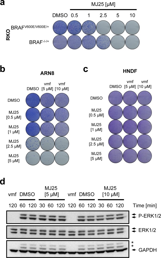
Malignant melanoma is the most dangerous type of skin cancer. Although recent progress in treatment has been achieved, lack of response, drug resistance and relapse remain major problems. The tumor suppressor p53 is rarely mutated in melanoma, yet it is inactive in the majority of cases due to dysregulation of upstream pathways. Thus, we screened for compounds that can activate p53 in melanoma cells. Here we describe effects of the small molecule MJ25 (2-{[2-(1,3-benzothiazol-2-ylsulfonyl)ethyl]thio}-1,3-benzoxazole), which increased the level of p53-dependent transactivation both as a single agent and in combination with nutlin-3. Furthermore, MJ25 showed potent cytotoxicity towards melanoma cell lines, whilst having weaker effects against human normal cells. MJ25 was also identified in an independent screen as an inhibitor of thioredoxin reductase 1 (TrxR1), an important selenoenzyme in the control of oxidative stress and redox regulation. The well-characterized TrxR inhibitor auranofin, which is FDA-approved and currently in clinical trials against leukemia and a number of solid cancers, displayed effects comparable with MJ25 on cells and led to eradication of cultured melanoma cells at low micromolar concentrations. In conclusion, auranofin, MJ25 or other inhibitors of TrxR1 should be evaluated as candidate compounds or leads for targeted therapy of malignant melanoma.

Keywords: auranofin; malignant melanoma; p53; thioredoxin reductase 1; vemurafenib.


Conflict of interest statement

DISCLOSURE OF POTENTIAL CONFLICTS OF INTEREST

The authors declare no conflict of interest.

Figures

Figure 1
Figure 1. MJ25 activates p53 and eradicates melanoma cells in culture
a. Chemical structure of MJ25. b. ARN8 cells, which express wt p53 and have been stably transfected with a p53-dependent β-galactosidase expression vector (RGC-ΔFos-LacZ reporter plasmid), were co-treated with vehicle (−) (DMSO) or nutlin-3 [2 μM] (+) and vehicle (0) (DMSO) or MJ25 at the indicated concentrations for 16 hours. p53-dependent transcription was assessed by measurement of β-galactosidase activity, taking protein levels into consideration. Error bars represent standard deviation. **, p < 0.01; ****, p < 0.001 (unpaired one-tailed Student's t-test; n = 4). c. ARN8 cells and human normal dermal fibroblasts (HNDFs) were treated with MJ25 at increasing concentrations for 9 hours. Protein levels were determined by Western blotting. GAPDH served as loading control. d. Cell growth and viability were measured in a number of melanoma cell lines, HNDFs and human normal epithelial melanocytes (HNEMs) by sulforhodamine B (SRB) assay after treatment with MJ25 at the indicated concentrations for 72 hours. Error bars represent standard deviation. (e and f) The effect of MJ25 on cell viability and colony-forming capacity was studied in e. RKO p53+/+ and p53def/def cells as well as f. HCT116 p53+/+ and p53def/def cells. g. H1299 cells (p53 null; top panel) and H1299 cells stably transfected with mutant p53 (R175H; bottom panel) were treated with MJ25 at the indicated concentrations for each 6 or 24 hours, respectively. p21 levels were determined by WB. GAPDH was used as loading control.
Figure 2
Figure 2. MJ25 appears to be non-genotoxic
a. MJ25's DNA alkylating capacity was assessed in an in vitro DNA alkylation assay. Form I (lower band) represents supercoiled (unaffected) plasmids and form II (upper band) open circular plasmids, which appear upon DNA alkylation. b. ARN8 cells and HNDFs were treated with MJ25 at various concentrations for 9 hours. Changes in levels of γ-H2AX were determined by Western blotting. GAPDH served as loading control.
Figure 3
Figure 3. MJ25's cytotoxic effect is enhanced by mutant BRAF
a. RKO BRAFV600E/V600E/+ and BRAF−/−/+ cells were treated for 72 hours with MJ25 as indicated, and cell viability and clonogenic capacity were determined. (b and c) The effect of MJ25 either alone or in combination with vemurafenib (vmf) on cell viability and clonogenic capacity was determined in b. ARN8 cells and c. HNDFs. DMSO served as vehicle control. d. ARN8 cells were treated with vemurafenib (vmf) [5 μM], DMSO or MJ25, respectively, at the indicated concentrations for the indicated periods of time. Changes in protein levels were determined by Western blotting. Stars indicate bands representing phospho-ERK subunits that remained when re-using the membrane for blotting against GAPDH.
Figure 4
Figure 4. MJ25 is an inhibitor of thioredoxin reductase 1 (TrxR1)
a. The capability of MJ25 and auranofin to inhibit recombinant, rat-derived TrxR1 in vitro was measured by an NADPH dependent 5,5′-dithiobis-[2-nitrobenzoic acid] (DTNB) assay. b. ARN8 cells were treated with MJ25, auranofin or DMSO, respectively, for the indicated periods of time. TrxR1 inhibition was subsequently assessed in cell lysates by an NADPH and Trx dependent insulin reduction endpoint assay, measuring thiol formation using DTNB. Ratios between MJ25 and DMSO as well as auranofin and DMSO were determined for each point in time. (c and d) ARN8 cells were treated with c. MJ25 or d. auranofin, while in each half of the samples growth media were supplemented with sodium selenite [75 nM] three days prior to seeding as well as during seeding and treatment for 72 hours. Cell viability and clonogenic capacity were determined. e. ROS levels were determined in ARN8 cells 3 hours after the indicated treatment by measuring fluorescence of 2′,7′-dichlorofluorescein (DCF). f. Induction of anti-oxidative proteins by MJ25 and auranofin was investigated in ARN8 cells at the indicated points in time by Western blotting. DMSO served as vehicle control (0 μM). g. ARN8 cells were pre-treated with L-buthionine sulfoximine (BSO) or vehicle (H2O) for 72 hours, upon which cells were re-plated in BSO- and vehicle-free growth medium. Cell viability was assessed by SRB assay after 72 hours in the presence of vehicle (DMSO), MJ25 (left panel) or auranofin (middle panel), respectively. Intracellular glutathione (GSH) levels were determined 72 hours after BSO / vehicle treatment (right panel). h. Inhibition of yeast-derived glutathione reductase by MJ25 and auranofin was determined in vitro by measurement of glutathione disulfide (GSSG) reduction.
Figure 5
Figure 5. Inhibition of TrxR1 by MJ25 and auranofin is irreversible and likely occurs at its Sec-dependent active site
a. NADPH-reduced TrxR1 was incubated with compounds as indicated, and Sec-dependent enzyme activity was subsequently measured by an NADPH dependent DTNB reduction assay (left panel). Reversibility of inhibition was investigated by desalting of the enzyme and subsequent determination of enzyme activity by the NADPH dependent DTNB assay (right panel). b. Sec-independent activity of TrxR1 was determined with an aliquot of TrxR1 incubated with compounds as indicated, followed by measurement of NADPH dependent juglone reduction (left panel). To ensure complete inhibition of the Sec-dependent active site, DTNB activity was tested with the same master mixes of enzyme and compounds (right panel).
Figure 6
Figure 6. Effects of auranofin on melanoma cell viability and p53
a. A number of melanoma cell lines as well as HNDFs and HNEMs were treated with auranofin at various concentrations for 72 hours. Cell growth and viability were measured by SRB assay. Data are representative of four independent experiments; error bars represent standard deviation. b. ARN8 cells and HNDFs were treated with auranofin at increasing concentrations for 9 hours. Changes in protein levels were determined by Western blotting. GAPDH was used as loading control. c. H1299 cells (p53 null; top panel) and H1299 cells stably transfected with mutant p53 (R175H; bottom panel) were treated with auranofin at the indicated concentrations for each 6 or 24 hours, respectively. p21 levels were determined by Western blotting. GAPDH served as loading control. (d and e) The effect of MJ25 on cell viability and colony-forming capacity was studied in d. RKO p53+/+ and p53def/def as well as e. HCT116 p53+/+ and p53def/def cells. f. ARN8 cells were treated with DMSO (−) or nutlin-3 [2 μM] (+) and DMSO (0) or auranofin (aur) at the indicated concentrations for 16 hours. p53-dependent transcription was assessed by measurement of β-galactosidase activity under consideration of protein levels. Error bars represent standard deviation. **, p < 0.01; ****, p < 0.001 (unpaired two-tailed Student's t-test; n = 4). g. ARN8 cells and HNDFs were treated and analyzed as in b..
Figure 7
Figure 7. Auranofin's cytotoxic effect in relation to mutant BRAF
a. RKO BRAFV600E/V600E/+ and BRAF−/−/+ cells were treated for 72 hours with auranofin as indicated, and cell viability and clonogenic capacity were determined. b. ARN8 cells were treated with vemurafenib (vmf) [5 μM], DMSO or auranofin, respectively, at the indicated concentrations for the indicated periods of time. Changes in protein levels were determined by Western blotting. GAPDH was used as loading control. Stars indicate bands representing phospho-ERK subunits that remained when re-using the membrane for blotting against GAPDH. (c and d) ARN8 cells c. and HNDFs d. were treated with auranofin at increasing concentrations in combination with DMSO or vemurafenib (vmf) as indicated. Viability and clonogenic potential were assessed.

References

    1. MacKie RM, Hauschild A, Eggermont AM. Epidemiology of invasive cutaneous melanoma. Ann Oncol. 2009;20(Suppl 6vi):1–7. - PMC - PubMed
    1. Siegel R, Naishadham D, Jemal A. Cancer statistics, 2013. CA Cancer J Clin. 2013;63:11–30. - PubMed
    1. Bollag G, Hirth P, Tsai J, Zhang J, Ibrahim PN, Cho H, Spevak W, Zhang C, Zhang Y, Habets G, Burton EA, Wong B, Tsang G, et al. Clinical efficacy of a RAF inhibitor needs broad target blockade in BRAF-mutant melanoma. Nature. 2010;467:596–599. - PMC - PubMed
    1. Sharma A, Shah SR, Illum H, Dowell J. Vemurafenib: targeted inhibition of mutated BRAF for treatment of advanced melanoma and its potential in other malignancies. Drugs. 2012;72:2207–2222. - PubMed
    1. Aplin AE, Kaplan FM, Shao Y. Mechanisms of resistance to RAF inhibitors in melanoma. J Invest Dermatol. 2011;131:1817–1820. - PMC - PubMed

Publication types

MeSH terms