Review
. 2015;7(1):e1024405.
doi: 10.1080/19382014.2015.1024405.

Structural similarities and differences between the human and the mouse pancreas

Affiliations
Review

Structural similarities and differences between the human and the mouse pancreas

Jurij Dolenšek et al. Islets. 2015.

Abstract

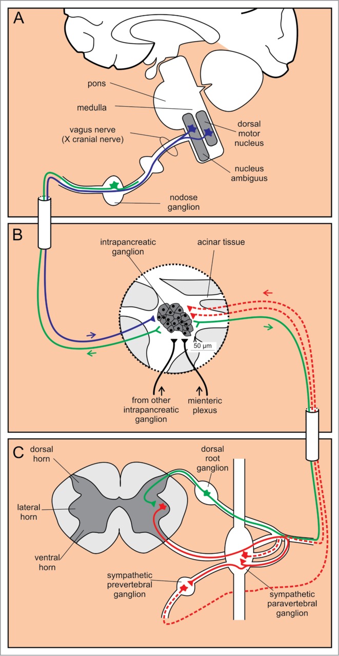
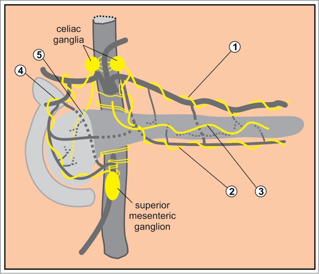
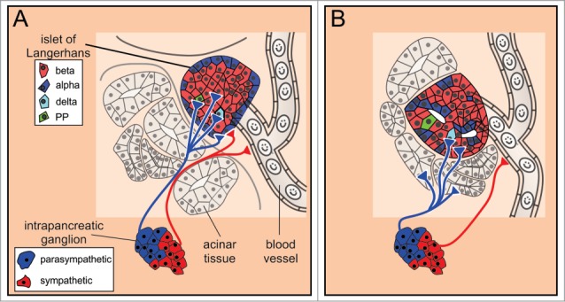
Mice remain the most studied animal model in pancreas research. Since the findings of this research are typically extrapolated to humans, it is important to understand both similarities and differences between the 2 species. Beside the apparent difference in size and macroscopic organization of the organ in the 2 species, there are a number of less evident and only recently described differences in organization of the acinar and ductal exocrine tissue, as well as in the distribution, composition, and architecture of the endocrine islets of Langerhans. Furthermore, the differences in arterial, venous, and lymphatic vessels, as well as innervation are potentially important. In this article, the structure of the human and the mouse pancreas, together with the similarities and differences between them are reviewed in detail in the light of conceivable repercussions for basic research and clinical application.

Keywords: anatomy; endocrine; exocrine; human pancreas; innervation; islet of Langerhans; mouse pancreas; structure; vasculature.


Figures

Figure 1.
Figure 1.
Macroscopic anatomy of the human and the mouse pancreas. (A) Human pancreas consists of the head, the body, and the tail. (B) Mouse pancreas has 3 lobes that are less well defined: the duodenal, the gastric, and the splenic lobe. Note the 10-fold difference in linear dimension (which accounts for the 1000-fold difference in the size of the organ as well as the organism). The color coding indicates homologous parts.
Figure 2.
Figure 2.
Microscopic anatomy of the human (A, C, and E) and the mouse (B, D, and F) pancreas. (A and B) Macroscopic anatomy of the human and the mouse pancreas, respectively. (C and D) Magnifying a portion of pancreas reveals larger lobules in humans when compared to mouse, whereas the islets of Langerhans are of fairly comparable size in humans and mice. (E and F) Cell composition and location of the islets of Langerhans within the pancreas are markedly different in the 2 species. Note the more diffusely distributed endocrine cells in humans (E) and the mantle-core pattern in mice (F).
Figure 3.
Figure 3.
Major blood vessels of the pancreas. (A) Arteries. 1 abdominal aorta, 2 celiac artery, 3 superior mesenteric artery, 4 common hepatic artery, 5 gastroduodenal artery. (B) Veins. 1 portal vein, 2 superior mesenteric vein.
Figure 4.
Figure 4.
Three possible patterns of islet microcirculation. (A) From periphery to center. (B) From center to periphery, blood reaches the β cells first. (C) From one pole to the other. Red color indicates arterial blood and blue color venous blood.
Figure 5.
Figure 5.
Lymph nodes of the pancreas. (A) Groups of lymph nodes that outline the gland. (B) Groups of nodes at the aorta. 8: hepatic nodes. 9: celiac nodes. 10: splenic and gastrosplenic nodes. 11: suprapancreatic nodes. 12: hepatoduodenal nodes. 13a: superior posterior nodes. 13b: inferior posterior nodes. 14: superior mesenteric nodes. 15: middle colic nodes. 16: paraaortic nodes. 17a: superior anterior nodes. 17b: inferior anterior nodes. 18: infrapancreatic nodes. Red dots on the top and at the bottom of lymph nodes indicate the nodes most frequently involved in carcinoma of the body or the tail, and the head, respectively.
Figure 6.
Figure 6.
Autonomic innervation of intrapancreatic ganglia. (A) Cell bodies of efferent parasympathetic fibers are located in 2 nuclei in the medulla, whereas the afferent sensory cell bodies are positioned in the nodose ganglion. (B) Intrapancreatic ganglia receive input from sympathetic (red) and parasympathetic (blue) fibers, from other intrapancreatic ganglia and from the myenteric plexus (black). Additionally, sensory fibers (green) project from intrapancreatic ganglia. (C) Preganglionic efferent sympathetic fibers (red solid line) project from cell bodies in the lateral horn of the spinal cord to paravertebral and prevertebral sympathetic ganglia. From here, the postganglionic sympathetic fibers (red dashed lines) project to the intrapancreatic ganglia. Sensory afferent fibers (green) have their cell bodies in the dorsal root ganglia.
Figure 7.
Figure 7.
Macroscopic autonomic innervation of the pancreas. 1: the splenic plexus accompanying the splenic artery; 2: the plexus accompanying the transverse pancreatic artery; 3: the plexus around the main pancreatic duct; 4: the anterior hepatic plexus; 5: the posterior hepatic plexus.
Figure 8.
Figure 8.
The autonomic innervation of islets of Langerhans. (A) In mice, the postganglionic sympathetic fibers contact α cells and smooth muscle cells of the blood vessels, whereas the parasympathetic fibers contact all endocrine cell types. (B) In humans, the islets are sparsely innervated. The sympathetic and parasympathetic postganglionic fibers preferentially contact vascular smooth muscle cells and exocrine tissue, respectively.

References

    1. Case RM. Is the rat pancreas an appropriate model of the human pancreas? Pancreatology 2006; 6:180-90; PMID:16534243; http://dx.doi.org/ 10.1159/000091849 - DOI - PubMed
    1. Levetan CS, Pierce SM. Distinctions between the islets of mice and men: implications for new therapies for type 1 and 2 diabetes. Endocr Pract 2013; 19:301-12; PMID:23186955; http://dx.doi.org/ 10.4158/EP12138.RA - DOI - PubMed
    1. Grouwels G, Vasylovska S, Olerud J, Leuckx G, Ngamjariyawat A, Yuchi Y, Jansson L, Van de Casteele M, Kozlova EN, Heimberg H. Differentiating neural crest stem cells induce proliferation of cultured rodent islet beta cells. Diabetologia 2012; 55:2016-25; PMID:22618811; http://dx.doi.org/ 10.1007/s00125-012-2542-0 - DOI - PubMed
    1. Rutter GA, Hodson DJ. Minireview: intraislet regulation of insulin secretion in humans. Mol Endocrinol 2013; 27:1984-95; PMID:24243488; http://dx.doi.org/ 10.1210/me.2013-1278 - DOI - PMC - PubMed
    1. Farnsworth NL, Benninger RKP. New insights into the role of connexins in pancreatic islet function and diabetes. FEBS Letters 2014; 588:1278-87; PMID:24583073; http://dx.doi.org/ 10.1016/j.febslet.2014.02.035 - DOI - PMC - PubMed

Publication types

LinkOut - more resources