. 2015 Jun 1;10(6):e0128561.
doi: 10.1371/journal.pone.0128561. eCollection 2015.

Cardiac metabolic pathways affected in the mouse model of barth syndrome

Affiliations

Cardiac metabolic pathways affected in the mouse model of barth syndrome

Yan Huang et al. PLoS One. .

Abstract

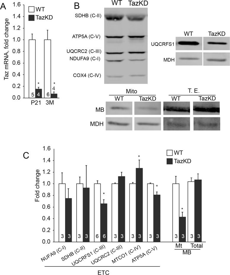
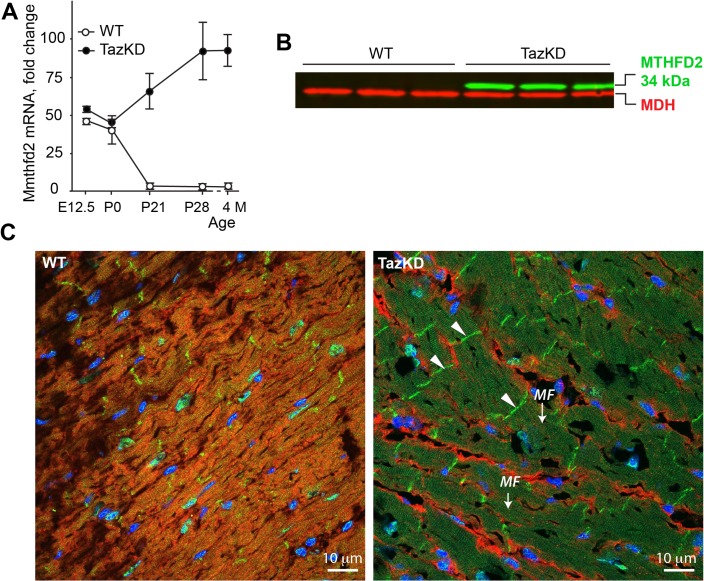
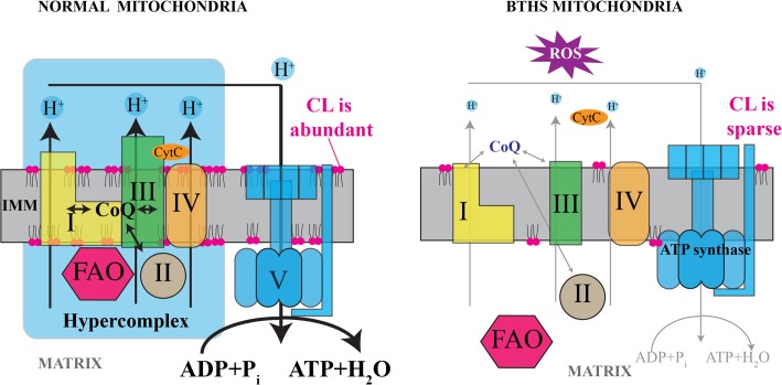
Cardiolipin (CL) is a mitochondrial phospholipid essential for electron transport chain (ETC) integrity. CL-deficiency in humans is caused by mutations in the tafazzin (Taz) gene and results in a multisystem pediatric disorder, Barth syndrome (BTHS). It has been reported that tafazzin deficiency destabilizes mitochondrial respiratory chain complexes and affects supercomplex assembly. The aim of this study was to investigate the impact of Taz-knockdown on the mitochondrial proteomic landscape and metabolic processes, such as stability of respiratory chain supercomplexes and their interactions with fatty acid oxidation enzymes in cardiac muscle. Proteomic analysis demonstrated reduction of several polypeptides of the mitochondrial respiratory chain, including Rieske and cytochrome c1 subunits of complex III, NADH dehydrogenase alpha subunit 5 of complex I and the catalytic core-forming subunit of F0F1-ATP synthase. Taz gene knockdown resulted in upregulation of enzymes of folate and amino acid metabolic pathways in heart mitochondria, demonstrating that Taz-deficiency causes substantive metabolic remodeling in cardiac muscle. Mitochondrial respiratory chain supercomplexes are destabilized in CL-depleted mitochondria from Taz knockdown hearts resulting in disruption of the interactions between ETC and the fatty acid oxidation enzymes, very long-chain acyl-CoA dehydrogenase and long-chain 3-hydroxyacyl-CoA dehydrogenase, potentially affecting the metabolic channeling of reducing equivalents between these two metabolic pathways. Mitochondria-bound myoglobin was significantly reduced in Taz-knockdown hearts, potentially disrupting intracellular oxygen delivery to the oxidative phosphorylation system. Our results identify the critical pathways affected by the Taz-deficiency in mitochondria and establish a future framework for development of therapeutic options for BTHS.


Conflict of interest statement

Competing Interests: The authors have declared that no competing interests exist.

Figures

Fig 1
Fig 1. iTRAQ workflow for differential protein profiling in mitochondria from 3-month-old wild type and TazKD mice.
(A) Sample preparation workflow from a preparative SDS-PAGE gel, trypsin digestion, iTRAQ labeling, fractionation by strong cation exchange, followed by nanoLC-MS/MS to produce both protein identification by peptide fragmentation data and relative quantitation from the iTRAQ reporter fragment ions. (B) The preparative SDS-PAGE gel with pre-stained molecular weight markers showing the gel regions collected for trypsin digestion. (C) Overlay of the nano-LC-MS total ion current (TIC) from each of the 20 SCX fractions.
Fig 2
Fig 2. (A) Representative 2D-DIGE image of differentially labeled proteins in mitochondria from 3-month-old wild type and TazKD mice.
Proteins with changes in expression levels of at least 1.5-fold are marked with red (decreased) and green (increased) circles. Spot numbers correspond to those in Table 1. (B) Top-rated network of proteins involved in lipid metabolism and associated with altered mitochondrial proteins in TazKD hearts. Shapes denote functional classes of proteins. Shapes in green depict down-regulated proteins; red-colored symbols indicate up-regulated proteins. Proteins in white are theoretically identified by Ingenuity Pathway Knowledge Base analysis. Solid and dashed lines depict direct and indirect interactions, respectively.
Fig 3
Fig 3. Quantitative analysis of mitochondrial ETC enzymes, myoglobin (MB) and MTHFD2 in cardiac mitochondria of TazKD mice.
(A) Quantification of Taz mRNA levels in hearts of 3 weeks old (P21) and 3 months old (3M) WT and TazKD mice. The expression levels were determined by quantitative RT-PCR and represented as relative normalized expression. (B) Representative Western blots of mitochondrial proteins from TazKD and WT hearts. Mitochondrial proteins (30 μg) were separated by SDS-electrophoresis and subjected to Western blot analysis using a cocktail of monoclonal antibodies specific to individual polypeptides of ETC complexes NDUFA9 (C-I), SDHB (C-II), UQCRC (C-III), COX4 (C-IV) and ATP5A (C-V). Additionally, blots were probed with antibodies specific to Rieske Fe-S cluster protein (UQCRFS1) and myoglobin (MB). Myoglobin content was analyzed in mitochondrial fraction (Mito) and the total cardiac tissue protein extract (T.E.) fractions. Antibodies specific to mitochondrial malate dehydrogenase (MDH) were used as a loading control. The entire (uncropped) images of Western blots are shown in S1 Fig. (C) Quantitative assessments of Western blot analyses. Bars represent fold-changes in band intensities in TazKD samples compared to WT controls. For A and C: Data shown are means ± standard error of mean (SEM). Number of experiments per group is denoted in the corresponding bar. Asterisks indicate significant differences of P≤0.05 (Student’s t-test) between WT and TazKD.
Fig 4
Fig 4. Evidence of destabilization of mitochondrial supercomplexes in cardiolipin-depleted cardiac mitochondria from TazKD mice.
(A and D) Mitochondria from WT and TazKD hearts were solubilized with digitonin and subjected to blue-native gel electrophoresis (BNGE, first dimension) and then to SDS-gel electrophoresis under denaturing conditions (SDS-PAGE, second dimension). Gels were subjected to western blot analyses with an antibody cocktail specific to subunits of mitochondrial ETC complexes (Anti-OxPhos Cocktail). Bands representing the individual polypeptides that correspond to ETC complexes are marked. In a separate set of experiments, mitochondria from WT (B and C) and TazKD (E and F) hearts were solubilized with lauryl maltoside and subjected to sucrose gradient ultracentrifugation. Individual fractions of the gradient were subjected to western blot analyses using Anti-OxPhos Cocktail (B and E) or FAO enzyme-specific antibodies LCHAD, VLCAD and MCAD (C and F). P—pellet; I—input material (i.e. the solubilized mitochondria preparation that was applied to the sucrose gradient). The scales on the right side depict molecular masses in kilodaltons (kDa).
Fig 5
Fig 5. Altered Mthfd2 expression in adult TazKD heart.
(A) Quantification of Mthfd2 mRNA levels in hearts of WT and TazKD mice during development. (B) Immunoblots of total protein extracts from 3 month-old WT and TazKD hearts with anti-MTHFD2 antibodies (green). Antibodies specific to mitochondrial malate dehydrogenase (MDH) were used as a loading control (red). (C) Immunofluorescent staining of MTHFD2 in LV sections of 3 month-old WT and TazKD mice revealing localization of MTHFD2 protein (green) in intercalated disks (arrowheads) and myofilaments (MF). Mitochondria were visualized with antibodies specific to complex I subunit NDUFA9 (red). Nuclei were stained with DAPI (blue).
Fig 6
Fig 6. Structures of mitochondrial respiratory chain complexes that are affected in TazKD mice.
Polypeptides that are quantitatively reduced in TazKD cardiac mitochondria are highlighted with colors. Structurally integrated cardiolipin (CL) molecules within complex III are highlighted in yellow.
Fig 7
Fig 7. Hypothetical pathophysiological mechanism of Barth syndrome.
In normal mitochondria with high content of CL in the IMM, respiratory complexes are assembled into supercomplexes and form even higher-order structures (hypercomplexes) by interacting with FAO enzymes. In contrast, in CL-depleted mitochondria from BTHS hearts, supercomplexes are destabilized, and the relative quantities of NDUFA5 (C-I), SDHA (C-II), UQCRFS1 and CYC1 (C-III) and ATPA (C-V) are reduced. Defects in respiratory chain complexes would likely induce overproduction of reactive oxygen species (ROS). In CL-deficient mitochondria, disassociation of FAO enzymes from RC complexes would reduce the efficiency of metabolic channeling through these pathways. Reduced oxygen availability at the outer mitochondrial membrane (OMM) due to low myoglobin binding to mitochondria would further diminish aerobic ATP synthesis, but might also reduce generation of ROS.

References

    1. Schlame M, Ren M, Xu Y, Greenberg ML, Haller I. Molecular symmetry in mitochondrial cardiolipins. Chemistry and physics of lipids. 2005;138(1–2):38–49. . - PubMed
    1. Chicco AJ, Sparagna GC. Role of cardiolipin alterations in mitochondrial dysfunction and disease. Am J Physiol Cell Physiol. 2007;292(1):C33–44. . - PubMed
    1. Bolhuis PA, Hensels GW, Hulsebos TJ, Baas F, Barth PG. Mapping of the locus for X-linked cardioskeletal myopathy with neutropenia and abnormal mitochondria (Barth syndrome) to Xq28. American journal of human genetics. 1991;48(3):481–5. . - PMC - PubMed
    1. Vreken P, Valianpour F, Nijtmans LG, Grivell LA, Plecko B, Wanders RJ, et al. Defective remodeling of cardiolipin and phosphatidylglycerol in Barth syndrome. Biochem Biophys Res Commun. 2000;279(2):378–82. . - PubMed
    1. Houtkooper RH, Rodenburg RJ, Thiels C, van Lenthe H, Stet F, Poll-The BT, et al. Cardiolipin and monolysocardiolipin analysis in fibroblasts, lymphocytes, and tissues using high-performance liquid chromatography-mass spectrometry as a diagnostic test for Barth syndrome. Anal Biochem. 2009;387(2):230–7. Epub 2009/05/21. doi: S0003-2697(09)00060-8 [pii] 10.1016/j.ab.2009.01.032 . - DOI - PubMed

Publication types

MeSH terms

LinkOut - more resources