. 2015 Jun 1;10(6):e0128431.
doi: 10.1371/journal.pone.0128431. eCollection 2015.

Group B streptococcal infection and activation of human astrocytes

Affiliations

Group B streptococcal infection and activation of human astrocytes

Terri D Stoner et al. PLoS One. .

Abstract

Background: Streptococcus agalactiae (Group B Streptococcus, GBS) is the leading cause of life-threatening meningitis in human newborns in industrialized countries. Meningitis results from neonatal infection that occurs when GBS leaves the bloodstream (bacteremia), crosses the blood-brain barrier (BBB), and enters the central nervous system (CNS), where the bacteria contact the meninges. Although GBS is known to invade the BBB, subsequent interaction with astrocytes that physically associate with brain endothelium has not been well studied.

Methodology/principal findings: We hypothesize that human astrocytes play a unique role in GBS infection and contribute to the development of meningitis. To address this, we used a well- characterized human fetal astrocyte cell line, SVG-A, and examined GBS infection in vitro. We observed that all GBS strains of representative clinically dominant serotypes (Ia, Ib, III, and V) were able to adhere to and invade astrocytes. Cellular invasion was dependent on host actin cytoskeleton rearrangements, and was specific to GBS as Streptococcus gordonii failed to enter astrocytes. Analysis of isogenic mutant GBS strains deficient in various cell surface organelles showed that anchored LTA, serine-rich repeat protein (Srr1) and fibronectin binding (SfbA) proteins all contribute to host cell internalization. Wild-type GBS also displayed an ability to persist and survive within an intracellular compartment for at least 12 h following invasion. Moreover, GBS infection resulted in increased astrocyte transcription of interleukin (IL)-1β, IL-6 and VEGF.

Conclusions/significance: This study has further characterized the interaction of GBS with human astrocytes, and has identified the importance of specific virulence factors in these interactions. Understanding the role of astrocytes during GBS infection will provide important information regarding BBB disruption and the development of neonatal meningitis.


Conflict of interest statement

Competing Interests: The authors have declared that no competing interests exist.

Figures

Fig 1
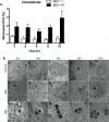
Fig 1. Interaction of GBS with human SVG-A astrocytes.
A, confluent monolayers of SVG-A cells were infected with GBS strain COH1 at the indicated MOI for 30 minutes for adhesion and 2 h for invasion assays and the extent of bacterial adherence and invasion were quantified as described in Materials and Methods. The data are expressed as the percentage of recovered CFU/initial inoculum. All experiments were performed in triplicate and repeated in at least three independent experiments. B, SVG-A cells were infected with COH1 at an MOI of 1 and 10 for 24 hours and percent cell viability determined by trypan blue dye exclusion and compared to an uninfected control. C, SVG-A cells were pretreated in media alone, DMSO vehicle control or cytochalasin D (Cyt D) at 1 to 5 μg/ml for 30 min at 37°C and 5% CO2. Cells were then incubated with COH1 at an MOI of 1 for 2 h and intracellular GBS was quantified. The data shown represents the percentage of bacterial uptake after cytochalasin D treatment relative to untreated control. Significance was determined by one-way ANOVA using tukeys multiple comparison test, P < 0.0001 (****). D, Transmission electron microscopic (TEM) images of SVG-A cells that were either uninfected (control) or infected with GBS COH1 at an MOI of 10. For TEM scale bars for panels A, B, D, and E are 2um and for C and F are 500nm.
Fig 2
Fig 2. GBS adhesion, invasion and intracellular survival of SVG-A cells.
A and B, Confluent monolayers of SVG-A cells were incubated with GBS, COH1 wild-type strain, at an MOI of 1 for various times at 37°C, 5% CO2 and adhesion and invasion was examined as described in Materials and Methods. The data shown are a representative of 3 independent experiments performed in triplicate. Incubation at shorter and longer times resulted in a similar level of adhesion and invasion of COH1 to SVG-A cells and was not statistically significant as determined by one-way ANOVA and Tukey's multiple comparison tests. C, SVG-A cells were infected with COH1 expressing GFP at an MOI of 10 and incubated for 8 h at 37°C. Cells were fixed and processed for fluorescence microscopy and stained with Cell Mask (red) to visualize the plasma membrane or with DAPI (blue) to visualize the nucleus. The image is representative of many images from 2 independent experiments.
Fig 3
Fig 3. Clinically relevant isolates of GBS adhere and invade, but non-GBS, bacteria fail to invade human astrocytes.
A and B, Wild-type GBS serotypes (Ia, Ib, III, and V), were incubated with confluent SVG-A cells at an MOI of 1 for 30 minutes for adhesion and 2 h for invasion assays and the levels of both surface-adherence and invasion were quantified. These data are expressed as the relative percentage of clinical isolates compared to COH1, a representative experiment of at least 3 independent experiments performed in triplicate is shown. C, SVG-A cells grown to confluence were infected with GBS COH1 at an MOI of 1 or the non-GBS strain, S. gordonii for 30 min for adhesion or 2 h for invasion assays. An unpaired, two tailed t-test with Welch’s correction was used to compare S. gordonii to COH1. Data are expressed as the relative percent adherence or invasion and are representative of 3 independent experiments performed in triplicate or quadruplicate.
Fig 4
Fig 4. Role of GBS surface determinants in SVG-A interaction.
A and B, SVG-A cells grown to confluence were infected at an MOI of 1 with wild type strains COH1 or NCTC10/84 and respective isogenic mutants. SVG-A cells were infected for 30 min for adhesion or 2 h for invasion assays, bacteria were recovered and quantified as described in Materials and Methods. A representative experiment of at least 3 independent experiments performed in triplicate is shown. Data are expressed as the percentage of recovered CFU for GBS mutants relative to that of the wild type parental strain. Data were analyzed by unpaired, two-tailed t-test. (*) = P< 0.05, (**), P< 0.01 and (****), P< 0.0001.
Fig 5
Fig 5. Intracellular survival of wild-type GBS in human astrocytes.
A, Wild-type GBS, COH1, intracellular survival assays in SVG-A cells were carried out for up to 12 hr at MOI of 1 and 10. To assess intracellular survival in SVG-A cells over time, intracellular bacteria were enumerated after the addition of antibiotics (gentamicin (50 μg/ml) and penicillin (25 μg/ml) from 2 to 12 h time points. B, Transmission electron micrographs demonstrating GBS (COH1) intracellular survival over time in SVG-A cells at magnifications (6,000X for A-J, and 30,000X for K-O). All assays were performed in triplicate or quadruplet and repeated at least three times.
Fig 6
Fig 6. Activation of human astrocytes following GBS infection.
A-D, Transcript abundance of IL-1β, IL-6, VEGF, and IL-8 in SVG-A cells was determined using quantitative RT-PCR following infection with GBS COH1 (MOI = 50). Fold change was calculated using GAPDH and then normalized to media controls as described in Materials and Methods. E, Protein production of IL-8 was determined by ELISA as described in Materials and Methods. Data is one representative experiment of at least 2 independent experiments performed in a minimum of 4 replicates. Data was analyzed by two-way ANOVA with Bonferroni’s multiple comparisons post-test. (*) = P< 0.05, (***) = P< 0.001, (****) = P< 0.0001.

References

    1. Heath PT, Balfour G, Weisner AM, Efstratiou A, Lamagni TL, Tighe H, et al. Group B streptococcal disease in UK and Irish infants younger than 90 days. The Lancet. 2004;363(9405):292–4. 10.1016/s0140-6736(03)15389-5 - DOI - PubMed
    1. Thigpen MC. Bacterial Meningitis in the United States, 1998–2007. The New England Journal of Medicine. 2011;364(21):2016–25. 10.1056/NEJMoa1005384 - DOI - PubMed
    1. Nizet V, Kim KS, Stins M, Jonas M, Chi EY, Nguyen D, et al. Invasion of brain microvascular endothelial cells by group B streptococci. Infection and Immunity. 1997;65(12):5074–81. - PMC - PubMed
    1. Chin K, Fitzhhardinge P. Sequelae of early-onset group B hemolytic streptococcal neonatal meningitis. J Pediatr. 1985;106(5):819–22. - PubMed
    1. Edwards MS RM, Haffar AA, Murphy MA, Desmond MM, Baker CJ. Long-term sequelae of group B streptococcal meningitis in infants. J Pediatr 1985;106(5):717–22. - PubMed

Publication types

MeSH terms