. 2015 Jul;18(7):1051-7.
doi: 10.1038/nn.4035. Epub 2015 Jun 1.

β-amyloid disrupts human NREM slow waves and related hippocampus-dependent memory consolidation

Affiliations

β-amyloid disrupts human NREM slow waves and related hippocampus-dependent memory consolidation

Bryce A Mander et al. Nat Neurosci. 2015 Jul.

Abstract

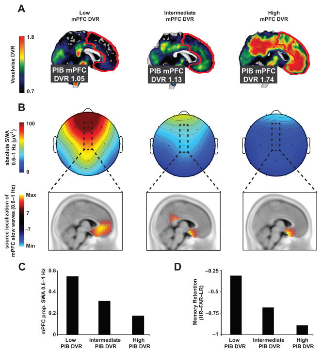
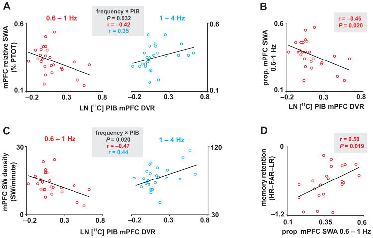
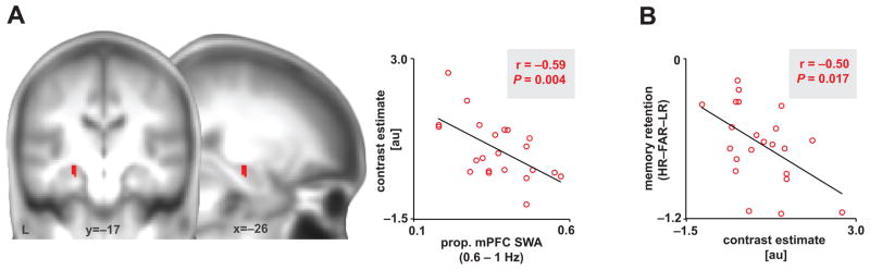
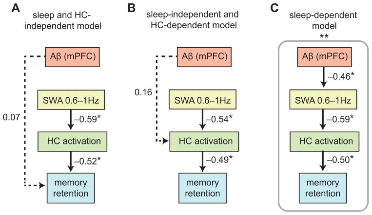
Independent evidence associates β-amyloid pathology with both non-rapid eye movement (NREM) sleep disruption and memory impairment in older adults. However, whether the influence of β-amyloid pathology on hippocampus-dependent memory is, in part, driven by impairments of NREM slow wave activity (SWA) and associated overnight memory consolidation is unknown. Here we show that β-amyloid burden in medial prefrontal cortex (mPFC) correlates significantly with the severity of impairment in NREM SWA generation. Moreover, reduced NREM SWA generation was further associated with impaired overnight memory consolidation and impoverished hippocampal-neocortical memory transformation. Furthermore, structural equation models revealed that the association between mPFC β-amyloid pathology and impaired hippocampus-dependent memory consolidation was not direct, but instead statistically depended on the intermediary factor of diminished NREM SWA. By linking β-amyloid pathology with impaired NREM SWA, these data implicate sleep disruption as a mechanistic pathway through which β-amyloid pathology may contribute to hippocampus-dependent cognitive decline in the elderly.


Figures

Figure 1
Figure 1. Aβ, NREM SWA, and memory retention measures in three sample subjects
[11C]PIB-PET DVR images demonstrating Aβ deposition (a), NREM SWA and associated localized slow wave source (b), proportion of NREM SWA 0.6–1Hz at FZ&CZ derivations (c), and overnight memory retention (long delay recognition testing – short delay recognition testing; d) measures in three sample participants with low mPFC PIB DVR (left column), intermediate mPFC PIB DVR (middle column), and high mPFC PIB DVR (right column). PIB-PET mPFC region of interest (ROI) is outlined in white (a) and the mPFC EEG derivations are outlined in black (b), with accompanying source analysis (thresholded at ±7) verifying mPFC overlap across PIB-PET and EEG ROIs (see Methods and Supplementary Fig. 2). Prop. denotes proportion, PIB denotes Pittsburgh compound B, PET denotes positron emission tomography, DVR denotes distribution volume ratio referenced against the whole cerebellum, mPFC denotes medial prefrontal cortex, and [HR–FAR–LR] denotes [hit rate to originally studied word pairs – false alarm rate to new, unstudied words – false alarm rate to originally studied word pairs].
Figure 2
Figure 2. Associations between Aβ, NREM SWA, and memory retention measures
Associations between LN transformed [11C]PIB-PET DVR measured mPFC Aβ deposition, mPFC relative SWA, mPFC SW density, and overnight memory retention. Interaction plots of two-way, repeated measures ANCOVAs, which revealed that Aβ burden was associated with lower relative mPFC NREM SWA and SW density 0.6–1Hz and higher mPFC NREM SWA and SW density at 1–4Hz (parameter estimates for each frequency bin plotted in a) for SWA and c) for SW density. mPFC Aβ burden was also negatively associated with proportion of mPFC NREM SWA 0.6–1Hz (b). mPFC NREM SWA 0.6–1Hz, in turn, positively predicted overnight memory retention (d). %PTOT denotes percentage of total spectral power (0.6–50Hz), Prop. denotes proportion, PIB denotes Pittsburgh compound B, PET denotes positron emission tomography, DVR denotes distribution volume ratio referenced against the cerebellum, mPFC denotes medial prefrontal cortex, and [HR–FAR–LR] denotes [hit rate to originally studied word pairs – false alarm rate to new, unstudied words – false alarm rate to originally studied word pairs].
Figure 3
Figure 3. Associations between NREM SWA, retrieval-related hippocampus activation, and memory retention
(a) Negative association between proportion of mPFC SWA 0.6–1Hz and left hippocampal activation greater during successful associative episodic retrieval than correct rejection of novel words (Hits-Correct Rejections); 8mm-sphere ROI: [x=−22, y=−14, z=−12; x=−23, y=−15, z=−16 in mni coordinates]. Activations were inclusively masked by hippocampal anatomy and displayed and considered significant at the voxel level of P<0.05 family-wise error (FWE) corrected for multiple comparisons within the a priori hippocampal region of interest. Peak effects were detected at [x=−24, y=−16, z=−14]. Hot colors represent the extent of the negative association between hippocampal activation and proportion of SWA 0.6–1Hz. (b) Negative association between overnight memory retention and the average contrast estimate of significant hippocampal voxels, extracted using marsbar. au denotes arbitrary units, prop. denotes proportion, and [HR–FAR–LR] denotes [hit rate to originally studied word pairs – false alarm rate to new, unstudied words – false alarm rate to originally studied word pairs].
Figure 4
Figure 4. Path models linking Aβ, NREM SWA, retrieval-related hippocampus activation, and memory retention
Path analysis models examining the relative contributions of [11C]PIB-PET DVR measured mPFC β-amyloid (Aβ) deposition, proportion of mPFC NREM SWA 0.6–1Hz, and retrieval-related hippocampal (HC) activation to overnight memory retention (long delay recognition testing — short delay recognition testing) in three hypothesized models (a–c). Values represent standardized regression weights. Models were estimated and model fit for the sleep and HC-independent model (a, BIC = 29.640; RMR = 0.021; GFI = 0.858), the sleep-independent and HC-dependent model (b, BIC = 29.131; RMR = 0.021; GFI = 0.873), and the sleep-dependent model (c, BIC = 24.676; RMR = 0.006; GFI = 0.931) were compared against a saturated model (BIC = 30.910; RMR = 0.000; GFI = 1.000), and an independence model (BIC = 30.747; RMR = 0.046; GFI = 0.617). * denotes path significance at P<0.05.

Comment in

References

    1. Boyle PA, et al. Much of late life cognitive decline is not due to common neurodegenerative pathologies. Ann Neurol. 2013 - PMC - PubMed
    1. Buckner RL, et al. Molecular, structural, and functional characterization of Alzheimer’s disease: evidence for a relationship between default activity, amyloid, and memory. J Neurosci. 2005;25:7709–7717. - PMC - PubMed
    1. Jack CR, Jr, et al. Lancet Neurol. 2010;9:119–128. - PMC - PubMed
    1. Sepulcre J, Sabuncu MR, Becker A, Sperling R, Johnson KA. In vivo characterization of the early states of the amyloid-beta network. Brain. 2013;136:2239–2252. - PMC - PubMed
    1. Spires-Jones TL, Hyman BT. The intersection of amyloid beta and tau at synapses in Alzheimer’s disease. Neuron. 2014;82:756–771. - PMC - PubMed

Publication types

Substances