. 2015 Sep:86:191-9.
doi: 10.1016/j.freeradbiomed.2015.05.029. Epub 2015 May 29.

Carbon monoxide modulates cytochrome oxidase activity and oxidative stress in the developing murine brain during isoflurane exposure

Affiliations

Carbon monoxide modulates cytochrome oxidase activity and oxidative stress in the developing murine brain during isoflurane exposure

Ying Cheng et al. Free Radic Biol Med. 2015 Sep.

Abstract

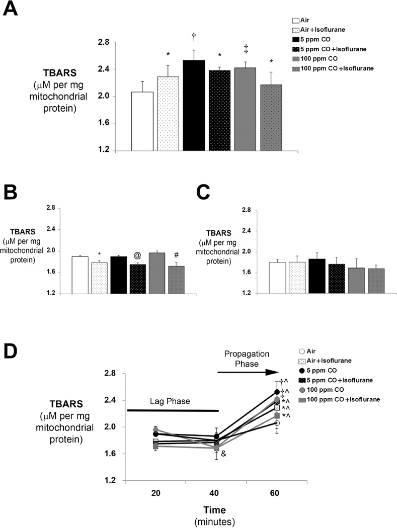
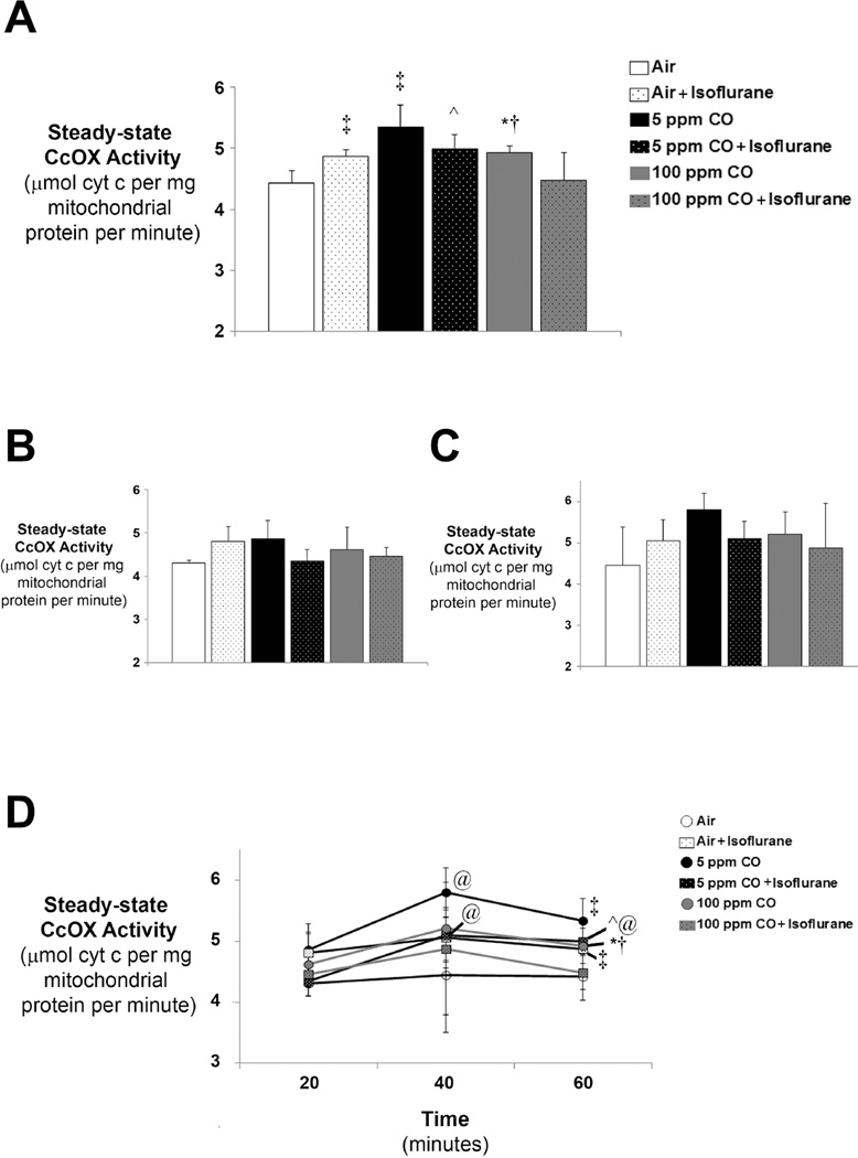
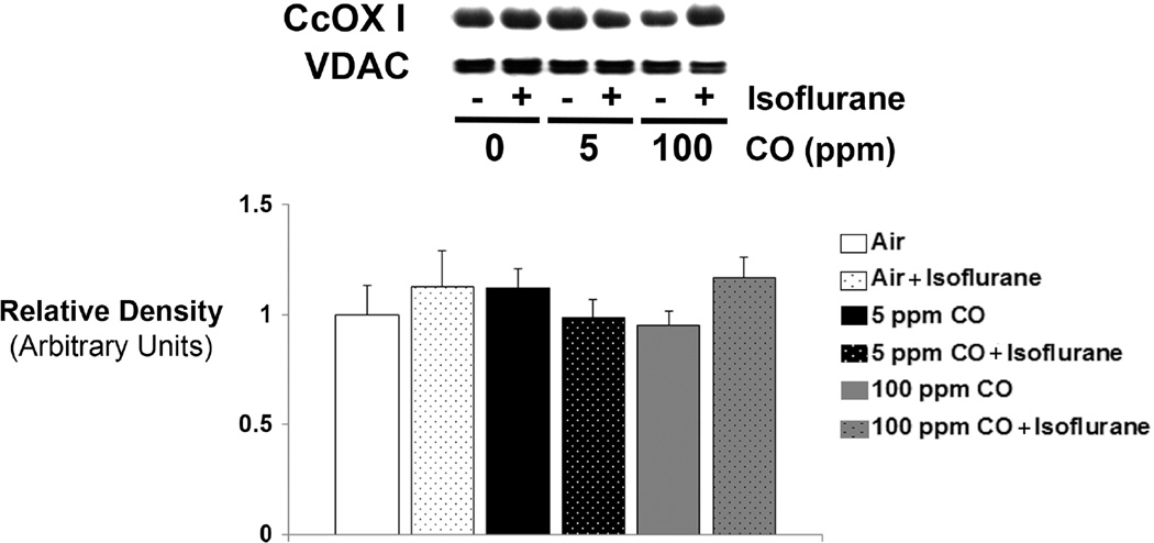
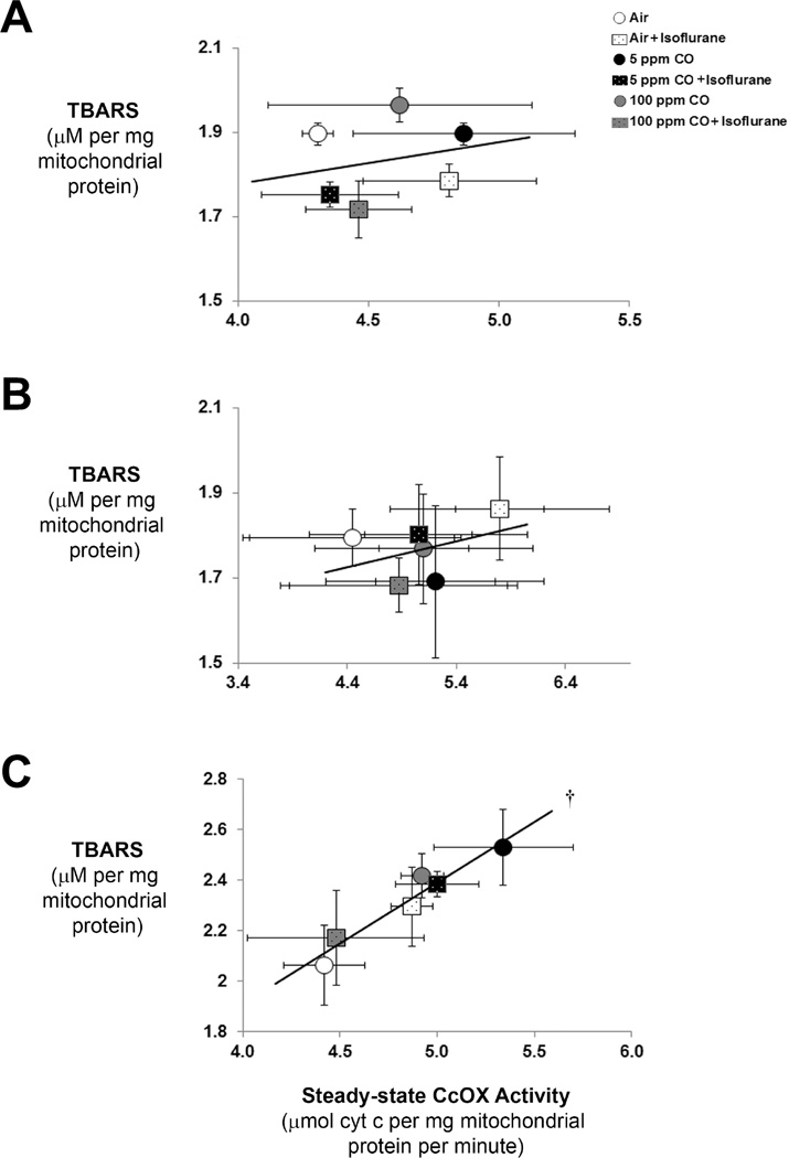
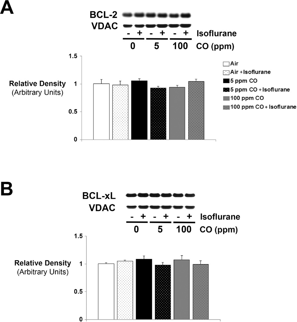
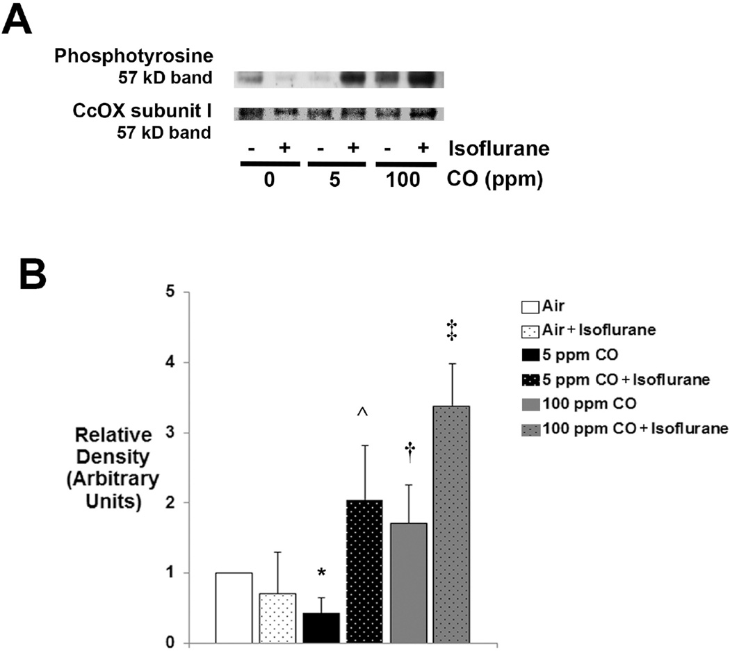
Commonly used anesthetics induce widespread neuronal degeneration in the developing mammalian brain via the oxidative-stress-associated mitochondrial apoptosis pathway. Dysregulation of cytochrome oxidase (CcOX), the terminal oxidase of the electron transport chain, can result in reactive oxygen species (ROS) formation. Isoflurane has previously been shown to activate this enzyme. Carbon monoxide (CO), as a modulator of CcOX, is of interest because infants and children are routinely exposed to CO during low-flow anesthesia. We have recently demonstrated that low concentrations of CO limit and prevent isoflurane-induced neurotoxicity in the forebrains of newborn mice in a dose-dependent manner. However, the effect of CO on CcOX in the context of anesthetic-induced oxidative stress is unknown. Seven-day-old male CD-1 mice underwent 1h exposure to 0 (air), 5, or 100ppm CO in air with or without isoflurane. Exposure to isoflurane or CO independently increased CcOX kinetic activity and increased ROS within forebrain mitochondria. However, exposure to CO combined with isoflurane paradoxically limited CcOX activation and oxidative stress. There were no changes seen in steady-state levels of CcOX I protein, indicating post-translational modification of CcOX as an etiology for changes in enzyme activity. CO exposure led to differential effects on CcOX subunit I tyrosine phosphorylation depending on concentration, while combined exposure to isoflurane with CO markedly increased the enzyme phosphorylation state. Phosphorylation of tyrosine 304 of CcOX subunit I has been shown to result in strong enzyme inhibition, and the relative reduction in CcOX kinetics following exposure to CO combined with isoflurane may have been due, in part, to such phosphorylation. Taken together, the data suggest that CO modulates CcOX in the developing brain during isoflurane exposure, thereby limiting oxidative stress. These CO-mediated effects could have implications for the development of low-flow anesthesia in infants and children to prevent anesthesia-induced oxidative stress.

Keywords: Anesthesia; Brain; Carbon monoxide; Cytochrome oxidase; Development; Isoflurane; Neurotoxicity; Oxidative stress; Phosphorylation; Reactive oxygen species.


Figures

Figure 1
Figure 1. Thiobarbituric acid reactive substances (TBARS) in forebrain mitochondria following exposure to carbon monoxide (CO) with or without isoflurane
Levels of TBARS were determined with a colorimetric assay in mitochondria isolated immediately following exposure. Values from the six experimental cohorts are expressed as means plus standard deviation. (A) TBARS following 1-hour exposure. N = 6 animals per cohort. (B) TBARS following 20 minute exposure. N = 3 animals per cohort. (C) TBARS following 40 minute exposure. N = 3 animals per cohort. (D) TBARS over time. Statistical significance was determined at each time point with two-way ANOVA using post hoc Tukey’s test. One-way ANOVA was utilized to assess significance within each experimental cohort over time. *P < 0.05 vs. CO (or air) matched exposure without isoflurane at same time point. †P < 0.001 vs. air-exposed control at same time point. ‡P < 0.005 vs. air-exposed control at same time point. @P < 0.005 vs. CO matched exposure without isoflurane at same time point. #P < 0.01 vs. CO matched exposure without isoflurane at same time point. ^P < 0.01 vs. cohort matched exposure at 20 and 40 minutes. &P < 0.05 100 ppm CO at 40 minutes vs. cohort matched exposure at 20 minutes.
Figure 2
Figure 2. Cytochrome oxidase (CcOX) kinetic activity following exposure to CO with or without isoflurane
Steady-state CcOX specific activity was measured in isolated mitochondria immediately after exposure. Values are expressed as means plus standard deviation. (A) CcOX kinetics following 1-hour exposure. N = 6 animals per cohort. (B) CcOX kinetics following 20 minute exposure. N = 3 animals per cohort. (C) CcOX kinetics following 40 minute exposure. N = 3 animals per cohort. Statistical significance was determined at each time point with two-way ANOVA using post hoc Tukey’s test. One-way ANOVA was utilized to assess significance within each experimental cohort over time. *P < 0.05 vs. 100 ppm CO + isoflurane, 5 ppm CO at the same time point. ^P < 0.05 vs. 100 ppm CO + isoflurane at the same time point. †P < 0.01 vs. air-exposed control at the same time point. ‡P < 0.001 vs. air-exposed control at the same time point. @P < 0.05 vs. cohort matched exposure at 20 minutes.
Figure 3
Figure 3. Cytochrome oxidase (CcOX) expression following 1-hour exposure to CO with or without isoflurane
A representative immunoblot of steady-state levels CcOX subunit I protein is depicted. Concentration of CO exposure (or air [0 ppm]) with (+) or without (−) isoflurane is indicated. VDAC was used as the mitochondrial protein loading control. Graphical representation of relative densities is shown below the blot. Values were normalized to VDAC density and are expressed as means plus standard deviation. Air-exposed control values were set arbitrarily to 1. N = 3 animals per cohort. Statistical significance was determined with two-way ANOVA using post hoc Tukey’s test.
Figure 4
Figure 4. Correlation between thiobarbituric acid reactive substances (TBARS) and cytochrome oxidase (CcOX) kinetic activity
Linear regression analysis of level of TBARS with CcOX specific activity was performed. Pearson correlation coefficients and r-values were calculated. Values are expressed as means +/− standard deviation. (A) Correlation following 20 minute exposure. N = 3 animals per cohort. Pearson correlation coefficient is 0.06 and r-value is 0.24. P = 0.65. (B) Correlation following 40 minute exposure. N = 3 animals per cohort. Pearson correlation coefficient is 0.15 and r-value is 0.38. P = 0.45. (C) Correlation following 1-hour exposure. N = 6 animals per cohort. Pearson correlation coefficient is 0.94 and r-value is 0.97. †P < 0.002.
Figure 5
Figure 5. Expression of BCL-2 and BCL-xL following exposure to CO with or without isoflurane
Steady-state levels of (A) BCL-2 and (B) BCL-xL protein were determined. Representative immunoblots are depicted. Concentration of CO exposure (or air [0 ppm]) with (+) or without (−) isoflurane is indicated. VDAC was used as the mitochondrial protein loading control. Graphical representations of relative densities are shown below the blots. Values were normalized to VDAC density and are expressed as means plus standard deviation. Air-exposed control values were set arbitrarily to 1. N = 3 animals per cohort. Statistical significance was determined with two-way ANOVA using post hoc Tukey’s test.
Figure 6
Figure 6. Tyrosine phosphorylation of cytochrome oxidase (CcOX) subunit I
CcOX was extracted from isolated mitochondria and steady-state levels of phosphotyrosine were determined. (A) A representative immunoblot of the 57 kD band is depicted. Concentration of CO exposure (or air [0 ppm]) with (+) or without (−) isoflurane is indicated. CcOX subunit I (57 kD) staining with Coomassie dye was used as the loading control. (B) Graphical representation of relative densities is shown. Values are expressed as means plus standard deviation. Air-exposed control values were set arbitrarily to 1. N = 3 animals per cohort. Statistical significance was determined with two-way ANOVA using post hoc Tukey’s test. *P < 0.05 vs. air-exposed control. ^P < 0.025 vs. 5 ppm CO. †P < 0.005 vs. 5 ppm CO. ‡P < 0.005 vs. air + isoflurane.

References

    1. Jevtovic-Todorovic V, Hartman RE, Izumi Y, Benshoff ND, Dikranian K, Zorumski CF, Olney JW, Wozniak DF. Early exposure to common anesthetic agents causes widespread neurodegeneration in the developing rat brain and persistent learning deficits. J Neurosci. 2003;23:876–882. - PMC - PubMed
    1. Stefovska VG, Uckermann O, Czuczwar M, Smitka M, Czuczwar P, Kis J, Kaindl AM, Turski L, Turski WA, Ikonomidou C. Sedative and anticonvulsant drugs suppress postnatal neurogenesis. Ann Neurol. 2008;64:434–445. - PubMed
    1. Istaphanous GK, Loepke AW. General anesthetics and the developing brain. Curr Opin Anaesthesiol. 2009;22:368–373. - PubMed
    1. Brambrink AM, Evers AS, Avidan MS, Farber NB, Smith DJ, Zhang X, Dissen GA, Creeley CE, Olney JW. Isoflurane-induced Neuroapoptosis in the Neonatal Rhesus Macaque Brain. Anesthesiology. 2010;112:834–841. - PMC - PubMed
    1. Istaphanous GK, Howard J, Nan X, Hughes EA, McCann JC, McAuliffe JJ, Danzer SC, Loepke AW. Comparison of the Neuroapoptotic Properties of Equipotent Anesthetic Concentrations of Desflurane, Isoflurane, or Sevoflurane in Neonatal Mice. Anesthesiology. 2011;114:578–587. - PubMed

Publication types

MeSH terms

LinkOut - more resources