. 2015 Sep;146(1):100-12.
doi: 10.1111/imm.12486.

Interleukin-10 plays a key role in the modulation of neutrophils recruitment and lung inflammation during infection by Streptococcus pneumoniae

Affiliations

Interleukin-10 plays a key role in the modulation of neutrophils recruitment and lung inflammation during infection by Streptococcus pneumoniae

Hernán F Peñaloza et al. Immunology. 2015 Sep.

Abstract

Streptococcus pneumoniae is a major aetiological agent of pneumonia worldwide, as well as otitis media, sinusitis, meningitis and sepsis. Recent reports have suggested that inflammation of lungs due to S. pneumoniae infection promotes bacterial dissemination and severe disease. However, the contribution of anti-inflammatory molecules to the pathogenesis of S. pneumoniae remains unknown. To elucidate whether the production of the anti-inflammatory cytokine interleukin-10 (IL-10) is beneficial or detrimental for the host during pneumococcal pneumonia, we performed S. pneumoniae infections in mice lacking IL-10 (IL-10(-/-) mice). The IL-10(-/-) mice showed increased mortality, higher expression of pro-inflammatory cytokines, and an exacerbated recruitment of neutrophils into the lungs after S. pneumoniae infection. However, IL-10(-/-) mice showed significantly lower bacterial loads in lungs, spleen, brain and blood, when compared with mice that produced this cytokine. Our results support the notion that production of IL-10 during S. pneumoniae infection modulates the expression of pro-inflammatory cytokines and the infiltration of neutrophils into the lungs. This feature of IL-10 is important to avoid excessive inflammation of tissues and to improve host survival, even though bacterial dissemination is less efficient in the absence of this cytokine.

Keywords: bacterial infection; cytokines; lung inflammation; neutrophils; systemic bacterial infection.


Figures

Figure 1
Figure 1
S. pneumoniae infection induces IL-10 production in lungs at 48 hr post-infection (hpi) (a) Wild-type (WT) mice were intranasally infected with 3 × 107 CFU of S. pneumoniae D39, 24 and 48 hpi total lung RNA and proteins were obtained. Quantification of Il10 mRNA was performed by quantitative PCR, using Taqman probes. The β2-microglobulin gene was used as endogenous control. Results are relative to uninfected WT mice. *P < 0·05 by Kruskal–Wallis test with a posteriori Dunn multiple comparison test. (b) IL-10 in lungs was detected before infection and at 24 and 48 hpi in WT mice by the Mouse ELISA Duoset (R&D Systems, Minneapolis, MN). *P < 0·05 by one-way analysis of variance test with a posteriori Tukey multiple comparison test.
Figure 2
Figure 2
Interleukin-10 knock out mice (IL-10−/−) are more susceptible to suffer a severe S. pneumoniae infection in a dose-dependent manner. Wild-type (WT) and IL-10−/− mice were intranasally infected with (a) 3 × 105, (b) 3 × 106 and (c) 3 × 107 CFU of S. pneumoniae D39, and survival rate (left panels), body weight (middle panels) and clinical score (right panels) were recorded on a daily basis for 10 days. For dead mice, maximum score value was considered. *P < 0·05, log rank test.
Figure 3
Figure 3
Pro-inflammatory cytokine expression in lungs of wild-type (WT) and interleukin-10-knock out mice (IL-10−/−) after S. pneumoniae infection. WT and IL-10−/− mice were intranasally infected with 3 × 107 CFU of S. pneumoniae D39. RNA from lungs and proteins from lungs and bronchoalveolar lavage fluid (BALF) were obtained at 24 hr and 48 hr. Transcripts and protein quantification of (a) Tumour Necrosis Factor-α (TNF-α), (b) Interferon-γ (IFN-γ), (c) IL-6 and (d) IL-1β mRNA was performed by quantitative PCR, using Taqman probes and ELISA kits, respectively. In transcripts analyses, the the β2-microglobulin gene was used as endogenous control. The results are shown as relative expression, compared with WT uninfected mice (ΔΔCT). Data show mean + SEM. *P < 0·05, Student’s t-test between IL-10−/− and WT mice at each time-point; two-way analysis of variance test with a posteriori Tukey multiple comparison test was performed to test the expression change across time; (a) P < 0·05 between uninfected and 24 hr post-infection (hpi); (c) P < 0·05 between 24 hpi and 48 hpi and; (b) P < 0·05 between uninfected and 48 hpi.
Figure 4
Figure 4
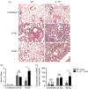
Lung damage after S. pneumoniae infection is more severe in interleukin-10-knock out mice (IL-10−/−). Wild-type (WT) and IL-10−/− mice were intranasally infected with 3 × 107 CFU of S. pneumoniae D39. At 24 and 48 hr post-infection (hpi) lungs and bronchoalveolar lavage fluid (BALF) were recovered. Lungs were removed, embedded in paraffin, 5-μm lung sections were stained with haematoxylin & eosin. (a) Representative lung tissue photographies observed in an optical microscope at 20 × magnification. (b) Lung histopathological score (c) and total proteins in BALF were measured at 24 and 48 hpi, using the Bradford methodology. Data shown mean + SEM *P < 0·05, Student’s t-test between IL-10−/− and WT mice at each time-point; two-way analysis of variance tests with a posteriori Tukey multiple comparison test were performed to test the expression change across time; (a) P < 0·05 between uninfected and 24 hpi; (c) P < 0·05 between 24 hpi and 48 hpi and; (b) P < 0·05 between uninfected and 48 hpi.
Figure 5
Figure 5
Interleukin-10 knock out mice (IL-10−/−) have higher numbers of neutrophils in lungs after S. pneumoniae infection. As a gate strategy, singlets were selected from total FSC/SSC events; then total CD45+ leucocytes were gated and Cd11b+ cells were selected. Finally, Ly6G and Ly6C antibodies were used to discriminate between neutrophils, Inflammatory monocytes and Ly6C- myeloid cells (see Supplementary material, Fig. S1). At 24 and 48 hr post-infection (hpi), absolute numbers of (a) neutrophils (CD45+ CD11b+ Ly6G+ Ly6Cmed), (b) inflammatory monocytes (CD45+ CD11b+ Ly6Glow Ly6C+) and (c) Ly6C- myeloid cells (CD45+ CD11b+ Ly6G Ly6C) were measured by flow cytometry in lungs of wild-type (WT) and IL-10−/−, using Countbright absolute counting beads (Life Technology, Grand Island, NY). Data shown are mean + SEM. *P < 0·05, Student’s t-test between IL-10−/− and WT mice at each time-point; two-way analysis of variance test with a posteriori Tukey multiple comparison test were performed to test the expression change across time; (a) P < 0·05 between uninfected and 24 hpi; (c) P < 0·05 between 24 hpi and 48 hpi and; (b) P < 0·05 between uninfected and 48 hpi.
Figure 6
Figure 6
Interleukin-10 knock out mice (IL-10–/–) show reduced S. pneumoniae dissemination after infection. Bacterial loads of (a) lungs, (b) brain, (c) spleen and (d) blood were quantified at 24 and 48 hr post-infection (hpi) by seeding organs onto blood agar, as described in the Materials and methods. *P < 0·05, Student’s t test between IL-10−/− and wild-type (WT) mice at each time-point; two-way analysis of variance test with a posteriori Sidak’s multiple comparison test were performed to test the expression change across time, (a) P < 0·05 between 24 hpi and 48 hpi.
Figure 7
Figure 7
Etanercept neutralizes tumour necrosis factor-α (TNF-α) in vivo but does not improve the survival rate of interleukin-10 knock out mice (IL-10−/−) after an infection with S. pneumoniae. (a) IL-10−/− mice were treated intraperitoneally daily with 200 μg/500 μl of Etanercept from 1 day before the infection. At day 0, mice were intranasally infected with 3 × 107 CFU of S. pneumoniae D39, at 48 hr post-infection (hpi) TNF-α neutralization was measured in bronchoalveolar lavage fluid (BALF) by ELISA, *P < 0·05 by one-way analysis of variance test with Tukey’s multiple comparisons test. (b) Survival rate was followed by 10 days. *P < 0·05, log rank test. Neutrophils (CD45 + CD11b + Ly6G+ Ly6Cmed) and inflammatory monocytes (CD45 + CD11b + Ly6Glow Ly6C+) absolute numbers in (c) lungs and (d) BALF were measured at 48 hpi. (e) Neutrophils/Ly6C+ monocytes ratio in lungs at 48 hpi. *P < 0·05, one-way analysis of variance test and with Holm–Sidak multiple comparisons test.

References

    1. Nieto PA, Riquelme SA, Riedel CA, Kalergis AM, Bueno SM. Gene elements that regulate Streptococcus pneumoniae virulence and immunity evasion. Curr Gene Ther. 2013;13:51–64. - PubMed
    1. Kadioglu A, Weiser JN, Paton JC, Andrew PW. The role of Streptococcus pneumoniae virulence factors in host respiratory colonization and disease. Nat Rev Microbiol. 2008;6:288–301. - PubMed
    1. O’Brien KL, Wolfson LJ, Watt JP, et al. Burden of disease caused by Streptococcus pneumoniae in children younger than 5 years: global estimates. Lancet. 2009;374:893–902. - PubMed
    1. Drijkoningen JJ, Rohde GG. Pneumococcal infection in adults: burden of disease. Clin Microbiol Infect. 2014;20(Suppl. 5):45–51. - PubMed
    1. Jackson ML, Neuzil KM, Thompson WW, Shay DK, Yu O, Hanson CA, Jackson LA. The burden of community-acquired pneumonia in seniors: results of a population-based study. Clin Infect Dis. 2004;39:1642–50. - PMC - PubMed

Publication types

MeSH terms

LinkOut - more resources