

An Unusual Cause of a Breast Mass in a Patient from China
- PMID: 26033021
- PMCID: PMC4530759
- DOI: 10.4269/ajtmh.15-0235
An Unusual Cause of a Breast Mass in a Patient from China
Abstract
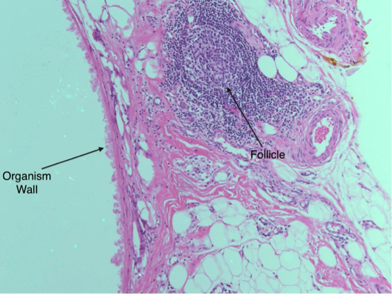
Sparganosis is a parasitic infection caused by Spirometra spp. and often presents as a subcutaneous swelling, most commonly noticed in the abdominal wall or extremities. Amphibians such as frogs ingest infected copepods (crustaceans that have ingested coracidia, i.e., Spirometra spp. embryos) and serve as a secondary intermediate host. Complete surgical excision is recommended for definitive diagnosis and treatment. Granulomatous inflammation is the most common histologic finding. Although dissemination can occur, most cases are localized. Serum enzyme-linked immunosorbent assay (ELISA) has been suggested as a potential surveillance tool. Medical therapy with antiparasitic agents, such as praziquantel, is not typically recommended but may be effective at high doses. Preventing recurrence thus depends on adequate surgical removal of the parasite. We report a case of a breast mass caused by sparganosis infection in a Chinese female whose likely exposure was due to frog consumption. The diagnosis was confirmed on surgical excision and no systemic antiparasitic therapy was required.
© The American Society of Tropical Medicine and Hygiene.
Figures


References
-
- Koo M, Kim JH, Kim JS, Lee JE, Nam SJ, Yang JH. Cases and literature review of breast sparganosis. World J Surg. 2011;35:573–579. - PubMed
-
- Chung SY, Park KS, Lee Y, Park CK. Breast sparganosis: mammographic and ultrasound features. JCU. 1995;23:447–451. - PubMed
-
- Palmer PES. Reeder MM. The Imaging of Tropical Diseases. 2nd edition. Baltimore/London: The Williams & Wilkins Company, 1st edition 1981; 2000. Springer. December 12, 2000.
Publication types
MeSH terms
Substances
LinkOut - more resources
Full Text Sources
Other Literature Sources
Medical
Miscellaneous
