. 2015 Jun 1;308(11):L1178-88.
doi: 10.1152/ajplung.00290.2014. Epub 2015 Apr 10.

Influenza induces IL-8 and GM-CSF secretion by human alveolar epithelial cells through HGF/c-Met and TGF-α/EGFR signaling

Affiliations

Influenza induces IL-8 and GM-CSF secretion by human alveolar epithelial cells through HGF/c-Met and TGF-α/EGFR signaling

Yoko Ito et al. Am J Physiol Lung Cell Mol Physiol. .

Abstract

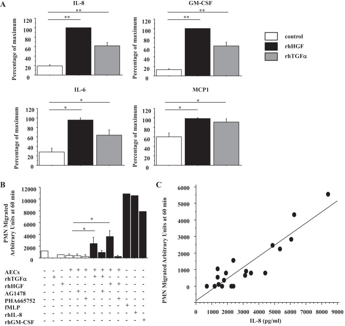
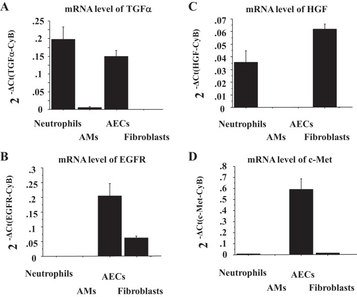
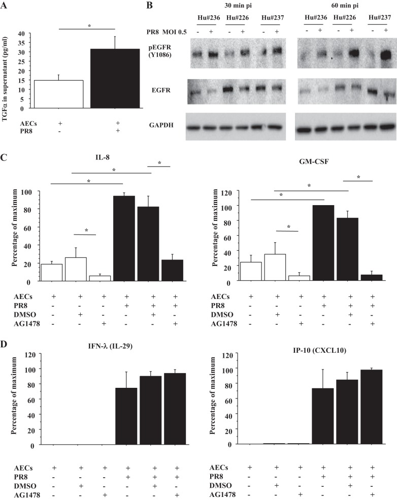
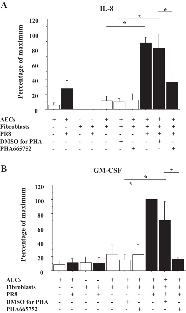
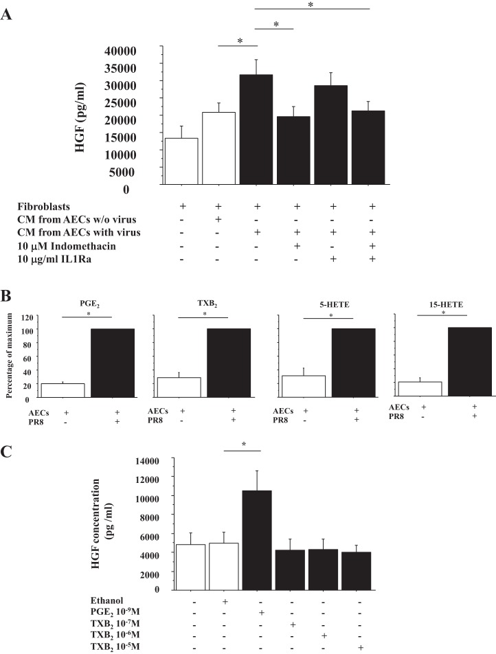
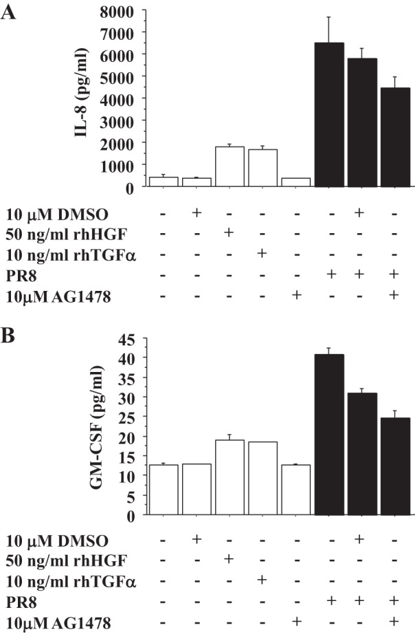
The most severe complication of influenza is viral pneumonia, which can lead to the acute respiratory distress syndrome. Alveolar epithelial cells (AECs) are the first cells that influenza virus encounters upon entering the alveolus. Infected epithelial cells produce cytokines that attract and activate neutrophils and macrophages, which in turn induce damage to the epithelial-endothelial barrier. Hepatocyte growth factor (HGF)/c-Met and transforming growth factor-α (TGF-α)/epidermal growth factor receptor (EGFR) are well known to regulate repair of damaged alveolar epithelium by stimulating cell migration and proliferation. Recently, TGF-α/EGFR signaling has also been shown to regulate innate immune responses in bronchial epithelial cells. However, little is known about whether HGF/c-Met signaling alters the innate immune responses and whether the innate immune responses in AECs are regulated by HGF/c-Met and TGF-α/EGFR. We hypothesized that HGF/c-Met and TGF-α/EGFR would regulate innate immune responses to influenza A virus infection in human AECs. We found that recombinant human HGF (rhHGF) and rhTGF-α stimulated primary human AECs to secrete IL-8 and granulocyte macrophage colony-stimulating factor (GM-CSF) strongly and IL-6 and monocyte chemotactic protein 1 moderately. Influenza infection stimulated the secretion of IL-8 and GM-CSF by AECs plated on rat-tail collagen through EGFR activation likely by TGF-α released from AECs and through c-Met activated by HGF secreted from lung fibroblasts. HGF secretion by fibroblasts was stimulated by AEC production of prostaglandin E2 during influenza infection. We conclude that HGF/c-Met and TGF-α/EGFR signaling enhances the innate immune responses by human AECs during influenza infections.

Keywords: alveolar epithelial cells; hepatocyte growth factor/c-Met; influenza; innate immune responses; transforming growth factor-α/epidermal growth factor receptor.


Figures

Fig. 1.
Fig. 1.
Recombinant human hepatocyte growth factor (rhHGF) and rh transforming growth factor (TGF)-α induce human alveolar epithelial cells (AECs) to secrete IL-8 and granulocyte macrophage colony-stimulating factor (GM-CSF) strongly, and IL-6 and monocyte chemotactic protein-1 (MCP-1) moderately, which attract neutrophils. A: IL-8, GM-CSF, IL-6, and MCP-1 protein concentrations in culture medium of human AECs stimulated by 50 ng/ml rhHGF or 10 ng/ml rhTGF-α for 24 h were measured by MILLPLEX; n = 6. Open bar, no growth factor; solid bar, rhHGF; shaded bar, rhTGF-α. The range of maximal stimulation: IL-8, 4,792.3–20,136.3 pg/ml; GM-CSF, 58.6–295.5 pg/ml; IL-6, 11.6–497.5 pg/ml; MCP-1, 26,184.5–104,165.1 pg/ml. B: degree of neutrophil migration was measured in vitro using conditioned medium AECs stimulated by rhTGF-α or rhHGF ±2.5 μM AG1478, ±500 nM PHA665752; n = 3. 1 μM N-Formyl-Met-Leu-Phe (fMLP), 10 ng/ml rhIL-8, and 10 ng/ml rhGM-CSF are positive controls. PMN, polymorphonuclear leukocytes. C: correlation diagram between IL-8 protein concentration in the same samples as B and the degree of neutrophil migration (y = −883.184 + 0.670 * x; R2 = 0.831) *P < 0.0001, **P < 0.05.
Fig. 2.
Fig. 2.
Human AECs express TGF-α, epidermal growth factor (EGFR), c-Met, but not HGF. The mRNA levels of TGF-α (A), EGFR (B), HGF (C), and c-Met (D) were measured by real-time PCR. These levels were normalized to the constitutive probe cyclophilin B (CyB). Values were means ± SE (n = 3 for each cell type). AMs, alveolar macrophages.
Fig. 3.
Fig. 3.
Influenza virus infection of AECs induces TGF-α secretion and activates EGFR, which stimulates secretion of IL-8 and GM-CSF by AECs. A: TGF-α concentration of culture medium from AECs with or without influenza A virus/Puerto Rico/8/1934 (PR8) infection (multiplicity of infection, MOI = 0.5) for 24 h was measured by ELISA. Values were means ± SE for 5 different human donors. *P < 0.05. B: human (Hu) AECs were infected by PR8 (MOI = 0.5). The AECs were harvested at 30 and 60 min after infection, and protein levels of phospho-EGFR and EGFR normalized by GAPDH were measured by immunoblotting. This blot is from 3 different human donors. pi, postinfection. C: IL-8 and GM-CSF concentration of culture medium of AECs with or without PR8 infection (MOI = 0.5) and with or without AG1478, an EGFR inhibitor, for 24 h was measured by ELISA (n = 5 for IL-8, n = 4 for GM-CSF, as 1 human sample was below limits of quantitation). DMSO was a vehicle control for AG1478. The range of maximal stimulation: IL-8, 1,250.4–7,089.6 pg/ml; GM-CSF, 6.9–95.0 pg/ml. D: IFN-λ (IL-29) and CXCL10 (IFN-γ-inducible protein of 10, IP-10) concentration of culture medium of AECs with or without PR8 infection (MOI = 0.5) and with or without AG1478 for 24 h was measured by ELISA (n = 3). DMSO was a vehicle control for AG1478. The range of maximal stimulation: IFN-λ, 753.6-5,933.4 pg/ml; IP-10, 1,174.8–5,278.9 pg/ml.
Fig. 4.
Fig. 4.
HGF/c-Met signaling induces IL-8 and GM-CSF secretion by AECs cocultured with lung fibroblasts (FBs) during influenza infection. AECs plated on the inserts were preincubated by PHA665752, a c-Met inhibitor, or vehicle control at 2 h before infection and then infected with PR8 (MOI = 0.5). At 1 h after infection, the AECs were placed over lung FBs plated on the plates to start coculture, and the culture medium was harvested at 48 h after starting the coculture. A: IL-8 concentration of these culture medium was measured by ELISA (n = 3). DMSO was a vehicle control for PHA665752. The range of maximal stimulation: IL-8, 4,188.1–26,022.0 pg/ml. B: GM-CSF concentration of same culture medium as A was measured by ELISA (n = 3). DMSO was a vehicle control for PHA665752. The range of maximal stimulation: GM-CSF, 44.0–953.5 pg/ml.
Fig. 5.
Fig. 5.
HGF secretion by FBs is enhanced by prostaglandin E2 (PGE2) secreted by influenza virus-infected AECs, which is inhibited by indomethacin. A: lung FBs were stimulated by conditioned medium from AECs with or without PR8 (MOI = 0.5) and with or without 10 μM indomethacin and with or without 10 μg/ml IL-1Ra, and culture medium was harvested at 48 h after stimulation. The HGF concentration of this culture medium was measured by ELISA (n = 3). B: arachidonic acid metabolites secreted by influenza virus-infected AECs were analyzed by mass spectrometry. The range of maximal stimulation: PGE2, 17.1–22.5 ng/sample; range of thromboxane B2 (TXB2), 0.26–2.16 ng/sample; range of 5-hydroxyeicosatetraenoic acid (5-HETE), 0.16–0.35 ng/sample; 15-HETE, 3.51–6.06 ng/sample; n = 3. C: lung FBs were stimulated by 10−9 M PGE2, 10−7 M TXB2, 10−6 M TXB2, or 10−5 M TXB2, and culture medium was harvested at 48 h after stimulation. The HGF concentration of this culture medium was measured by ELISA (n = 3). Ethanol was a vehicle control for PGE2 and TXB2.
Fig. 6.
Fig. 6.
rhHGF and rhTGF-α stimulate IL-8 and GM-CSF production of alveolar type II (ATII) cells, but the basal levels of IL-8 and the level of IL-8 and GM-CSF stimulated by PR8 are not reduced by the EGFR inhibitor. A: concentration of IL-8 in the culture medium of ATII cells with or without rhHGF or rhTGF-α and with or without PR8 infection (MOI = 0.5) and with or without AG1478, an EGFR inhibitor, for 24 h was measured by ELISA. This is the representative data from 3 independent experiments for IL-8. DMSO was a vehicle control for AG1478. B: GM-CSF concentration of culture medium of ATII cells with or without rhHGF or rhTGF-α and with or without PR8 infection (MOI = 0.5) and with or without 10 μM AG1478, an EGFR inhibitor, for 24 h was measured by ELISA. This is the representative data from 3 independent experiments for GM-CSF. DMSO was a vehicle control for AG1478.

References

    1. Aggarwal A, Baker CS, Evans TW, Haslam PL. G-CSF and IL-8 but not GM-CSF correlate with severity of pulmonary neutrophilia in acute respiratory distress syndrome. Eur Respir J 15: 895–901, 2000. - PubMed
    1. Arndt PG, Young SK, Poch KR, Nick JA, Falk S, Schrier RW, Worthen GS. Systemic inhibition of the angiotensin-converting enzyme limits lipopolysaccharide-induced lung neutrophil recruitment through both bradykinin and angiotensin II-regulated pathways. J Immunol 177: 7233–7241, 2006. - PubMed
    1. Bradley-Stewart A, Jolly L, Adamson W, Gunson R, Frew-Gillespie C, Templeton K, Aitken C, Carman W, Cameron S, McSharry C. Cytokine responses in patients with mild or severe influenza A(H1N1)pdm09. J Clin Virol 58: 100–107, 2013. - PubMed
    1. Burgel PR, Nadel JA. Epidermal growth factor receptor-mediated innate immune responses and their roles in airway diseases. Eur Respir J 32: 1068–1081, 2008. - PubMed
    1. Falenta K, Rodaway A, Jones GE, Wells CM. Imaging haematopoietic cells recruitment to an acute wound in vivo identifies a role for c-Met signalling. J Microsc 250: 200–209, 2013. - PubMed

Publication types

MeSH terms