. 2015 Jun 2:5:10689.
doi: 10.1038/srep10689.

Subcellular localization of the sigma-1 receptor in retinal neurons - an electron microscopy study

Affiliations

Subcellular localization of the sigma-1 receptor in retinal neurons - an electron microscopy study

Timur A Mavlyutov et al. Sci Rep. .

Abstract

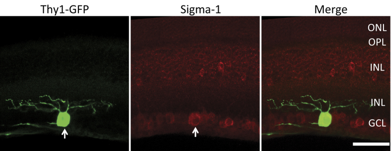
The Sigma-1 receptor (S1R) is known to play a protective role in the central nervous system including the retina. A major barrier for understanding the underlying mechanism is an ambiguity of S1R subcellular localizations. We thus conducted the first electron microscopy (EM) study of S1R subcellular distribution in the mouse retina. Immuno-EM imaging showed previously under-appreciated S1R presence in photoreceptor cells. Unlike in other cell types in previous reports, in photoreceptor cells S1R was found in the nuclear envelope but not localized in the endoplasmic reticulum (ER), raising a possibility of S1R-mediated modulatory mechanisms different than conventionally thought. While in bipolar cells S1R was detected only in the nuclear envelope, in ganglion cells S1R was identified predominantly in the nuclear envelope and found in the ER as well. A predominant localization of S1R in the nuclear envelope in all three retinal neurons implicates a potential role of S1R in modulating nuclear activities. Moreover, its absence in the plasma membrane and presence in the subsurface ER cisternae that are juxtaposed to the plasma membrane in ganglion cells may lend mechanistic insights generally important for frequently reported S1R modulations of ion channels in neurons.


Figures

Figure 1
Figure 1. Immunostaining of S1R on mouse retinal sections at different postnatal developmental stages.
Green, S1R; red, synaptophysin. OS, outer segment; IS, inner segment; ONL, outer nuclear layer; OPL, outer plexiform layer; INL, inner nuclear layer; IPL, inner plexiform layer; GCL, ganglion cell layer. Scale = 50 μm.
Figure 2
Figure 2. Presence of S1R in the bovine photoreceptor synaptic terminal.
(a), Confocal image of bovine retina immunostained for S1R (green) and a presynaptic marker, synaptophysin (red). Note positive S1R staining in the OPL. (b), Magnifed image of the boxed area in (a). Arrows indicate localization of S1R in synaptophysin-labeled synaptic terminals. (c), Electron microscopy image showing S1R in the photoreceptor synaptic terminal. Note the characteristic ribbon and vesicles in the boxed area. (d), Magnified image of the boxed area in (c). Arrows indicate S1R in presynaptic invagination. Arrowheads point to S1R localization in large vesicles. *, synaptic ribbon. Scales: (a), 50 μm; (b), 5 μm; (c), 0.5 μm; (d), 0.1 μm.
Figure 3
Figure 3. Electron microscopy images showing S1R distribution in the mouse photoreceptor subcellular compartments.
(a), Schematic of the compartments in the photoreceptor. (b), Ultrastructure of outer and inner segment. Asterisks label mitochondria. (c)–(e), Magnified images of the boxed areas in (b), showing the outer segment containing membrane discs, the connecting cilium (asterisk), and the inner segment (including ER), respectively. (f)–(h), Localization of S1R in the nuclear envelope. (f), nuclear region of several photoreceptor cells; (g), nuclear envelope of a single cell; (h), magnified box area in (g) showing S1R localization in the outer and inner membranes of the nuclear envelope (pointed to by arrows). (i) and (j), Photoreceptor synaptic terminal. The image in (j) is a magnified box area in (i) revealing the characteristic ribbon (asterisks) and vesicles. Scales: (b)–(e) and (g), 1 μm; (f), 3 μm; (h), 0.2 μm; (i), 0.5 μm; (j), 0.1 μm.
Figure 4
Figure 4. Subcelluar localization of S1R in bipolar and ganglion cells of the mouse retina.
(a)–(c), Bipolar cells. (b) shows magnification of the boxed area in (a). Arrows point to S1R immunolabeling in the inner and outer membranes of the nuclear envelope. Arrowheads mark the plasma membrane. (c) shows S1R localization in the ER membrane (star) connected to the nuclear envelope (arrows). (d)–(f), Ganglion cells. (d) shows predominant S1R localization in the nuclear envelope (arrows) but not in the plasma membrane (arrow heads). (e) highlights the presence of S1R in the ER (boxed area). (f) shows the magnification of the boxed area in (e), revealing S1R localization in the ER cisternae (asterisks) that are adjacent to the plasma membrane (arrow heads). Scales: (a), (c)–(e), 2 μm; (f), 1 μm; (b), 0.25 μm.
Figure 5
Figure 5. Presence of S1R in the soma and its absence in the dendrites of mouse retinal ganglion cells.
A retinal ganglion cell (green) is illuminated by Thy-1 driven GFP expression in the Thy-1/GFP mouse retina. Immunostaining of S1R is shown in red. Arrow points to the GFP positive ganglion cell. Note that while S1R positive staining is seen in the soma it is not evidently detected in the dendrites. Scale = 50 μm.

References

    1. Su T. P., Hayashi T., Maurice T., Buch S. & Ruoho A. E.. The sigma-1 receptor chaperone as an inter-organelle signaling modulator. Trends Pharmacol Sci. 31, 557–566; S0165-6147(10)00153-7/j.tips.2010.08.007 (2010). - PMC - PubMed
    1. Hayashi T. & Su T. P. Sigma-1 receptor chaperones at the ER-mitochondrion interface regulate Ca(2+) signaling and cell survival. Cell 131, 596–610; S0092-8674(07)01099-9/j.cell.2007.08.036 (2007). - PubMed
    1. Pabba M. et al. NMDA receptors are upregulated and trafficked to the plasma membrane after sigma-1 receptor activation in the rat hippocampus. J Neurosci. 34, 11325–11338; 10.1523/JNEUROSCI.0458-14.2014 (2014). - DOI - PMC - PubMed
    1. Xu J. et al. Identification of the PGRMC1 protein complex as the putative sigma-2 receptor binding site. Nat. Commun. 2, 380; 10.1038/ncomms1386 (2011). - DOI - PMC - PubMed
    1. Luty A. A. et al. Sigma nonopioid intracellular receptor 1 mutations cause frontotemporal lobar degeneration-motor neuron disease. Ann. Neurol. 68, 639–649; 10.1002/ana.22274 (2010). - DOI - PubMed

Publication types

MeSH terms

Substances