. 2015 Aug;59(8):4826-34.
doi: 10.1128/AAC.00331-15. Epub 2015 Jun 1.

Gallium Compounds Exhibit Potential as New Therapeutic Agents against Mycobacterium abscessus

Affiliations

Gallium Compounds Exhibit Potential as New Therapeutic Agents against Mycobacterium abscessus

Maher Y Abdalla et al. Antimicrob Agents Chemother. 2015 Aug.

Abstract

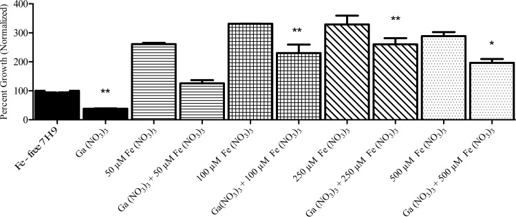
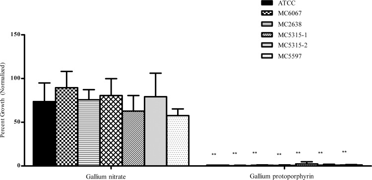
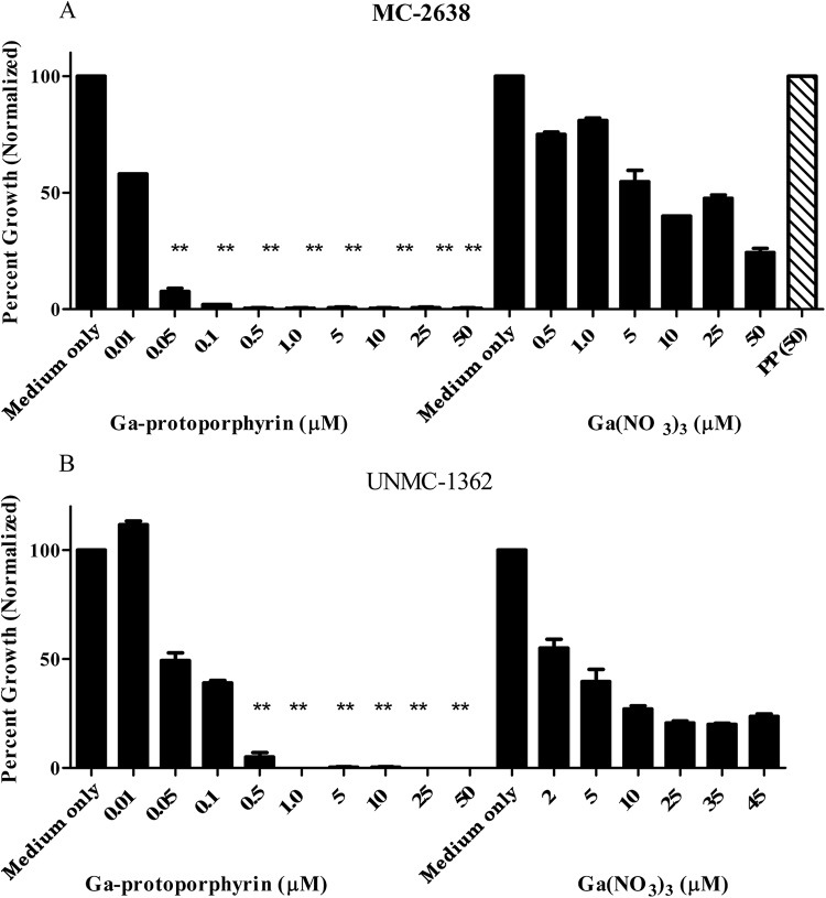
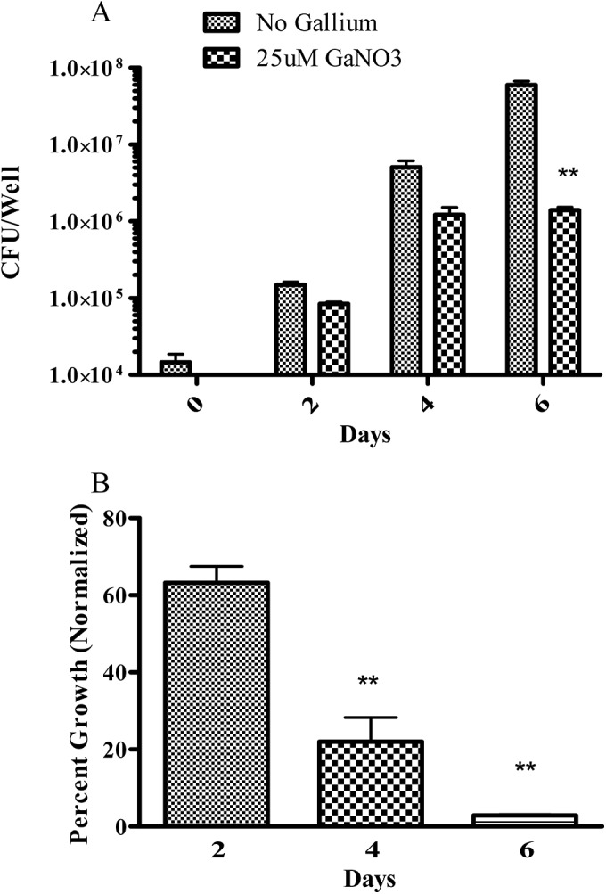
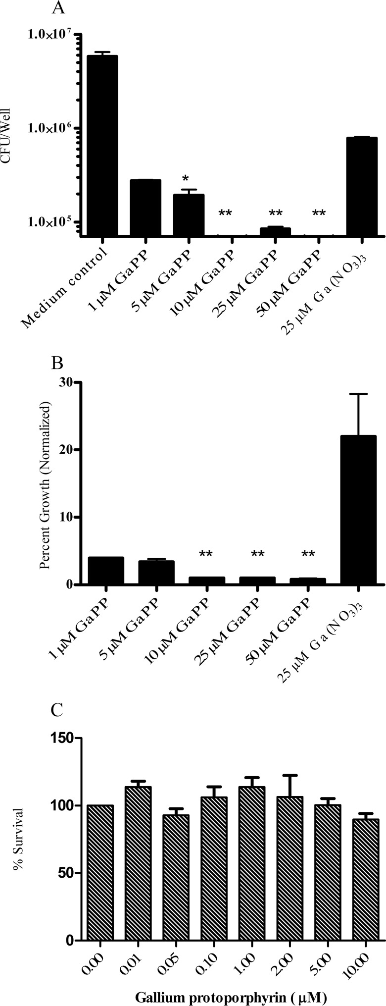
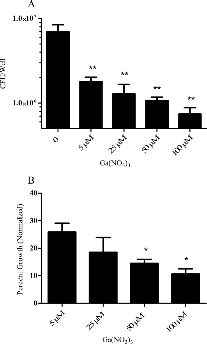
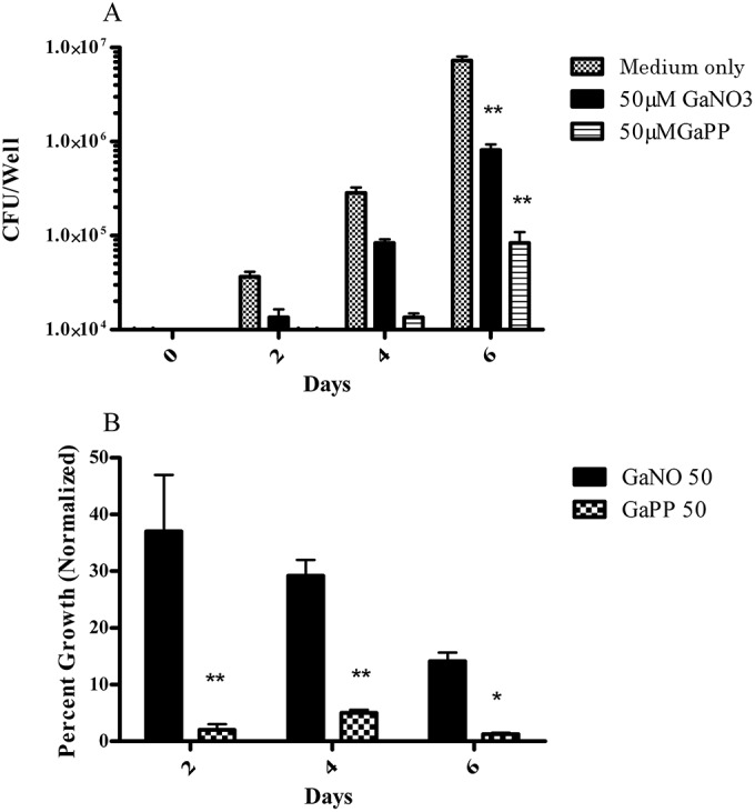
The rapidly growing nontuberculous mycobacterial species Mycobacterium abscessus has recently emerged as an important pathogen in patients with cystic fibrosis (CF). Treatment options are limited because of the organism's innate resistance to standard antituberculous antibiotics, as well as other currently available antibiotics. New antibiotic approaches to the treatment of M. abscessus are urgently needed. The goal of the present study was to assess the growth-inhibitory activity of different Ga compounds against an American Type Culture Collection (ATCC) strain and clinical isolates of M. abscessus obtained from CF and other patients. In our results, using Ga(NO3)3 and all of the other Ga compounds tested inhibited the growth of ATCC 19977 and clinical isolates of M. abscessus. Inhibition was mediated by disrupting iron uptake, as the addition of exogenous iron (Fe) restored basal growth. There were modest differences in inhibition among the isolates for the same Ga chelates, and for most Ga chelates there was only a slight difference in potency from Ga(NO3)3. In contrast, Ga-protoporphyrin completely and significantly inhibited the ATCC strain and clinical isolates of M. abscessus at much lower concentrations than Ga(NO3)3. In in vitro broth culture, Ga-protoporphyrin was more potent than Ga(NO3)3. When M. abscessus growth inside the human macrophage THP-1 cell line was assessed, Ga-protoporphyrin was >20 times more active than Ga(NO3)3. The present work suggests that Ga exhibits potent growth-inhibitory capacity against the ATCC strain, as well as against antibiotic-resistant clinical isolates of M. abscessus, including the highly antibiotic-resistant strain MC2638. Ga-based therapy offers the potential for further development as a novel therapy against M. abscessus.


Figures

FIG 1
FIG 1
Ga(NO3)3-mediated growth inhibition is reversed by Fe. Strain ATCC 19977 was cultured in the presence of 50 μM Ga(NO3)3 and increasing concentrations of Fe(NO3)3 for up to 7 days. Addition of equimolar or greater amounts of Fe relative to Ga increased M. abscessus growth significantly. *, P < 0.05; **, P < 0.01; n = 3 replicates.
FIG 2
FIG 2
Ga-protoporphyrin shows the greatest inhibition among Ga chelates. M. abscessus strains were cultured in the presence of 10 μM Ga-protoporphyrin for up to 7 days. Results show inhibition of all strains normalized to the control. Statistical analysis was done by comparing growth inhibition to that observed with Ga(NO3)3. **, P < 0.001; n = 3 replicates.
FIG 3
FIG 3
Ga-protoporphyrin inhibits clinical isolates of M. abscessus. Clinical isolates MC2638 (A) and UNMC-1362 (B) were cultured in the presence of different concentrations of Ga-protoporphyrin and Ga(NO3)3 for up to 7 days. Results show inhibition by each compound normalized to the control. PP(50) shows bacterial growth in the presence of protoporphyrin only at 50 μM. Statistical analysis was done by comparing Ga-protoporphyrin to Ga(NO3)3. **, P < 0.001; n = 3 replicates.
FIG 4
FIG 4
Ga(NO3)3 inhibits intracellular M. abscessus growth. THP-1 cells were TPA activated into macrophage-like cells. Cells were then infected with M. abscessus ATCC 19977 and cultured for up to 6 days with or without 25 μM Ga(NO3)3. Panel A shows results expressed in CFU counts, and panel B shows results normalized to the control (no Ga). Growth of intracellular M. abscessus was significantly inhibited in the presence of 25 μM Ga(NO3)3. **, P < 0.01; n = 3 replicates.
FIG 5
FIG 5
Ga-protoporphyrin inhibits intracellular M. abscessus growth. THP-1 cells were transformed and infected with M. abscessus ATCC 19977 for 1 h, washed, and cultured for up to 4 days in the presence of different concentrations of Ga-protoporphyrin as described in Materials and Methods. Ga-protoporphyrin significantly inhibited intracellular M. abscessus relative to the control (no Ga) or Ga(NO3)3 treated when expressed as the number of CFU/well (panel A) (P < 0.001) or when the data were expressed normalized to the control (no Ga). The time zero CFU count was 2.9 × 10⁴. No toxic effects of Ga-protoporphyrin against THP-1 cells were seen (panel C), as determined by MTT assay. *, P < 0.01; **, P < 0.001; n = 4 replicates.
FIG 6
FIG 6
Ga(NO3)3 inhibits intracellular growth of clinical isolate MC2638 at higher concentrations. THP-1 cells were transformed and infected with clinical isolate MC2638 for 1 h, washed, and cultured for up to 6 days in the presence of different concentrations of Ga(NO3)3 as described in Materials and Methods. Ga(NO3)3 significantly inhibited intracellular MC2638 (**, P < 0.001) as measured by CFU counting (A) or when results were normalized to the control (B) (*, P < 0.05; n = 4 replicates). The time zero CFU count was 2.2 × 10⁴.
FIG 7
FIG 7
Ga-protoporphyrin inhibits intracellular growth of clinical isolate MC2638. THP-1 cells were transformed and infected with clinical isolate MC2638 for 1 h, washed, and cultured for up to 6 days in the presence of different concentrations of Ga-protoporphyrin as described in Materials and Methods. Ga-protoporphyrin significantly inhibited intracellular MC2638 (**, P < 0.001) as determined by CFU counting (A) or when data were normalized to the control (B) (*, P < 0.05; n = 4 replicates).

References

    1. Hauser AR, Jain M, Bar-Meir M, McColley SA. 2011. Clinical significance of microbial infection and adaptation in cystic fibrosis. Clin Microbiol Rev 24:29–70. doi:10.1128/CMR.00036-10. - DOI - PMC - PubMed
    1. Razvi S, Quittell L, Sewall A, Quinton H, Marshall B, Saiman L. 2009. Respiratory microbiology of patients with cystic fibrosis in the United States, 1995 to 2005. Chest 136:1554–1560. doi:10.1378/chest.09-0132. - DOI - PubMed
    1. Petrini B. 2006. Mycobacterium abscessus: an emerging rapid-growing potential pathogen. APMIS 114:319–328. doi:10.1111/j.1600-0463.2006.apm_390.x. - DOI - PubMed
    1. Griffith DE, Girard WM, Wallace RJ. 1993. Clinical features of pulmonary disease caused by rapidly growing mycobacteria: an analysis of 154 patients. Am Rev Respir Dis 147:1271–1278. doi:10.1164/ajrccm/147.5.1271. - DOI - PubMed
    1. Chan ED, Bai X, Kartalija M, Orme IM, Ordway DJ. 2010. Host immune response to rapidly growing mycobacteria, an emerging cause of chronic lung disease. Am J Respir Cell Mol Biol 43:387–393. doi:10.1165/rcmb.2009-0276TR. - DOI - PubMed

Publication types

MeSH terms

LinkOut - more resources