. 2015 Jun;56 Suppl 4(0 4):39S-45S.
doi: 10.2967/jnumed.114.142729.

Role of Cardiac MR Imaging in Cardiomyopathies

Affiliations

Role of Cardiac MR Imaging in Cardiomyopathies

Christopher M Kramer. J Nucl Med. 2015 Jun.

Abstract

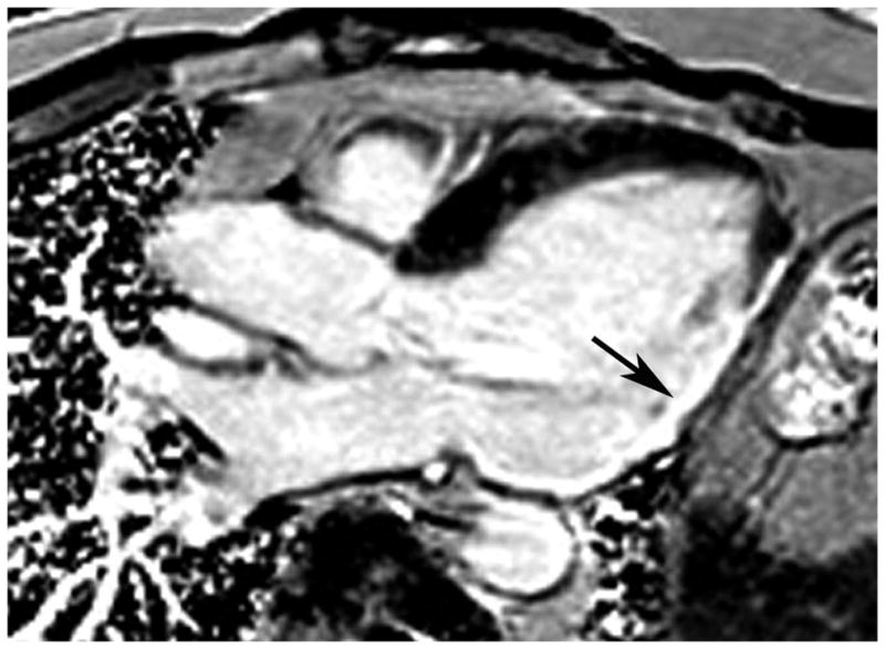
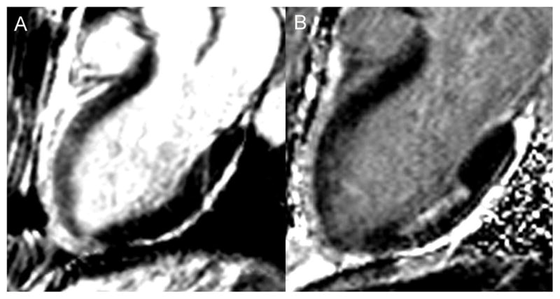
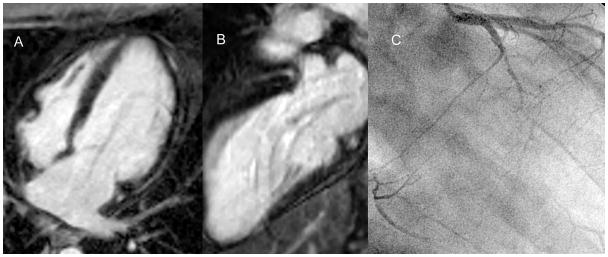
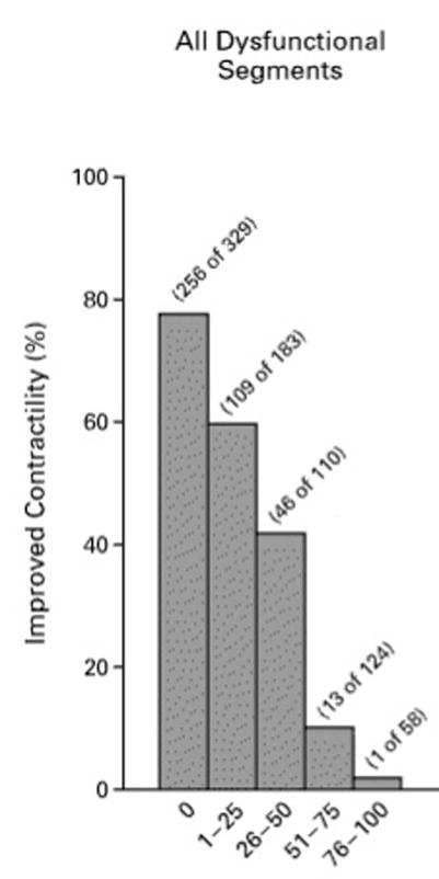
Cardiac MR imaging has made major inroads in the new millennium in the diagnosis and assessment of prognosis for patients with cardiomyopathies. Imaging of left and right ventricular structure and function and tissue characterization with late gadolinium enhancement (LGE) as well as T1 and T2 mapping enable accurate diagnosis of the underlying etiology. In the setting of coronary artery disease, either transmurality of LGE or contractile reserve in response to dobutamine can assess the likelihood of recovery of function after revascularization. The presence of scar reduces the likelihood of a response to medical therapy and to cardiac resynchronization therapy in heart failure. The presence and extent of LGE relate to overall cardiovascular outcome in cardiomyopathies. A major role for cardiac MR imaging in cardiomyopathies is to identify myocardial scar for diagnostic and prognostic purposes.

Keywords: cardiac MR imaging; coronary artery disease; infiltrative cardiomyopathy; ischemic cardiomyopathy; late gadolinium enhancement; nonischemic cardiomyopathy.


Figures

Figure 1
Figure 1
3-chamber long axis phase sensitive inversion recovery LGE image in a patient with heart failure and coronary artery disease. There is transmural LGE in the inferolateral wall (arrow).
Figure 2
Figure 2
A – 3-chamber long axis bright blood T2-W image in a patient with a spontaneous LAD dissection demonstrating bright signal in the anterior wall consistent with edema. B – 3-chamber long axis phase sensitive inversion recovery late gadolinium enhanced image in the same patient showing absence of LGE in the anterior wall, but evidence of a prior 50% transmural infarct in the mid inferior wall. Together these images demonstrate evidence of myocardial stunning without infarction in the anterior wall.
Figure 3
Figure 3
A - 4-chamber long axis phase sensitive inversion recovery LGE image in a patient with heart failure and EF 20% which shows no LGE. B - 2-chamber long axis phase sensitive inversion recovery LGE image in the same patient which shows no LGE. C – Right anterior oblique x-ray angiogram in this patient which demonstrates 3-vessel CAD with an occluded left circumflex, an occluded right coronary that fills through collaterals, and significant LAD disease. Thus, this patient demonstrates severe LV dysfunction in the setting of 3-vessel CAD without LGE signifying extensive hibernating myocardium.
Figure 4
Figure 4
Inverse relationship between transmurality of LGE on the x-axis and likelihood of improved function after revascularization. Adapted from (29) Kim et al, New England Journal of Medicine; 2000;43:1445–53.
Figure 5
Figure 5
Patterns of late gadolinium enhancement seen in ischemic and non-ischemic cardiomyopathies. From Edelman RR, et al., eds. Clinical Magnetic Resonance Imaging, 3rd ed. New York: Elsevier Press; 2005 (75).
Figure 6
Figure 6
A – Basal short axis phase sensitive inversion recovery LGE image in a patient with heart failure, global LV systolic dysfunction, and EF 25%. There is a midwall stripe of LGE in the septum. B - 4-chamber long axis phase sensitive inversion recovery LGE image in the same patient again showing the mid septal stripe of LGE.
Figure 7
Figure 7
A - 4-chamber long axis phase sensitive inversion recovery LGE image in a patient with heart failure and EF 30% demonstrating diffuse subendocardial LGE and signal nulling in the blood pool, all characteristic of amyloidosis, most likely the ATTR variety. B- Basal short axis phase sensitive inversion recovery LGE image in a different patient with heart failure, global LV systolic dysfunction, and EF 25%. In this patient, the LGE is patchy and in a noncoronary distribution, more consistent with AL amyloidosis which was subsequently diagnosed on fat pad biopsy.

References

    1. Miller S, Simonetti OP, Carr J, Kramer U, Finn JP. MR imaging of the heart with cine true fast imaging with steady-state precession: influence of spatial and temporal resolutions on left ventricular functional parameters. Radiology. 2002;223:263–269. - PubMed
    1. Isbell DC, Kramer CM. Cardiovascular magnetic resonance: structure, function, perfusion, and viability. J Nucl Cardiol. 2005;12:324–336. - PubMed
    1. Grothues F, Smith GC, Moon JCC, et al. Comparison of interstudy reproducibility of cardiovascular magnetic resonance with two-dimensional echocardiography in normal subjects and in patients with heart failure or left ventricular hypertrophy. Am J Cardiol. 2002;90:29–34. - PubMed
    1. Bellenger NG, Davies LC, Francis JM, Coats AJS, Pennell DJ. Reduction in sample size for studies of remodeling in heart failure by the use of cardiovascular magnetic resonance. J Cardiovasc Magn Reson. 2000;2:271–278. - PubMed
    1. Zerhouni EA, Parish DM, Rogers WJ, Yang A, Shapiro EP. Human heart: tagging with MR imaging: a new method for noninvasive assessment of myocardial motion. Radiology. 1988;169:59–63. - PubMed

Publication types

LinkOut - more resources