Review
. 2015 Jul;72(7):305-39.
doi: 10.1002/cm.21226. Epub 2015 Aug 22.

Emerging roles of sumoylation in the regulation of actin, microtubules, intermediate filaments, and septins

Affiliations
Review

Emerging roles of sumoylation in the regulation of actin, microtubules, intermediate filaments, and septins

Annabel Alonso et al. Cytoskeleton (Hoboken). 2015 Jul.

Abstract

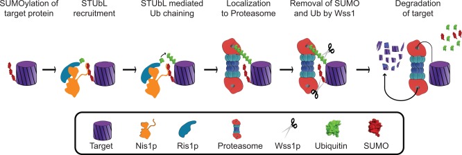
Sumoylation is a powerful regulatory system that controls many of the critical processes in the cell, including DNA repair, transcriptional regulation, nuclear transport, and DNA replication. Recently, new functions for SUMO have begun to emerge. SUMO is covalently attached to components of each of the four major cytoskeletal networks, including microtubule-associated proteins, septins, and intermediate filaments, in addition to nuclear actin and actin-regulatory proteins. However, knowledge of the mechanisms by which this signal transduction system controls the cytoskeleton is still in its infancy. One story that is beginning to unfold is that SUMO may regulate the microtubule motor protein dynein by modification of its adaptor Lis1. In other instances, cytoskeletal elements can both bind to SUMO non-covalently and also be conjugated by it. The molecular mechanisms for many of these new functions are not yet clear, but are under active investigation. One emerging model links the function of MAP sumoylation to protein degradation through SUMO-targeted ubiquitin ligases, also known as STUbL enzymes. Other possible functions for cytoskeletal sumoylation are also discussed.

Keywords: IF; MAPs; MT; SUMO; microfilaments; microtubule-associated proteins; septins.


Figures

Figure 1
Figure 1
Structure of ubiquitin and SUMO proteins. Ribbon drawing of ubiquitin, Smt3, SUMO1, and SUMO2. These molecules share a common secondary structure ββααββαβ that assembles into a ubiquitin‐like fold. Renderings were developed using the crystallography coordinates available from the Protein Data Bank with the following accession numbers: ubiquitin (1UBQ), Smt3 (3V60), SUMO1 (2UYZ), and SUMO2 (1WM3). The structures for the above molecules were analyzed using the PyMOL Molecular Graphics System, Version 1.3 Schrödinger, LLC.
Figure 2
Figure 2
SUMOylation Pathway: To portray each state in the sumoylation pathway, surface maps were developed using crystallography coordinates available from PDB with the following accession numbers: SUMO1 and Senp1 (2IY1), E1 complex (3KYC), E2 complex (2UYZ), E3 complex (3UIP), and sumoylated PCNA (3V60). The orientation of SUMO is maintained throughout the sumoylation processes depicted above.
Figure 3
Figure 3
Chemical bonds in the sumoylation pathway. (A) Isopeptide bond. SUMO is conjugated to the target protein via an isopeptide bond linkage between the terminal glycine residue of SUMO and the epsilon amino group of the lysine in the target. (B) Thioester Bond. Chemical linkage is highlighted between the terminal glycine carboxy group of SUMO and the active cysteine in the SUMO activating, and conjugating enzymes.
Figure 4
Figure 4
Structure of SUMO E1, E2, E3 enzymes. Tertiary ribbon structure of the SUMO activating enzyme dimers Sae1 and Sae2, SUMO conjugating enzyme Ubc9, and SUMO ligating enzymes Mms21, PIAS3, PIAS2, and Siz1. These renderings were developed using the crystallography coordinates associated with the following PDB accession numbers: Sae1 (1Y8Q), Sae2 (1Y8Q), Ubc9 (2GRR), Mms2 (3HTK), PIAS3 (4MVT), PIAS2 (4FO9), and Siz1 (3I2D).
Figure 5
Figure 5
STUbL Pathway Model. The Ris1p‐Nis1p STUbL complex can direct a sumoylated target protein to the proteasome for degradation (Adapted from Alanso et al. 2012).
Figure 6
Figure 6
Sumoylation of septins. During G1, a septin patch forms at the site of bud formation. As the bud emerges through the patch, the septins form a collar around the mother‐bud neck. The five septin proteins involved in this process are Cdc3p, Cdc10p, Cdc11p, Cdc12p, and Shs1p. Prior to cytokinesis, three of these septin proteins are sumoylated, but only on the mother side of the mother‐bud neck. Cdc3p is sumoylated at lysines 4, 11, 30, and 63. Cdc11p is sumoylated at lysine 412. Shs1p is sumoylated at lysines 426, and 437 [Johnson and Blobel, 1999; Takahashi et al., 1999]. The sumoylation event is color coded in red. During cytokinesis, the septin “hourglass” collar splits into two rings as the cells divide. After cytokinesis the septin rings dissociates, and the process starts again.

References

    1. Adhikari AS, Sridhar Rao K, Rangaraj N, Parnaik VK, Mohan Rao CH. 2004. Heat stress‐induced localization of small heat shock proteins in mouse myoblasts: intranuclear lamin A/C speckles as target for alphaB‐crystallin and Hsp25. Exp Cell Res 299:393–403. - PubMed
    1. Aebi U, Cohn J, Buhle L, Gerace L. 1986. The nuclear lamina is a meshwork of intermediate‐type filaments. Nature 323:560–564. - PubMed
    1. Akhmanova A, Steinmetz MO. 2008. Tracking the ends: a dynamic protein network controls the fate of microtubule tips. Nat Rev Mol Cell Biol 9:309–322. - PubMed
    1. Akyurek N, Ren Y, Rassidakis GZ, Schlette EJ, Medeiros LJ. 2006. Expression of inhibitor of apoptosis proteins in B‐cell non‐Hodgkin and Hodgkin lymphomas. Cancer 107:1844–1851. - PubMed
    1. Al‐Bassam J, Ozer RS, Safer D, Halpain S, Milligan RA. 2002. MAP2 and tau bind longitudinally along the outer ridges of microtubule protofilaments. J Cell Biol 157:1187–1196. - PMC - PubMed

Publication types

LinkOut - more resources