. 2015 Jun;72(6):282-91.
doi: 10.1002/cm.21227. Epub 2015 Jun 30.

Evidence for changes in beta- and gamma-actin proportions during inner ear hair cell life

Affiliations

Evidence for changes in beta- and gamma-actin proportions during inner ear hair cell life

Leonardo R Andrade. Cytoskeleton (Hoboken). 2015 Jun.

Abstract

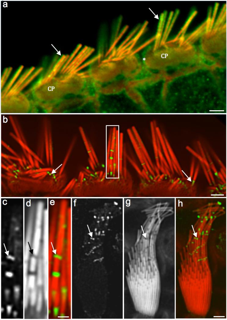
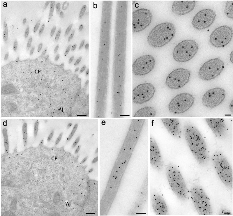
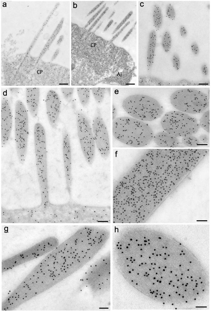
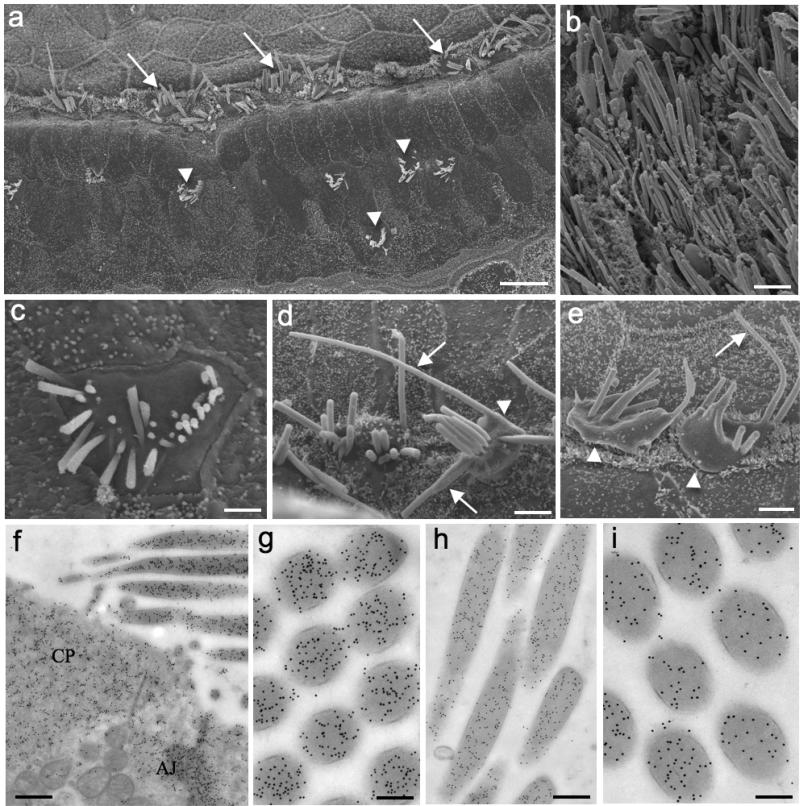
Cytoplasmic actin isoforms beta (β-) and gamma (γ-) perform crucial physiological roles in inner ear hair cells (HC). The stereocilium, which is structured by parallel actin filaments composed of both isoforms, is the responsive organelle to mechanical stimuli such as sound, gravity and head movements. Modifications in isoform proportions affect the function of the stereocilia as previously shown in genetic studies of mutant mice. Here, immunogold labeling TEM studies in mice showed that both β- and γ-actin isoforms colocalize throughout stereocilia actin filaments, adherens junctions and cuticular plates as early as embryonic stage 16.5. Gold-particle quantification indicated that there was 40% more γ- actin than β-actin at E16.5. In contrast, β- and γ-actin were equally concentrated in adult stereocilia of cochlear and vestibular HC. Interestingly, all actin-based structures presented almost five-fold more β-actin than γ-actin in 22 month- old mice, suggesting that γ-actin is probably under-expressed during the aging process. These data provide evidence of dynamic modifications of the actin isoforms in stereocilia, cuticular plates and cell junctions during the whole HC life.

Keywords: hair cells; immunogold TEM; stereocilia; β-actin; γ-actin.


Figures

Figure 1
Figure 1. Immunofluorescence shows a weak penetration of γ-actin antibody into the stereocilia shaft
Fig. 1a: Confocal microscopy image of adult rat IHCs showing the γ- actin labeling (green) at the stereocilia periphery (arrows), cuticular plates (CP) and junctional complex (*). Fig. 1b: Another adult rat organ of Corti with γ-actin labeling (green) with emphasis on the concentrated signals along and at the base of the stereocilia (arrows). Figs. 1c (γ-actin label), 1d (phalloidin) and 1e (composite) images (white rectangle in Fig. 1b) show a high magnification of the labeling into the stereocilia gaps (arrows). Figs. 1f (γ-actin labeling), 1g (phalloidin), and 1h (composite) show a rat utricle HC with concentrated fluorescence signals where the stereocilia bent (arrows). Absence of phalloidin labeling indicates the presence of actin monomers and not actin filaments within these gaps. Scale bars: 1a and 1b = 2 μm; 1c-e = 200 nm; 1f-h = 0.5 μm
Figure 2
Figure 2. Immunogold labeling reveals β- and γ-actin as early as E16.5 mouse in all actin-based structures
Fig. 2a: TEM image of cochlear OHCs showing the gold-particles labeling β-actin in stereocilia, cuticular plate (CP) area and adherens junctions (AJ). The same locations were labeled for γ-actin as shown in Fig. 2d. Fig. 2b: Utricle stereocilia longitudinally sectioned and labeled for β-actin. Fig. 2c: Tangential section of semicircular canal crista stereocilia labeled for β-actin. Fig 2e: Utricle stereocilia longitudinally sectioned and labeled for γ-actin.Fig. 2f: Tangential section of crista stereocilia labeled for γ-actin. Scale bars: 2a and d = 1 μm; 2b, c, e and f = 50 nm
Figure 3
Figure 3. β- and γ-actin seems to be equally expressed in adult hair cell stereocilia
Fig. 3a (β-actin) and Fig. 3b-c (γ-actin) show the colocalization of both isoforms in stereocilia, cuticular plate (CP), and adherens junctions (AJ) of P4 mouse cochlea HC. Figs. 3d-f show the γ-actin gold-particle distribution in stereocilia. Figs. 3g and h show the labeling for β-actin. Scale bars: 1a and 1b = 0.5 μm; 1c = 150 nm; 1d, 1e and f = 100 nm; 1g = 100 nm; 1h = 50 nm
Figure 4
Figure 4. The γ to γ-actin ratio changes considerably in aged mice
Fig. 4a: SEM image of old mouse organ of Corti showing altered IHCs (arrows) and few remaining OHCs (arrowheads) in the three rows of the sensory epithelium. Fig. 4b: SEM of utricle displaying the HC of the sensory epithelium. Fig. 4c: SEM of an OHC surface with shorter or missing stereocilia. Figs. 4d and e: SEM of IHCs exhibiting disorganized bundles, bulgy surfaces (arrowheads) and longer stereocilia (arrows). Figs. 4f and g (β- actin)and Figs. 2h and i (γ-actin) show immunogold labeling in the same regions described for the younger animals. Cuticular plate (CP), adherens junctions (AJ). Scale bars: 1a = 100 μm; 1b = 5 μm; 1c = 3 μm, 1d and e = 5 μm; 1f = 150 nm; 1g and i = 150 nm; h = 300 nm

References

    1. Belyantseva IA, Perrin BJ, Sonnemann KJ, Zhu M, Stepanyan R, McGee J, Frolenkov GI, Walsh EJ, Friderici KH, Friedman TB, et al. Gamma-actin is required for cytoskeletal maintenance but not development. Proc. Natl. Acad. Sci. USA. 2009;106:9703–9708. - PMC - PubMed
    1. Bergeron SE, Zhu M, Thiem SM, Friderici KH, Rubenstein PA. Ion dependent polymerization differences between mammalian beta- and gamma non muscle actin isoforms. J. Biol. Chem. 2010;285:16087–16095. - PMC - PubMed
    1. Bergersen LH, Storm-Mathisen J, Gundersen V. Immunogold quantification of amino acids and proteins in complex subcellular compartments. Nat. Protoc. 2008;3(1):144–52. - PubMed
    1. Bunnell TM, Ervasti JM. Structural and Functional Properties of the Actin Gene Family. Critical Reviews in Eukaryotic Gene Expression. 2011;21(3):255–266. - PubMed
    1. D'Amico F, Skarmoutsou E. Quantifying immunogold labelling in transmission electron microscopy. J. Microsc. 2008;230(1):9–15. - PubMed

LinkOut - more resources