. 2015 Sep 1;24(17):4780-91.
doi: 10.1093/hmg/ddv202. Epub 2015 Jun 1.

Loss of MyD88 alters neuroinflammatory response and attenuates early Purkinje cell loss in a spinocerebellar ataxia type 6 mouse model

Affiliations

Loss of MyD88 alters neuroinflammatory response and attenuates early Purkinje cell loss in a spinocerebellar ataxia type 6 mouse model

Tomonori Aikawa et al. Hum Mol Genet. .

Abstract

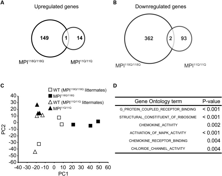
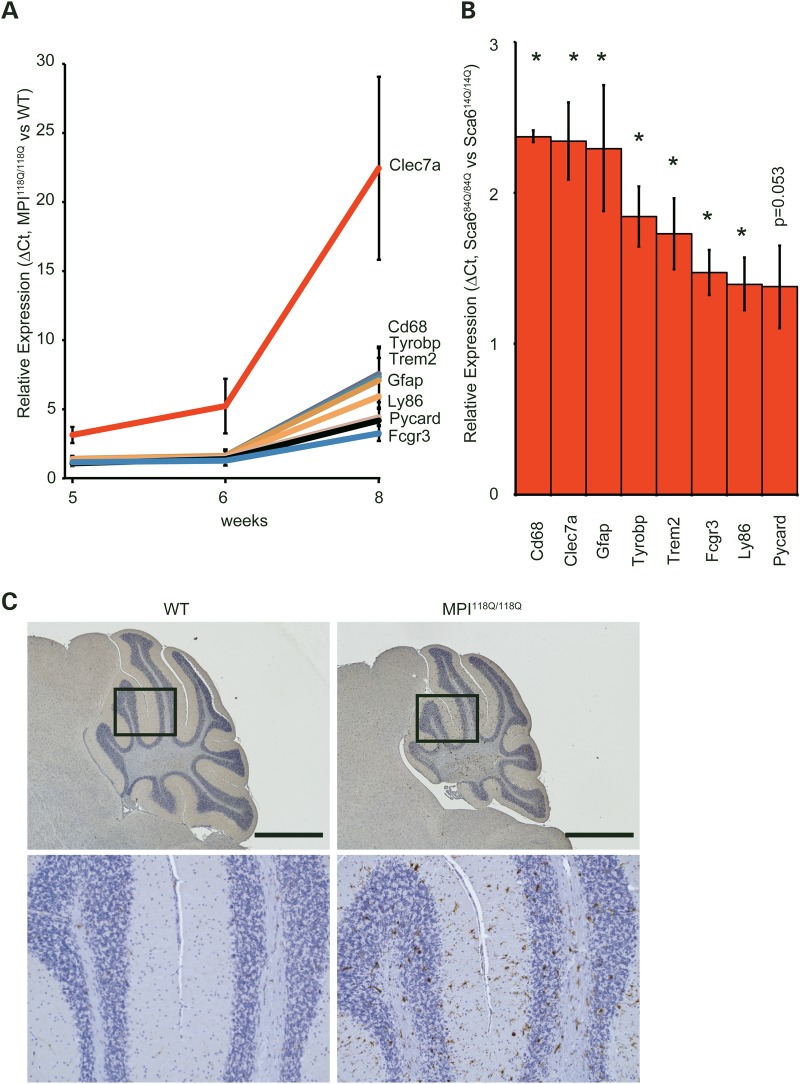
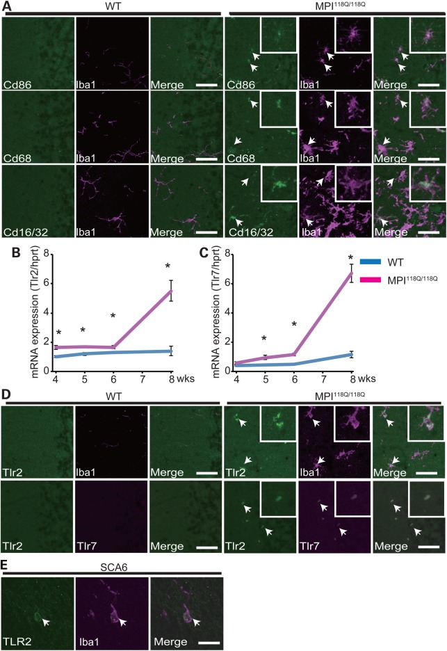
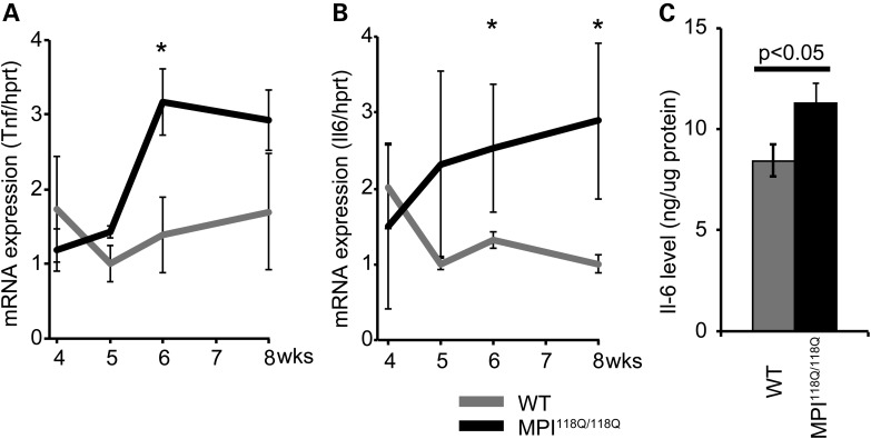
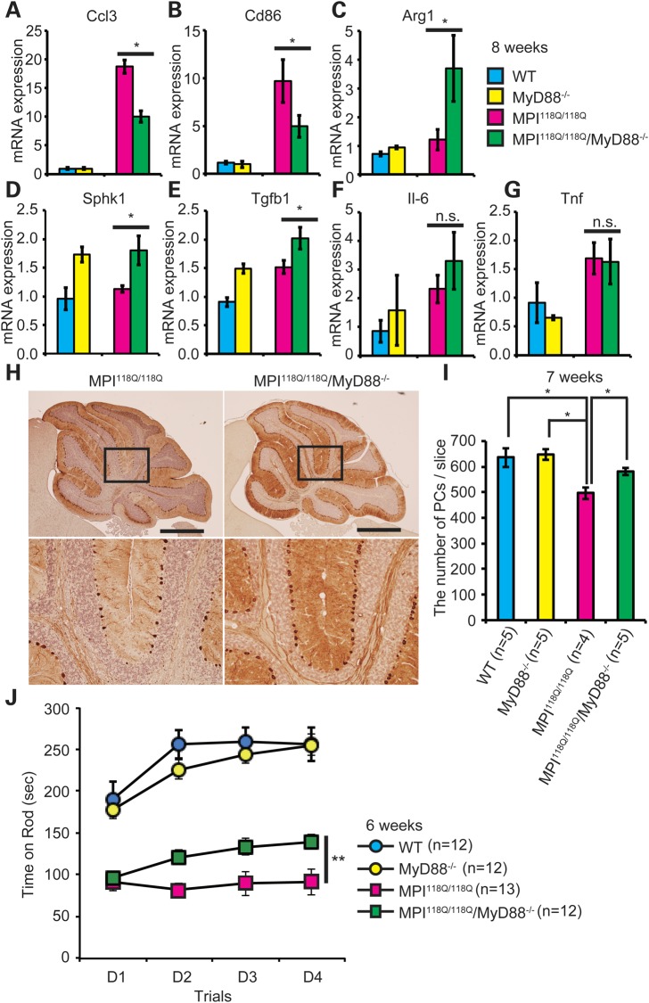
Spinocerebellar ataxia type 6 (SCA6) is dominantly inherited neurodegenerative disease, caused by an expansion of CAG repeat encoding a polyglutamine (PolyQ) tract in the Cav2.1 voltage-gated calcium channel. Its key pathological features include selective degeneration of the cerebellar Purkinje cells (PCs), a common target for PolyQ-induced toxicity in various SCAs. Mutant Cav2.1 confers toxicity primarily through a toxic gain-of-function mechanism; however, its molecular basis remains elusive. Here, we studied the cerebellar gene expression patterns of young Sca6-MPI(118Q/118Q) knockin (KI) mice, which expressed mutant Cav2.1 from an endogenous locus and recapitulated many phenotypic features of human SCA6. Transcriptional signatures in the MPI(118Q/118Q) mice were distinct from those in the Sca1(154Q/2Q) mice, a faithful SCA1 KI mouse model. Temporal expression profiles of the candidate genes revealed that the up-regulation of genes associated with microglial activation was initiated before PC degeneration and was augmented as the disease progressed. Histological analysis of the MPI(118Q/118Q) cerebellum showed the predominance of M1-like pro-inflammatory microglia and it was concomitant with elevated expression levels of tumor necrosis factor, interleukin-6, Toll-like receptor (TLR) 2 and 7. Genetic ablation of MyD88, a major adaptor protein conveying TLR signaling, altered expression patterns of M1/M2 microglial phenotypic markers in the MPI(118Q/118Q) cerebellum. More importantly, it ameliorated PC loss and partially rescued motor impairments in the early disease phase. These results suggest that early neuroinflammatory response may play an important role in the pathogenesis of SCA6 and its modulation could pave the way for slowing the disease progression during the early stage of the disease.


Figures

Figure 1.
Figure 1.
Gene expression profile of MPI118Q/118Q KI mice. (A) Venn diagram of genes that were significantly up-regulated in the MPI118Q/118Q mice but not in the MPI11Q/11Q mice. (B) Venn diagram of genes that were significantly down-regulated in the MPI118Q/118Q mice but not in the MPI11Q/11Q mice. (C) PCA of gene expression patterns in the MPI118Q/118Q and MPI11Q/11Q mice and their respective WT littermates. (D) GO enrichment analysis of the MPI118Q/118Q mice.
Figure 2.
Figure 2.
(A) Temporal expression patterns of eight neuroinflammatory genes in the MPI118Q/118Q cerebellum. These eight genes (Clec7a, Cd68, Tyrobp, Trem2, Gfap, Ly86, Pycard and Fcgr3) were selected from the microarray data of the MPI118Q/118Q KI mice. The qPCR data were evaluated using the ΔCt method. The statistically significant differences were detected for Clec7a (5, 6 and 8 weeks, P < 0.02, <0.05 and <0.02, respectively), Cd68 (5, 6 and 8 weeks, P < 0.05, <0.05 and <0.02, respectively), Gfap (5, 6 and 8 weeks, P < 0.05, <0.05 and <0.02, respectively), Fcgr3 (5, 6 and 8 weeks, P < 0.05, <0.05 and <0.01, respectively), Ly86 (6 and 8 weeks, P < 0.05 and <0.01, respectively), Tyrobp (8 weeks, P < 0.05), Trem2 (8 weeks, P < 0.01) and Pycard (8 weeks, P < 0.02) by Student's t-test. (B) The expression of eight neuroinflammatory genes (Clec7a, Cd68, Tyrobp, Trem2, Gfap, Ly86, Pycard and Fcgr3) in the Sca684Q/84Q KI cerebellum at 90 weeks of age. The error bars represent standard errors. The asterisks indicate a statistically significant difference (P < 0.05). (C) Immunohistochemical detection of Iba1-positive microglia in the 6-week-old MPI118Q/118Q (right) and WT (left) mice. The cerebellar sections were immunostained with Iba1 antibodies and counter-stained with hematoxylin (scale bars indicate 1 mm.)
Figure 3.
Figure 3.
Characterization of activated microglia in the cerebellum of the MPI118Q/118Q mice. (A) Double-immunofluorescence analysis of a microglial marker Iba1 and activated microglial marker (Cd86, Cd68 and Cd16/32) antibodies, stained cerebellar section of a 6-week-old MPI118Q/118Q mouse. (BD) The infiltrating microglia express Tlr2 and Tlr7 in the Sca6 model. The scale bars indicate 50 μm. (B) qPCR analysis of Tlr2 in the MPI118Q/118Q cerebellum at 4, 5, 6 and 8 weeks of age. (C) qPCR analysis of Tlr7 in the MPI118Q/118Q cerebellum at 4, 5, 6 and 8 weeks of age. The asterisks indicate a statistically significant difference (P < 0.05). The error bars represent standard errors. (D) Co-immunofluorescence studies of the microglial marker Iba1 with Tlr2 (top) and Tlr2 with Tlr7 (bottom). The scale bars indicate 50 μm. The arrows indicate doubly labeled cells. (E) Co-immunofluorescence studies of the microglial marker Iba1 with TLR2 in SCA6 postmortem cerebellum. The scale bars indicate 25 μm. The arrows indicate doubly labeled cells.
Figure 4.
Figure 4.
Expression analysis of two pro-inflammatory cytokines, Tnf and Il-6, in the MPI118Q/118Q cerebellum. The expression levels of the indicated pro-inflammatory cytokines, Tnf (A) and Il-6 (B) in the 4-, 5-, 6- and 8-week-MPI118Q/118Q cerebellum. The asterisks indicate P < 0.05. (C) The protein level of the cerebellum of the 8-week-MPI118Q/118Q mice measured using IL-6 ELISA. The error bars represent standard errors.
Figure 5.
Figure 5.
Deficiency of MyD88 ameliorated PC death and motor impairment in MPI118Q/118Q mice. (AF) qPCR analyses of Ccl3 (A), Cd86 (B), Arg1 (C), Sphk1 (D), Tgfb1 (E), Il-6 (F) and Tnf (G) in the WT (n = 4), MyD88−/− (n = 3), MPI118Q/118Q (n = 5) and MPI118Q/118Q; MyD88−/− (n = 4) cerebellum at 8 weeks of age. Statistically significant difference P < 0.05 by Student's t-test. (H) Anti-calbindin Abs immunohistochemistry on cerebellar sections of 7-week-old WT, MyD88−/−, MPI118Q/118Q and MPI118Q/118Q; MyD88−/− mice (scale bars indicate 1 mm). (I) Quantitative analysis showed that a decrease in the number of PCs was more pronounced in the MPI118Q/118Q cerebellum compared with the MPI118Q/118Q; MyD88−/− cerebellum at 7-week-old age (*P < 0.05 by Student's t-test). Error bars indicate SEM. (J) Rota-rod analysis of the DM mice. Mice were trained for four trials per day for 4 days (D1–D4). Error bars indicate SEM. MPI118Q/118Q versus MPI118Q/118Q/MyD88−/− (**repeated measures ANOVA, group effect; F = 11.608, P = 0.006, time effect; F = 0.296, P = 0.693, group and time interaction; F = 4.052, P = 0.045).

References

    1. Orr H.T., Zoghbi H.Y. (2007) Trinucleotide repeat disorders. Annu. Rev. Neurosci., 30, 575–621.2. - PubMed
    1. Gatchel J.R., Watase K., Thaller C., Carson J.P., Jafar-Nejad P., Shaw C., Zu T., Orr H.T., Zoghbi H.Y. (2008) The insulin-like growth factor pathway is altered in spinocerebellar ataxia type 1 and type 7. Proc. Natl Acad. Sci. U.S.A., 105, 1291–1296. - PMC - PubMed
    1. Zhuchenko O., Bailey J., Bonnen P., Ashizawa T., Stockton D.W., Amos C., Dobyns W.B., Subramony S.H., Zoghbi H.Y., Lee C.C. (1997) Autosomal dominant cerebellar ataxia (SCA6) associated with small polyglutamine expansions in the alpha(1A)-voltage-dependent calcium channel. Nat. Genet., 15, 62–69. - PubMed
    1. Ishikawa K., Tanaka H., Saito M., Ohkoshi N., Fujita T., Yoshizawa K., Ikeuchi T., Watanabe M., Hayashi A., Takiyama Y. et al. (1997) Japanese families with autosomal dominant pure cerebellar ataxia map to chromosome 19p13.1-p13.2 and are strongly associated with mild CAG expansions in the spinocerebellar ataxia type 6 gene in chromosome 19p13.1. Am. J. Hum. Genet., 61, 336–346. - PMC - PubMed
    1. Watase K., Barrett C.F., Miyazaki T., Ishiguro T., Ishikawa K., Hu Y., Unno T., Sun Y., Kasai S., Watanabe M. et al. (2008) Spinocerebellar ataxia type 6 knockin mice develop a progressive neuronal dysfunction with age-dependent accumulation of mutant Ca(v)2.1 channels. Proc. Natl Acad. Sci. U.S.A., 105, 11987–11992. - PMC - PubMed

Publication types

MeSH terms

Associated data