Review
. 2015:84:895-921.
doi: 10.1146/annurev-biochem-060614-033930.

A molecular description of cellulose biosynthesis

Affiliations
Review

A molecular description of cellulose biosynthesis

Joshua T McNamara et al. Annu Rev Biochem. 2015.

Abstract

Cellulose is the most abundant biopolymer on Earth, and certain organisms from bacteria to plants and animals synthesize cellulose as an extracellular polymer for various biological functions. Humans have used cellulose for millennia as a material and an energy source, and the advent of a lignocellulosic fuel industry will elevate it to the primary carbon source for the burgeoning renewable energy sector. Despite the biological and societal importance of cellulose, the molecular mechanism by which it is synthesized is now only beginning to emerge. On the basis of recent advances in structural and molecular biology on bacterial cellulose synthases, we review emerging concepts of how the enzymes polymerize glucose molecules, how the nascent polymer is transported across the plasma membrane, and how bacterial cellulose biosynthesis is regulated during biofilm formation. Additionally, we review evolutionary commonalities and differences between cellulose synthases that modulate the nature of the cellulose product formed.

Keywords: biofilm; cellulose synthase; cyclic di-GMP; exopolysaccharide biosynthesis; membrane transport; processive glycosyltransferase.


Figures

Figure 1
Figure 1
Cellulose is a linear, ribbon-shaped polymer of glucose molecules. The individual glucose units are connected via glycosidic bonds between their C1 and C4 positions, and the anomeric C1 carbon adopts the β-configuration. Each glucose unit is rotated by ~180° relative to its neighbors and forms two hydrogen bonds with each adjacent unit (dashed black lines). The unmodified C1- and C4-hydroxyl groups of cellulose form the polymer’s reducing and nonreducing ends, respectively. Cellulose is elongated at its nonreducing end via a nucleophilic substitution reaction that transfers a glucose unit from UDP-glucose to the polymer’s C4-hydroxyl group. The reaction is facilitated by deprotonation of the acceptor hydroxyl (its hydrogen atom is labeled H) by a general base (labeled B). For clarity, gray spheres indicating hydrogen atoms are shown only in the right panel.
Figure 2
Figure 2
Cellulose synthases (CeSs) from different kingdoms share similar features for cellulose synthesis and translocation. Membrane topologies were predicted for the indicated enzymes by the TOPCONS program suite (143), and the consensus predictions are shown as gray cartoons. Pc stands for Phytophthora capsici CesA3 (UniProt entry H6U2P7); Md, Micrasterias denticulata (gi|293413208|); At, Arabidopsis thaliana CesA3 (UniProt entry Q941L0); Cs, Ciona savignyi (UniProt entry Q6RCS2); Gh, Gossypium hirsutum CesA1 (UniProt entry I1T421); and Rs, Rhodobacter sphaeroides (UniProt entry Q3J125). The putative gating loop of each sequence is indicated by a wavy line and is labeled for Rs. The transmembrane (TM) helices contacting the cellulose polymer in Rs BcsA (bacterial cellulose synthase subunit A) are colored brown. In all enzymes, the TM helices frame an intracellular glycosyltransferase (GT) domain, whose architecture is represented by the Rs BcsA structure (Protein Data Bank entry 4P00) (center). Cellulose (cyan) and a UDP molecule (violet) are shown as sticks. Mg2+ is shown as a yellow sphere. Conserved motifs are labeled with Roman numerals and shown as consensus sequences obtained for the listed enzymes. For each motif, the Cαatom for the underlined residue is shown as a blue sphere. The insertions of the plant-conserved region (P-CR) and class-specific region (CSR), primarily found in plants, are represented as orange triangles.
Figure 3
Figure 3
The Rhodobacter sphaeroides bacterial cellulose synthase subunits A and B (BcsA and BcsB) form an inner membrane protein complex. The intracellular domain of the BcsA–B complex consists of a glycosyltransferase (GT) domain ( green) inserted between BcsA’s transmembrane (TM) helices 4 and 5, as well as a regulatory C-terminal PilZ domain (red ) that binds the allosteric activator cyclic di-GMP (c-di-GMP). The TM region is formed from BcsA’s eight TM helices (brown and yellow) and BcsB’s membrane anchor ( purple cylinder). TM3–8 of BcsA (brown) primarily contribute to forming the cellulose-conducting channel. BcsB’s periplasmic domain contains two copies of a repeating unit containing a carbohydrate-binding domain (CBD) (blue) linked to a flavodoxin-like domain (FD) ( gray). The translocating glucan is shown as cyan and red spheres, and UDP at the active site and c-di-GMP at the PilZ domain are shown in spheres colored according to elements. The putative membrane area is shaded gray. Coordinates were obtained from Protein Data Bank entry 4P00.
Figure 4
Figure 4
The cellulose synthase (CeS) donor- and acceptor-binding sites are formed from evolutionarily conserved sequences. Residues of Rhodobacter sphaeroides (Rs) bacterial cellulose synthase subunit A (BcsA) contacting either the UDP molecule or the acceptor glucose (sticks and spheres) are labeled with Roman numerals corresponding to the consensus sequences in Figure 2. Mg2+ coordinated by the DxD motif is shown as a yellow sphere. Asp343 of the TED motif hydrogen-bonds with the acceptor’s C4-hydroxyl group and likely functions as a general base during catalysis. The likely donor glucose–binding pocket is indicated by a dashed ellipsoid. The coordinates were derived from Protein Data Bank entry 4P00.
Figure 5
Figure 5
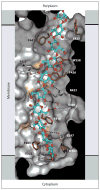
Cellulose synthase (CeS) forms a cellulose-conducting transmembrane channel. The membrane channel of bacterial cellulose synthase subunit A (BcsA) is lined with aromatic residues (brown sticks) that form CH–π stacking interactions with alternating faces of the cellulose polymer. Additionally, throughout the pore, the polymer’s equatorial hydroxyl groups form hydrogen bonds with hydrophilic BcsA residues ( gray sticks or yellow surfaces). One of two possible registers for the cellulose polymer is shown, as represented in Protein Data Bank entry 4P00.
Figure 6
Figure 6
The bacterial cellulose synthase (Bcs) contains multiple subunits at the cell envelope. The inner membrane (IM)-integrated BcsA–B complex may interact with the periplasmic domain of BcsC, thereby forming a transenvelope conduit for the nascent cellulose polymer (orange circles). The periplasmic cellulase BcsZ may cleave the translocating glucan stochastically or degrade mislocalized cellulose in the periplasm. C-di-GMP binds to the C-terminal PilZ domain of BcsA and is formed by the membrane-integrated diguanylate cyclase AdrA. The BcsD subunit is found primarily in cellulose microfibril–forming bacteria, and its biological function, as well as its cellular localization, is unclear. Abbreviations: c-di-GMP, cyclic-di-GMP; GT, glycosyltransferase; OM, outer membrane.

References

    1. Payen A. Mémoire sur la composition du tissu propre des plantes et du ligneux. C R Hebd Séances Acad Sci. 1838;7:1062–56.
    1. Klemm D, Heublein B, Fink HP, Bohn A. Cellulose: fascinating biopolymer and sustainable raw material. Angew Chem Int Ed. 2005;44:3358–93. - PubMed
    1. Staudinger H. Über Polymerisation. Ber Dtsch Chem Ges. 1920;53:1073–85.
    1. Wolfenden R, Lu X, Young G. Spontaneous hydrolysis of glycosides. J Am Chem Soc. 1998;120:6814–15.
    1. Pauly M, Keegstra K. Cell-wall carbohydrates and their modification as a resource for biofuels. Plant J. 2008;54:559–68. - PubMed

Publication types