. 2015 Aug 1;8(8):999-1009.
doi: 10.1242/dmm.020859. Epub 2015 May 28.

Phospholamban overexpression in mice causes a centronuclear myopathy-like phenotype

Affiliations

Phospholamban overexpression in mice causes a centronuclear myopathy-like phenotype

Val A Fajardo et al. Dis Model Mech. .

Abstract

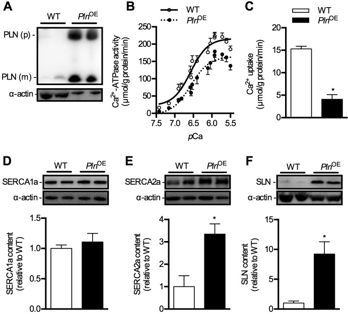
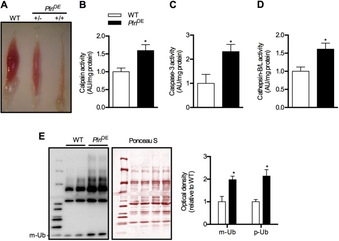
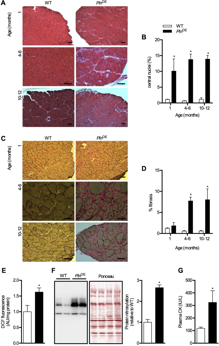
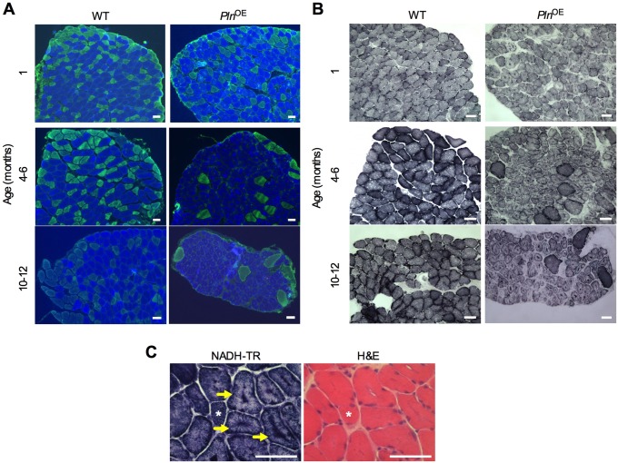
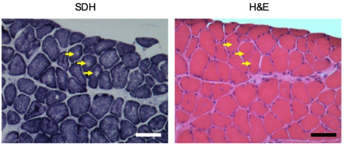
Centronuclear myopathy (CNM) is a congenital myopathy that is histopathologically characterized by centrally located nuclei, central aggregation of oxidative activity, and type I fiber predominance and hypotrophy. Here, we obtained commercially available mice overexpressing phospholamban (Pln(OE)), a well-known inhibitor of sarco(endo)plasmic reticulum Ca(2+)-ATPases (SERCAs), in their slow-twitch type I skeletal muscle fibers to determine the effects on SERCA function. As expected with a 6- to 7-fold overexpression of phospholamban, SERCA dysfunction was evident in Pln(OE) muscles, with marked reductions in rates of Ca(2+) uptake, maximal ATPase activity and the apparent affinity of SERCA for Ca(2+). However, our most significant discovery was that the soleus and gluteus minimus muscles from the Pln(OE) mice displayed overt signs of myopathy: they histopathologically resembled human CNM, with centrally located nuclei, central aggregation of oxidative activity, type I fiber predominance and hypotrophy, progressive fibrosis and muscle weakness. This phenotype is associated with significant upregulation of muscle sarcolipin and dynamin 2, increased Ca(2+)-activated proteolysis, oxidative stress and protein nitrosylation. Moreover, in our assessment of muscle biopsies from three human CNM patients, we found a significant 53% reduction in SERCA activity and increases in both total and monomeric PLN content compared with five healthy subjects, thereby justifying future studies with more CNM patients. Altogether, our results suggest that the commercially available Pln(OE) mouse phenotypically resembles human CNM and could be used as a model to test potential mechanisms and therapeutic strategies. To date, there is no cure for CNM and our results suggest that targeting SERCA function, which has already been shown to be an effective therapeutic target for murine muscular dystrophy and human cardiomyopathy, might represent a novel therapeutic strategy to combat CNM.

Keywords: Calcium regulation; Congenital myopathy; Dynamin 2; SERCA; Skeletal muscle.


Conflict of interest statement

Competing interests

The authors declare no competing or financial interests.

Figures

Fig. 1.
Fig. 1.
SERCA function in soleus muscles in PlnOE mice at 4-6 months of age. (A) Western blotting for PLN in WT and PlnOE mice from soleus muscle homogenates. For WT mice, 25 μg of total protein was loaded, whereas only 2.5 μg was required for PlnOE mice to detect PLN protein. (B) Ca2+-ATPase activity–pCa curves in WT (n=5) and PlnOE mice (n=6) in the presence of the Ca2+ ionophore. (C) Ca2+ uptake assessed in soleus muscles from WT (n=4) and PlnOE mice (n=5). (D-F) Western blotting for SERCA1a (D), SERCA2a (E) and SLN (F) in soleus muscle from WT and PlnOE mice (n=6 per genotype). Actin was used as a loading control and all values are expressed relative to WT. *P≤0.05 versus WT. All values are presented as means±s.e. PLN (p), PLN pentamer; PLN (m), PLN monomer.
Fig. 2.
Fig. 2.
Soleus muscles of PlnOE mice are atrophied at 4-6 months of age. (A) Representative images of soleus muscles extracted from WT and PlnOE mice. +/−, heterozygous; +/+, homozygous. (B) Calpain activity in soleus homogenates from WT (n=4) and PlnOE mice (n=5). (C) Caspase-3 activity in soleus muscle homogenates from WT (n=4) and PlnOE mice (n=7). (D) Cathepsin-B/L activity in soleus homogenates from WT and PlnOE mice (WT, n=4; PlnOE, n=6). Calpain activity, caspase-3 activity, and cathepsin-L activity are in arbitrary units normalized to mg protein and are presented relative to WT. (E) Total protein extracts from soleus muscles (15 μg) were immunoblotted with anti-ubiquitin (Ub) antibody. Ponceau stain was used as a loading control. The sum of optical densities from detectable ubiquitylated proteins (p-Ub) as well as the optical density of monomeric ubiquitin (m-Ub, 10 kDa) was measured and compared between genotypes (n=4 per group). Values were normalized to the sum of optical densities of bands visualized through Ponceau stain. *P≤0.05 versus WT. All values are presented as means±s.e.
Fig. 3.
Fig. 3.
Dystrophic features in soleus muscles from PlnOE mice. (A) H&E-stained sections of the soleus muscles from WT and PlnOE mice at 1 month, 4-6 months and 10-12 months of age. (B) Percentage of fibers containing central nuclei in the soleus at 1 month, 4-6 months and 10-12 months of age (n=3 per genotype at each age with 300-600 fibers counted per mouse). (C) Van-Geison-stained sections of the soleus muscles from WT and PlnOE mice at 1 month, 4-6 months and 10-12 months of age. (D) Quantification of fibrotic area in the soleus at 1 month, 4-6 months and 10-12 months of age (n=3-4 per group at each age). ImageJ software was used to quantify fibrotic area. (E) Reactive oxygen species generation in whole soleus determined using DCF assay at 4-6 months of age (WT, n=4; PlnOE, n=5). (F) Total protein extracts from soleus muscles (15 μg) were immunoblotted with anti-nitrotyrosine antibody. The sum of optical densities from detectable nitrosylated proteins (50-100 kDa) was measured and compared between genotypes at 4-6 months of age (n=6 per group). Values were normalized to the sum of optical densities of bands visualized through Ponceau stain. (G) Plasma CK levels in WT (n=5) and PlnOE mice (n=9) at 4-6 months of age. Scale bars: 50 μm (A,C). *P≤0.05 versus WT using two-way ANOVA and Tukey's post hoc analysis for % central nuclei and % fibrosis; Student's t-test for DCF, protein nitrosylation and plasma CK. All values are presented as means±s.e.
Fig. 4.
Fig. 4.
Centronuclear myopathy in the soleus muscles from PlnOE mice. (A) Representative images of soleus muscles showing type I fiber predominance and hypotrophy. MHC immunofluorescence stained sections of the soleus muscles at 1 month, 4-6 months and 10-12 months of age. Cross sections were stained with antibodies against MHC to identify type I (blue), type IIA (green), type IIB (red) and type IIX (unstained) fibers. (B) Representative images of soleus muscles showing central accumulation of oxidative activity. Succinate dehydrogenase (SDH)-stained sections display central aggregation of oxidative activity in the PlnOE mice at 1 month, 4-6 months and 10-12 months of age. (C) NADH-TR-stained cross sections demonstrating radiating SR strands in soleus muscles from PlnOE mice (arrows). Asterisks represent the same fiber across cryosections. Scale bars: 50 μm.
Fig. 5.
Fig. 5.
Potential core-like lesions in PlnOE mouse at 4-6 months of age. SDH-stained sections from the soleus muscle show areas devoid of oxidative staining representing potential core-like lesions (yellow arrows). Corresponding H&E-stained serial section shows that the lack of oxidative staining with SDH is not due to the presence of a vacuole or an artifact in the muscle fiber.

References

    1. Agrawal P. B., Pierson C. R., Joshi M., Liu X., Ravenscroft G., Moghadaszadeh B., Talabere T., Viola M., Swanson L. C., Haliloğlu G. et al. (2014). SPEG interacts with myotubularin, and its deficiency causes centronuclear myopathy with dilated cardiomyopathy. Am. J. Hum. Genet. 95, 218-226. 10.1016/j.ajhg.2014.07.004 - DOI - PMC - PubMed
    1. Al-Qusairi L. and Laporte J. (2011). T-tubule biogenesis and triad formation in skeletal muscle and implication in human diseases. Skelet. Muscle 1, 26 10.1186/2044-5040-1-26 - DOI - PMC - PubMed
    1. Al-Qusairi L., Weiss N., Toussaint A., Berbey C., Messaddeq N., Kretz C., Sanoudou D., Beggs A. H., Allard B., Mandel J.-L. et al. (2009). T-tubule disorganization and defective excitation-contraction coupling in muscle fibers lacking myotubularin lipid phosphatase. Proc. Natl. Acad. Sci. USA 106, 18763-18768. 10.1073/pnas.0900705106 - DOI - PMC - PubMed
    1. Altamirano F., Lopez J. R., Henriquez C., Molinski T., Allen P. D. and Jaimovich E. (2012). Increased resting intracellular calcium modulates NF-kappaB-dependent inducible nitric-oxide synthase gene expression in dystrophic mdx skeletal myotubes. J. Biol. Chem. 287, 20876-20887. 10.1074/jbc.M112.344929 - DOI - PMC - PubMed
    1. Anderson D. M., Anderson K. M., Chang C.-L., Makarewich C. A., Nelson B. R., McAnally J. R., Kasaragod P., Shelton J. M., Liou J., Bassel-Duby R. et al. (2015). A micropeptide encoded by a putative long noncoding RNA regulates muscle performance. Cell 160, 595-606. 10.1016/j.cell.2015.01.009 - DOI - PMC - PubMed

Publication types

MeSH terms