. 2015 Jun;8(6):527-41.
doi: 10.1242/dmm.019083. Epub 2015 Apr 7.

The Meckel-Gruber syndrome protein TMEM67 controls basal body positioning and epithelial branching morphogenesis in mice via the non-canonical Wnt pathway

Affiliations

The Meckel-Gruber syndrome protein TMEM67 controls basal body positioning and epithelial branching morphogenesis in mice via the non-canonical Wnt pathway

Zakia A Abdelhamed et al. Dis Model Mech. 2015 Jun.

Abstract

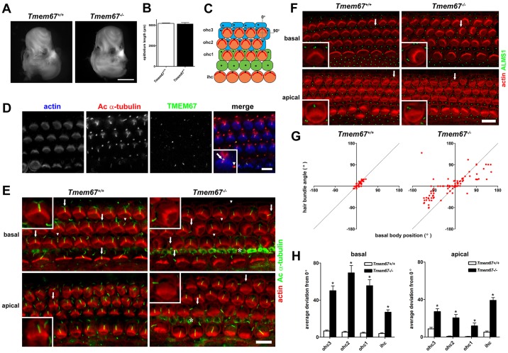
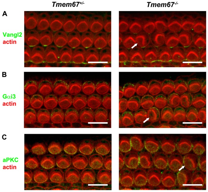
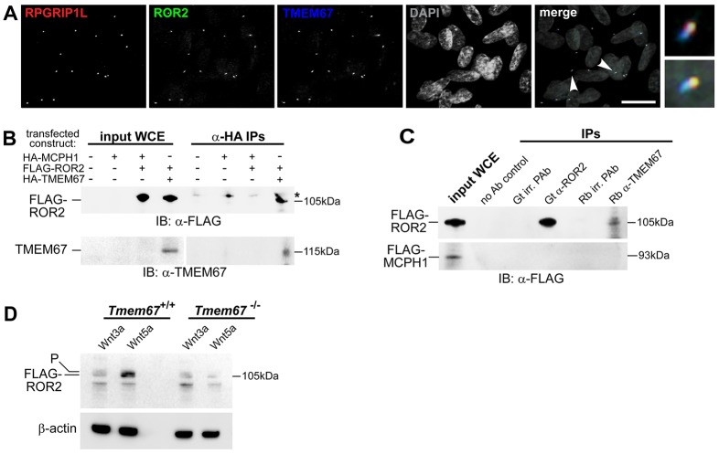
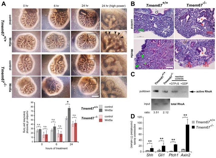
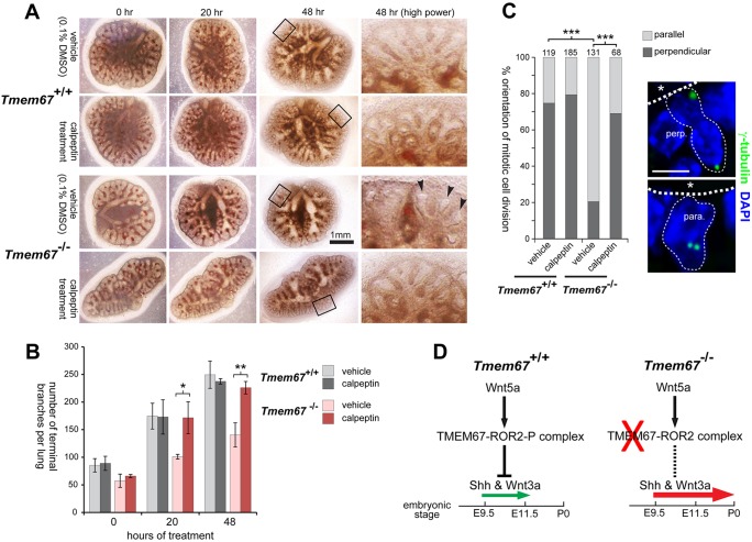
Ciliopathies are a group of developmental disorders that manifest with multi-organ anomalies. Mutations in TMEM67 (MKS3) cause a range of human ciliopathies, including Meckel-Gruber and Joubert syndromes. In this study we describe multi-organ developmental abnormalities in the Tmem67(tm1Dgen/H1) knockout mouse that closely resemble those seen in Wnt5a and Ror2 knockout mice. These include pulmonary hypoplasia, ventricular septal defects, shortening of the body longitudinal axis, limb abnormalities, and cochlear hair cell stereociliary bundle orientation and basal body/kinocilium positioning defects. The basal body/kinocilium complex was often uncoupled from the hair bundle, suggesting aberrant basal body migration, although planar cell polarity and apical planar asymmetry in the organ of Corti were normal. TMEM67 (meckelin) is essential for phosphorylation of the non-canonical Wnt receptor ROR2 (receptor-tyrosine-kinase-like orphan receptor 2) upon stimulation with Wnt5a-conditioned medium. ROR2 also colocalises and interacts with TMEM67 at the ciliary transition zone. Additionally, the extracellular N-terminal domain of TMEM67 preferentially binds to Wnt5a in an in vitro binding assay. Cultured lungs of Tmem67 mutant mice failed to respond to stimulation of epithelial branching morphogenesis by Wnt5a. Wnt5a also inhibited both the Shh and canonical Wnt/β-catenin signalling pathways in wild-type embryonic lung. Pulmonary hypoplasia phenotypes, including loss of correct epithelial branching morphogenesis and cell polarity, were rescued by stimulating the non-canonical Wnt pathway downstream of the Wnt5a-TMEM67-ROR2 axis by activating RhoA. We propose that TMEM67 is a receptor that has a main role in non-canonical Wnt signalling, mediated by Wnt5a and ROR2, and normally represses Shh signalling. Downstream therapeutic targeting of the Wnt5a-TMEM67-ROR2 axis might, therefore, reduce or prevent pulmonary hypoplasia in ciliopathies and other congenital conditions.

Keywords: Ciliopathy; Hair bundle; Kinocilia; MKS3; Meckelin; PCP; Planar cell polarity; Primary cilia; Stereocilia; TMEM67; Wnt signalling.


Conflict of interest statement

Competing interests

The authors declare no competing or financial interests.

Figures

Fig. 1.
Fig. 1.
Gross anatomical malformations, laterality defects, cardiac defects and pulmonary hypoplasia in Tmem67−/− mutant mouse embryos and pups. (A) Upper panels: whole-mount E11.5 embryos showing the earliest sign of laterality defects with inverted tail turning (arrowhead) in a Tmem67−/− mutant embryo. Whole-mount lungs of E15.5 embryos (middle panels) and P0 pups (lower panels). Tmem67−/− E15.5 mutant embryos had identical left (L) and right (R) lungs, indicating left lung isomerism. Lobes of the right lung in Tmem67+/+ are numbered as indicated. Scale bar: 1500 μm. (B) Upper panels: H&E-stained lung tissue section showing pulmonary hypoplasia, congested vessels and delayed development of the pulmonary alveoli in an E18.5 Tmem67−/− embryo. Lower panels: immunohistochemical staining for Ki-67 in E18.5 lung sections. Scale bars: 40 μm. (C) IF microscopy of E14.5 lung-tissue sections stained for primary cilia (acetylated α-tubulin; red), basal bodies (γ-tubulin; green) and for nuclei with DAPI (blue). Scale bar: 10 μm. Bar graphs show primary cilia length and number in Tmem67+/+ and Tmem67−/− tissues. Statistical significance of the pairwise comparison are **P<0.01 and ***P<0.001 (Student's two-tailed t-test). Error bars indicate s.e.m. (D) Upper panels: whole-mount E15.5 embryo images showing generally delayed development, underdeveloped limbs (white arrowheads) and omphalocele (red arrowhead) in Tmem67−/− embryos, with details of limb dysplasia shown below. Lower panels: whole-mount P1 pups, showing reduced body longitudinal axis in the Tmem67−/− pups. Scale bars: 1 cm. (E) Upper panels: H&E-stained horizontal section through the chest cavity of E12.5 Tmem67+/+ and Tmem67−/− animals showing a ventricular septal defect (VSD) (arrowhead) in the mutant. Scale bars: 100 μm. Lower panels: VSD (arrowhead) in an E15.5 sagittal heart section. Scale bar: 200 μm. (F) Horizontal sections through the thoracic cavity of the Tmem67−/− mutant and wild-type control showing aberrant lung lobulation, dextrocardia, major cardiac malformation and cardiac oedema or pericardial effusion (asterisk) in the Tmem67−/− embryo. Scale bars: 100 μm. (G) H&E- (upper panels) and IHC- (lower panels) stained E18.5 liver-tissue sections. H&E sections show a persistent double-layered ductal plate (black arrowheads) around the portal vein branches (pvb) and abnormally accumulating cells around the pvb in Tmem67−/− embryos (white arrowheads). IHC-stained liver sections for cytokeratin-19 show a double-layered ductal plate and multiple bile ducts in Tmem67−/− embryos. A normal bile duct in the Tmem67+/+ section is indicated (arrowhead). Scale bars: 50 μm.
Fig. 2.
Fig. 2.
Orientation defects in stereociliary hair bundles with uncoupling from kinocilium and basal body position of hair cells in the organ of Corti of neonatal Tmem67−/− mice. (A) Cochleae dissected from P0 Tmem67+/+ mice (control, left) were indistinguishable from those of Tmem67−/− littermates (right). Scale bar: 1 mm. (B) Total length measurements of phalloidin-stained organ of Corti were not significantly different between control and mutant animals (n=4 cochleae per genotype). (C) Schematic representation of cellular architecture of the neonatal organ of Corti. There is a single row of inner hair cells (ihc) located at the neural edge of the sensory epithelium, and three rows of outer hair cells (ohc1-3) spanning the abneural portion. The hair cell stereociliary bundles (red) are regularly oriented, with their vertices pointing towards the abneural pole, corresponding to an alignment of 0° (denoted by vertical dotted line). A line of alignment to 90° is also shown for reference. Ohc are surrounded by a mosaic of non-sensory supporting cells, including pillar cells (green) and Deiters' cells (blue). Primary cilia are represented as black dots. (D) Confocal projections of P0 Tmem67+/+ organ of Corti mid-turn region (50% of cochlear length) stained for actin using phalloidin to demarcate stereociliary hair bundles (blue), acetylated α-tubulin antibody (cilia; red) and TMEM67 (green). TMEM67 decorates the proximal regions of cilia in both hair-cell types and supporting cells. The magnified inset shows TMEM67 ciliary localisation in a single outer hair cell (arrow) and an adjacent Deiters' cell (arrowhead). Scale bar: 10 µm. (E) On the surface of the basal turn (10-20% of cochlear length) in the organ of Corti of a P0 Tmem67+/+ mouse (left), there was a regular arrangement of V-shaped stereociliary ohc hair bundles (phalloidin; red), with kinocilia (acetylated α-tubulin; green) positioned at the abneural pole (around 0°) of hair cells in all three rows (arrows; shown in magnified insets). Each kinocilium was in close apposition to the vertex of each hair bundle. Non-sensory supporting cells were also ciliated (arrowheads). In a Tmem67−/− littermate (right) kinocilia were often mislocalised from the abneural pole of the hair cell (arrows; shown in magnified insets), and in these cells the orientation of the hair bundle was uncoupled from the kinocilium position. Adjacent supporting cells were often not ciliated (arrowheads). Similar effects were seen in the apical turn region (∼70-80% cochlear length). Cytoskeletal staining of inner pillar cells is indicted by asterisks. Scale bar: 10 µm. (F) Basal body position and hair-bundle orientation were tightly coupled in basal and apical regions of the Tmem67+/+ organ of Corti (left). Uncoupling of hair-bundle orientation from basal body position was apparent in all rows of hair cells, in both basal and apical regions in Tmem67−/− cochleae (detail indicated by arrows is shown in magnified insets). Scale bar: 10 µm. (G) Scatter plots showing hair-bundle orientation versus position of the basal body for individual ohc in the basal region (corresponding to ∼10-20% of cochlear length) of a Tmem67+/+ mouse (left; n=230) and a Tmem67−/− littermate (right; n=165). Dashed lines indicate the position of perfect correlation (Pearson's coefficient of correlation, r=1). (H) Genotype-specific differences in basal body position for individual hair cell rows in basal (10-20%, left) and apical (70-80%, right) cochlear regions. Average deviations from 0° were significantly different between the genotypes for all rows (pairwise comparisons are *P<0.001; Student's unpaired t-test) in both basal and apical regions. Error bars indicate s.e.m.
Fig. 3.
Fig. 3.
Normal PCP and apical planar asymmetry in the organ of Corti of neonatal Tmem67−/− mice. Confocal projections of P0 Tmem67+/− (left panels) and Tmem67−/− (right panels) basal turn organ of Corti (corresponding to 10-20% of cochlear length) stained for actin to demarcate stereociliary hair bundles and cell borders (red). (A) In both genotypes, Vangl2 (green) localised to supporting cells at the adherens junction with hair cells. (B) Gαi3 (green) is enriched in the lateral ‘bare zone’ on the apical surface of outer hair cells. (C) aPKC (green) is enriched in the medial/neural compartment on the apical surface of outer hair cells. Misaligned hair bundles in Tmem67−/− cochleae (arrows) are adjacent to normally expressed Vangl2, or display the normal asymmetric expression of Gαi3 and aPKC. Scale bars: 10 µm.
Fig. 4.
Fig. 4.
Non-canonical Wnt signalling defects in Tmem67−/− cells and interaction of Wnt5a with the TMEM67 N-terminal domain. (A) Schematic diagram of conserved domains and structural motifs within the TMEM67 protein, comprising a signal peptide (yellow), a cysteine-rich domain (CRD, orange), regions of β-sheet periodicity (grey), seven predicted transmembrane helices (TM, black) and a coiled-coil domain (CC, blue). Locations are indicated by amino acid residue (aa), with pathogenic missense mutations highlighted in red. The approximate locations of the two epitopes used to raise N-terminal (Nt) and C-terminal (Ct) rabbit polyclonal antibodies (Ab) are indicated. The TMEM67 regions used for exogenous protein expression are indicated by grey boxes. (B) TOPFlash assays to quantify canonical Wnt signalling activity in Tmem67+/+ and Tmem67−/− MEFs, following treatment with either control L-cell or Wnt3a-conditioned media, as indicated, and co-transfection with empty vector control, wild-type HA-TMEM67, or HA-TMEM67 containing a series of pathogenic missense mutations. Wild-type HA-TMEM67 rescued de-regulated canonical Wnt signalling in Tmem67−/− cells, but missense constructs did not. (C) Tmem67−/− cells had a defective response to Wnt5a, expressed as the ratio of Wnt3a response:combined response to both Wnt3a and Wnt5a. The correct response to Wnt5a was only rescued with wild-type HA-TMEM67. Values shown are means of at least four independent replicates and error bars indicate ±s.e.m. The statistical significance of the pair-wise comparisons with wild-type HA-TMEM67 values (#) are represented as *P<0.05, **P<0.01 and ***P<0.001, Student's two-tailed t-test. (D) Left panel: Coomassie-stained SDS-PAGE analysis of fluorescence-labelled BSA (F-BSA), Wnt3a (F-Wnt3a) and Wnt5a (F-Wnt5a) proteins. Molecular masses of protein size standards (kDa) are indicated. Middle panel: the same gel photographed under UV light to show fluorescent labelling of BSA control, Wnt3a and Wnt5a proteins. Right panel: expression of TMEM67-Nt proteins (predicted molecular mass 48 kDa), containing the indicated missense mutations. (E) Preferential in vitro interaction of F-Wnt5a, but not F-Wnt3a or F-BSA negative control, with increasing amount of wild-type TMEM67-Nt. (F) Interaction of F-Wnt5a with wild-type TMEM67-Nt only, but not TMEM-Nt proteins containing the indicated missense mutations. Values shown are the means of three independent replicates and error bars indicate ±s.e.m. The statistical significance of the pair-wise comparisons with wild-type TMEM67-Nt values (#) are represented as *P<0.05 and **P<0.01, Student's two-tailed t-test.
Fig. 5.
Fig. 5.
The receptor tyrosine kinase-like orphan receptor ROR2 colocalises and interacts with TMEM67, and is dependent on this interaction for phosphorylation. (A) Four-colour IF imaging showing that endogenous ROR2 (green) colocalizes with TMEM67 (blue) and RPGRIP1L (red) at the ciliary transition zone. Arrowheads indicate regions shown in magnified insets. DAPI is pseudocoloured in grey. Scale bar: 10 μm. (B) Anti-HA co-immunoprecipitations (IPs) demonstrating interaction between full-length exogenous HA-tagged TMEM67 (size 115 kDa) and FLAG-tagged ROR2 (size 105 kDa). Input whole-cell extracts (WCE) for the indicated transfected constructs are on the left. IP of an irrelevant protein (HA-tagged MCPH1) was a negative control. Results are shown for immunoblotting (IB) for anti-FLAG (upper panel) and anti-TMEM67 (lower panel). * indicates a non-specific band in IPs; see supplementary material Fig. S6 for full unprocessed images. (C) Upper panel: IPs demonstrating interaction between FLAG-tagged ROR2 and endogenous TMEM67. Input WCE is shown on the left, and negative control IPs include a no antibody (Ab) control and goat (Gt) and rabbit (Rb) irrelevant (irr.) polyclonal antibodies (PAb). Immunoblotting (IB) for anti-FLAG shows pulldown of FLAG-ROR2 by Gt anti-ROR2 and Rb anti-TMEM67. Lower panel: IPs with irrelevant protein (FLAG-MCPH1, size 93 kDa). (E) Loss of the active phosphorylated ROR2 isoform (labelled P) in mutant Tmem67−/− cells following Wnt5a treatment, compared with strong induction of the active isoform (upper band, as indicated) in wild-type Tmem67+/+ cells. Loading control is for β-actin.
Fig. 6.
Fig. 6.
Loss of Wnt5a-induced branching morphogenesis during Tmem67−/− embryonic lung ex vivo organogenesis. (A) Embryonic (E12.5) lungs were explanted and treated for 0, 6 and 24 h with either control-conditioned medium or medium containing Wnt5a. Magnified insets (black frames) under high power are shown for 24-h treatments. Epithelial branching is significantly induced by Wnt5a in Tmem67+/+ lungs, but this response is absent in Tmem67−/− lungs. The bar graph shows quantification of the total number of branches in one lung for each genotype. Values shown are means of three independent replicates and error bars indicate ±s.e.m. The statistical significance of the pair-wise comparisons are represented as *P<0.05 and n.s. for non-significant, Student's two-tailed t-test. (B) H&E staining of ex-vivo-cultured embryonic lung sections, showing normal acini (ac) and mesenchymal tissue (ms, in green) for wild-type Tmem67+/+ lung, and the stimulation of normal epithelial branching by Wnt5a (green asterisk and arrowheads). In contrast, Tmem67−/− lungs have abnormal mesenchymal cell condensates (red arrowheads), suggesting defective epithelial-mesenchymal induction. The red asterisks indicate abnormal bronchiolar formation; cl indicates the direction of the central lung. (C) Rho activation pull-down assays of whole-cell extracts from wild-type Tmem67+/+ and mutant Tmem67−/− embryonic (E15.5) lungs. Total RhoA in input material is shown as the loading control, with the ratio indicating active:total RhoA levels. A positive control for the assay (+GTPγS; loading with non-hydrolyzable GTPγS) and a negative control (+GDP; loading with GDP) are also shown. (D) Quantitative real-time PCR assays of transcript expression levels in wild-type Tmem67+/+ and mutant Tmem67−/− embryonic (E15.5) lungs for Shh, downstream effectors of the Shh signalling pathway (Gli1 and Ptch1) and a downstream effector of the canonical Wnt signalling pathway (Axin2). Levels of transcripts were all significantly increased in Tmem67−/− embryonic lungs, with the indicated pair-wise comparisons represented as **P<0.01, Student's two-tailed t-test for n=3 independent assays. Error bars indicate ±s.e.m.
Fig. 7.
Fig. 7.
Rescue of normal embryonic lung-branching morphogenesis and polarity in mutant Tmem67−/− tissue by ex vivo treatment with the RhoA activator calpeptin. (A) Embryonic lungs (age E11.5) grown in culture for the indicated times after treatment with either vehicle control (0.1% DMSO) or calpeptin at final concentration 1 unit/ml for 3 h. Tmem67−/− lungs had abnormally dilated branches (arrowheads) surrounded by areas of condensed mesenchyme, in contrast to the fine distal branches visible in Tmem67+/+ lungs. Calpeptin treatment of mutant Tmem67−/−lungs resulted in more developed branch development and a general morphology that was similar to the wild-type lungs. Magnified insets are indicated by the black frames and shown on the right. (B) The bar graph shows the quantification of the total number of terminal branches per lung (total n=3) for each genotype and treatment condition. The statistical significance of the indicated pair-wise comparisons is *P<0.05 and **P<0.01, Student's two-tailed t-test. Error bars indicate ±s.e.m. (C) The polarity of mitotic cell division is rescued by treatment with calpeptin from predominantly parallel (para.) in mutant alveoli to predominantly perpendicular (perp.) divisions, as observed in wild-type epithelia. The statistical significance of the indicated pair-wise comparisons is ***P<0.001, chi-squared test, with the total number of cells counted in ten fields of view indicated above each bar. Representative examples of mitotic divisions, visualised by γ-tubulin (green) and indicated by the fine dotted lines, are shown on the right. Apical surfaces are highlighted by the broad dotted lines, with asterisks indicating the alveolar lumen. Scale bar: 20 μm. (D) Schematic in which signalling through the Wnt5a-TMEM67-ROR2 axis normally represses Shh and canonical Wnt (Wnt3a) signalling to moderate levels (small green arrow) between embryonic ages E9.5 and E11.5. Loss or mutation of any component in this axis (red cross) causes loss of repression (dashed line) with Shh and canonical Wnt pathway de-regulation and ectopic expression of Shh at later gestation ages (large red arrow). This contributes to pulmonary hypoplasia with condensed mesenchyme and impaired development of the alveolar system in the ciliopathy disease state.

References

    1. Abdelhamed Z. A., Wheway G., Szymanska K., Natarajan S., Toomes C., Inglehearn C. and Johnson C. A. (2013). Variable expressivity of ciliopathy neurological phenotypes that encompass Meckel–Gruber syndrome and Joubert syndrome is caused by complex de-regulated ciliogenesis, Shh and Wnt signalling defects. Hum. Mol. Genet. 22, 1358-1372. 10.1093/hmg/dds546 - DOI - PMC - PubMed
    1. Adams M., Smith U. M., Logan C. V. and Johnson C. A. (2008). Recent advances in the molecular pathology, cell biology and genetics of ciliopathies. J. Med. Genet. 45, 257-267. 10.1136/jmg.2007.054999 - DOI - PubMed
    1. Adams M., Simms R. J., Abdelhamed Z., Dawe H. R., Szymanska K., Logan C. V., Wheway G., Pitt E., Gull K., Knowles M. A. et al. (2012). A meckelin–filamin A interaction mediates ciliogenesis. Hum. Mol. Genet. 21, 1272-1286. 10.1093/hmg/ddr557 - DOI - PMC - PubMed
    1. Akbarzadeh S., Wheldon L. M., Sweet S. M. M., Talma S., Mardakheh F. K. and Heath J. K. (2008). The deleted in brachydactyly B domain of ROR2 is required for receptor activation by recruitment of Src. PLoS ONE 3, e1873 10.1371/journal.pone.0001873 - DOI - PMC - PubMed
    1. Arts H. H., Doherty D., van Beersum S. E. C., Parisi M. A., Letteboer S. J. F., Gorden N. T., Peters T. A., Märker T., Voesenek K., Kartono A. et al. (2007). Mutations in the gene encoding the basal body protein RPGRIP1L, a nephrocystin-4 interactor, cause Joubert syndrome. Nat. Genet. 39, 882-888. 10.1038/ng2069 - DOI - PubMed

Publication types

MeSH terms

Supplementary concepts