. 2015 Oct;1853(10 Pt A):2349-60.
doi: 10.1016/j.bbamcr.2015.05.031. Epub 2015 May 31.

PAK1 modulates a PPARγ/NF-κB cascade in intestinal inflammation

Affiliations

PAK1 modulates a PPARγ/NF-κB cascade in intestinal inflammation

Kyle Dammann et al. Biochim Biophys Acta. 2015 Oct.

Abstract

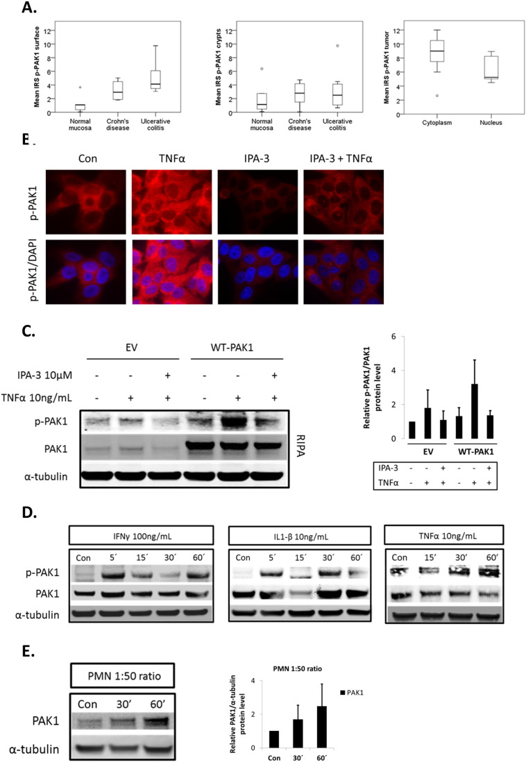
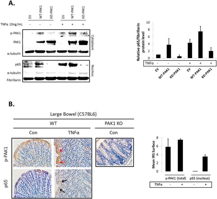
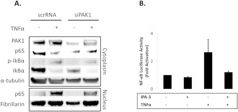
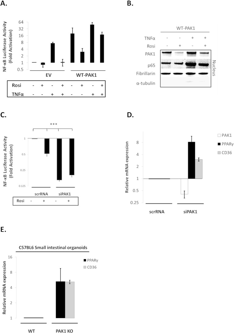
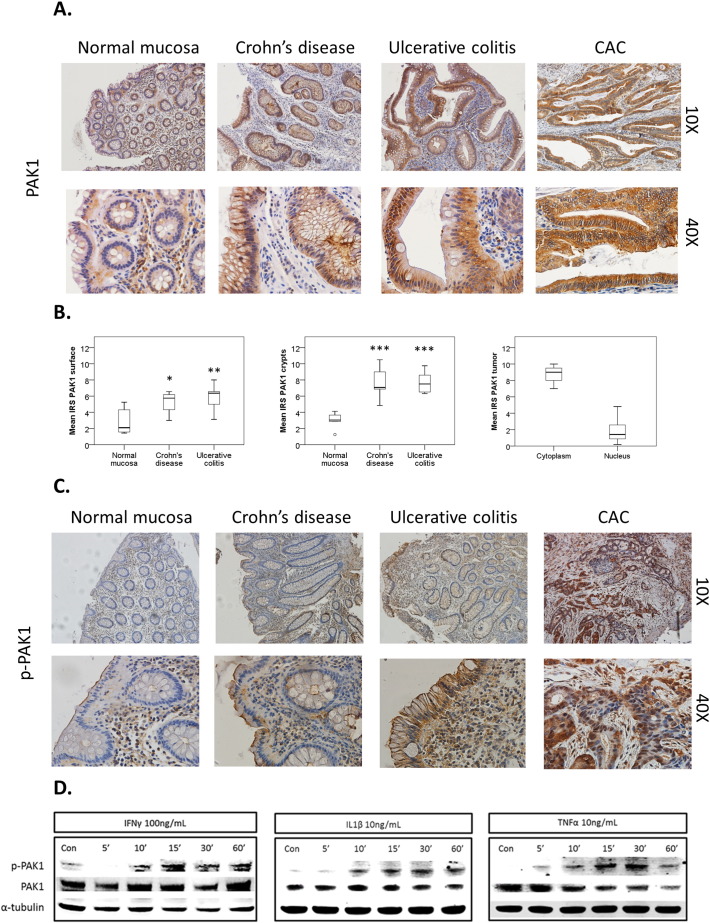
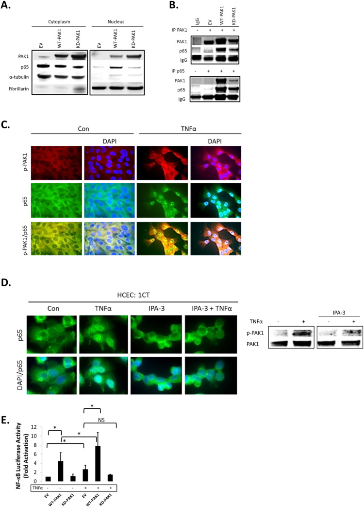
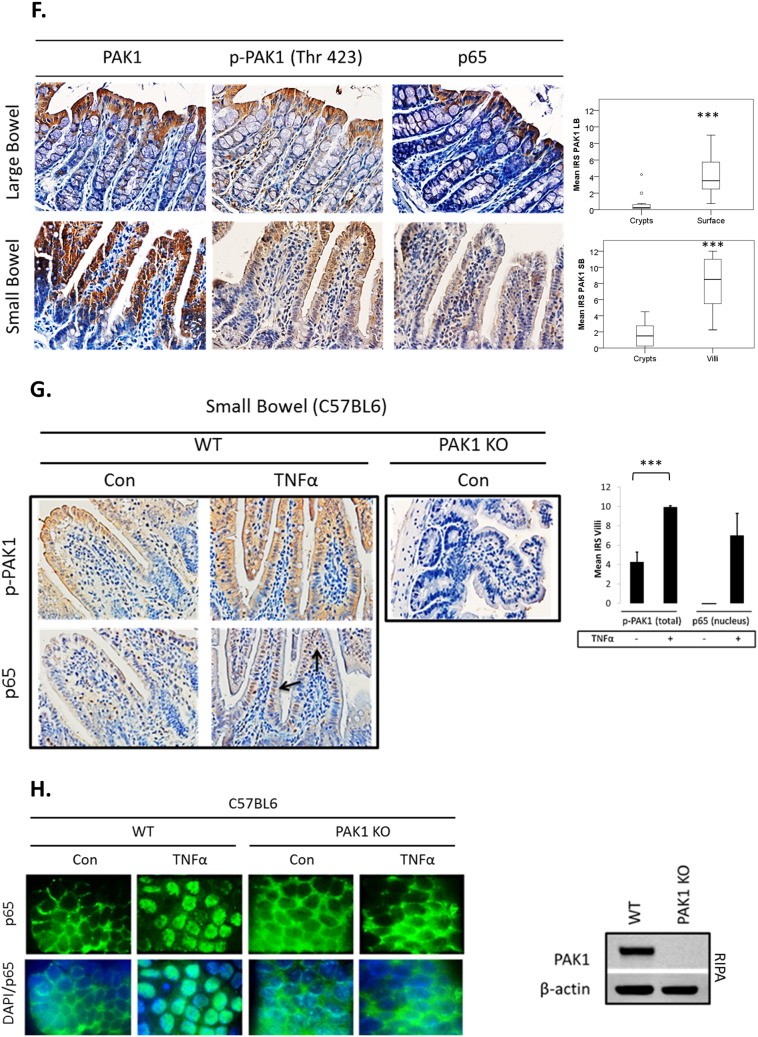
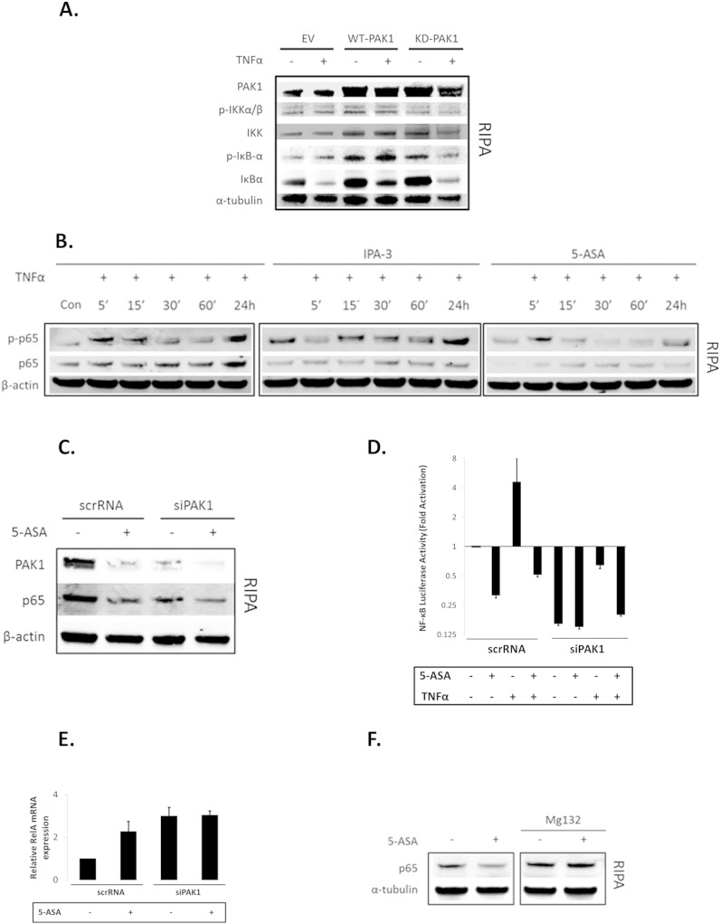
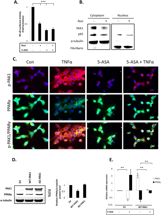
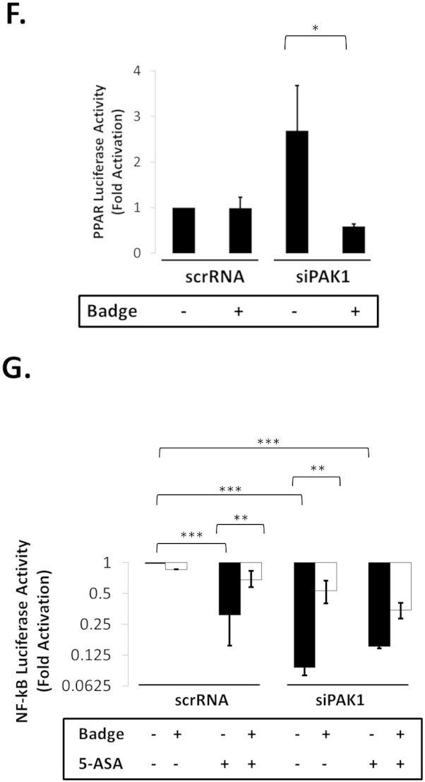
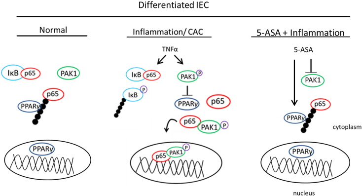
P21-activated kinases (PAKs) are multifunctional effectors of Rho GTPases with both kinase and scaffolding activity. Here, we investigated the effects of inflammation on PAK1 signaling and its role in colitis-driven carcinogenesis. PAK1 and p-PAK1 (Thr423) were assessed by immunohistochemistry, immunofluorescence, and Western blot. C57BL6/J wildtype mice were treated with a single intraperitoneal TNFα injection. Small intestinal organoids from these mice and from PAK1-KO mice were cultured with TNFα. NF-κB and PPARγ were analyzed upon PAK1 overexpression and silencing for transcriptional/translational regulation. PAK1 expression and activation was increased on the luminal intestinal epithelial surface in inflammatory bowel disease and colitis-associated cancer. PAK1 was phosphorylated upon treatment with IFNγ, IL-1β, and TNFα. In vivo, mice administered with TNFα showed increased p-PAK1 in intestinal villi, which was associated with nuclear p65 and NF-κB activation. p65 nuclear translocation downstream of TNFα was strongly inhibited in PAK1-KO small intestinal organoids. PAK1 overexpression induced a PAK1-p65 interaction as visualized by co-immunoprecipitation, nuclear translocation, and increased NF-κB transactivation, all of which were impeded by kinase-dead PAK1. Moreover, PAK1 overexpression downregulated PPARγ and mesalamine recovered PPARγ through PAK1 inhibition. On the other hand PAK1 silencing inhibited NF-κB, which was recovered using BADGE, a PPARγ antagonist. Altogether these data demonstrate that PAK1 overexpression and activation in inflammation and colitis-associated cancer promote NF-κB activity via suppression of PPARγ in intestinal epithelial cells.

Keywords: Colitis-associated cancer; Inflammation; NF-κB; PAK1; PPARγ; Ulcerative colitis.


Figures

Supplementary Fig. S1
(A–E). Inflammation promotes PAK1 activation. (A) Box plots compare PAK1 activation at the luminal intestinal surface and crypts in normal mucosa (n = 5), CD (n = 6), UC (n = 8), and also in CAC samples (n = 7). Tukey HSD; *p < 0.05, **p < 0.01, ***p < 0.001. (B) Immunofluorescence of p-PAK1 in TNFα-treated cells with DMSO (Con) or IPA-3 pretreatment. Upper panel shows p-PAK1; lower panel is p-PAK1 merged with DAPI staining. (C) WB of whole cell lysates (RIPA) in EV or WT-PAK1 overexpressing cells. TNFα activation of PAK1 was blocked upon IPA-3 pretreatment. Densitometry of relative p-PAK1 expression upon IPA-3 pretreatment, data are mean and SD from 2 independent experiments. (D) Inflammatory cytokines activate PAK1 in HCT116 cells. WB of RIPA lysates. Cells were left untreated (Con) or treated with IFNγ, IL-1β, or TNFα for 5–60 min and analyzed for PAK1 activation using p-PAK1 Thr 423 antibody. PAK1 activation increased from 5–60 min. Total PAK1 expression was also evaluated and α-tubulin was utilized as a loading control. (E) HCEC-1CT cells were co-cultured with PMNs in a 1:50 epithelial to neutrophil ratio and analyzed by WB for PAK1 expression. Total PAK1 expression increased from 30–60 min. Relative densitometry of PAK1 expression upon co-culture treatment. Data are mean and SD of 2 independent experiments.
Supplementary Fig. S2
(A–B). PAK1 overexpression regulates NF-kB activation downstream of TNFα in HCEC-1CT. (A) WB of HCEC-1CT cytoplasmic and nuclear lysates. Cells were transfected with EV, WT-PAK1 or KD-PAK1 then treated with TNFα and probed for p-PAK1, PAK1, and p65. WT-PAK1 but not KD-PAK1 overexpression increased nuclear translocation of p65. In the presence of TNFα, KD-PAK1 overexpression impedes nuclear accumulation of p65. Loading controls included α-tubulin (cytoplasm) and Fibrillarin (nucleus). Densitometry of nuclear p65 protein levels upon PAK1 overexpression in untreated or TNFα-treated cells. Data are mean and SD of 2 independent experiments. (B) IHC of p-PAK1 and p65 within large bowel tissue in TNFα injected mice. A modest increase in p-PAK1 expression (red arrows) and p65 (black arrows) nuclear translocation was observed at the luminal surface following TNFα treatment. PAK1 KO mice were stained for p-PAK1 as a negative control. Graphs show mean villi p-PAK1 and p65 IRSs (± SD) (n = 3) mice per group.
Supplementary Fig. S3
(A–B). PAK1 is required for complete activation of NF-κB downstream of IκB in HCEC-1CT. (A) The effect of PAK1 on p65 was evaluated using siPAK1 or scrRNA. Cells were treated with TNFα and analyzed for PAK1, p65, p-IκB-α, and IκBα expression in nuclear and cytoplasmic cell fractions by WB. α-Tubulin and Fibrillarin were utilized as loading controls. siPAK1 impeded nuclear accumulation of p65 on TNFα treatment independent of IκB degradation. (B) NF-kB luciferase assay of TNFα treated cells with or without IPA-3 pretreatment. IPA-3 blocked TNFα induction of NF-kB.
Supplementary Fig. S4
(A–E). PAK1 modulates a PPARγ/p65 cascade. (A) NF-κB luciferase assay of EV and WT-PAK1 overexpressing cells upon TNFα treatment (30 min) with or without Rosi pretreatment (12 h). Rosi recovered both the effect of TNFα and PAK1 overexpression on NF-kB. (B) WT-PAK1 overexpressing cells were pretreated with Rosi (12 h) with or without TNFα (30 min) and a nuclear fraction was harvested and analyzed by WB for PAK1 and p65. Fibrillarin (nuclear) and α-tubulin were used as loading controls. Rosi impeded PAK1 overexpression and reduced the effect of WT-PAK1 and TNFα on p65. (C) NF-κB luciferase assay performed in PAK1 knockdown cells using siPAK1 or scrRNA with or without Rosi. siPAK1 and Rosi blocked NF-kB transcriptional activity, Tukey HSD; ***p < 0.001. (D) RTPCR of PAK1, PPARγ, and CD36, in PAK1 knock down cells using siPAK1 or scrRNA. siPAK1 increased PPARγ transcription and its downstream target CD36. (E) RTPCR of PPARγ and CD36 in WT and PAK1 KO mouse SIO. PAK1 deletion increased mRNA levels of PPARγ and its downstream target CD36.
Fig. 1
Fig. 1
(A–D). PAK1 is overexpressed and activated in IBD. (A) Human colonic tissue stained for PAK1. PAK1 expression increases in comparison to normal mucosa at the luminal epithelial surface and crypts in both Crohn's disease (CD), ulcerative colitis (UC), and in tumors of colitis associated cancer (CAC). (B) Box plots compare mean PAK1 immunoreactivity scores (IRS) in normal mucosa (n = 6), CD (n = 7), and UC (n = 6) at the luminal surface and crypts. PAK1 is overexpressed in CAC (n = 8). The circle is the outlier from the boxplot. (C) Human colonic tissue stained for p-PAK1. In CD and UC, PAK1 activation in epithelial cells is found at the membrane, specifically at the luminal surface, but not in the crypts. In CAC, PAK1 phosphorylation is found within the nuclei and cytoplasm of tumor cells. (D) Inflammatory cytokines activate PAK1 in HCEC-1CT. WB of HCEC-1CT RIPA whole cell lysates using 50 μg protein. Cells were either untreated (Con) or treated with IFNγ, IL-1β, or TNFα for 5–60 min and analyzed for PAK1 activation with a p-PAK1 Thr 423 antibody. TNFα induced the most profound effect in PAK1 activation at 30 min. α-Tubulin was utilized as a loading control.
Fig. 2
Fig. 2
(A–H). PAK1 overexpression regulates NF-kB activation downstream of TNFα in HCEC-1CT. (A) WB of HCEC-1CT cytoplasmic and nuclear lysates. Cells were transfected with EV, WT-PAK1 or KD-PAK1 and probed for PAK1 and p65. WT-PAK1 but not KD-PAK1 overexpression increased nuclear translocation of p65. Loading controls included α-tubulin (cytoplasm) and Fibrillarin (nucleus). (B) Co-IP of PAK1 (top) and p65 (bottom) probed for PAK1 and p65 demonstrating a PAK1–p65 interaction. (C) Immunofluorescence (IF) of p-PAK1 and p65 co-staining in untreated (Con) and TNFα treated cells. Upper panel p-PAK1, middle panel p65, and lower panel merged p-PAK1/p65 images with or without merged DAPI channel. PAK1 and p65 colocalized upon TNFα. (D) IF of p65 or merged DAPI/p65 in HCEC-1CT. Cells were pre-treated with 10 μM IPA-3 (2 h) followed by 10 ng/mL TNFα for 30 min. Nuclear accumulation of p65 by TNFα is impeded by p-PAK1 inhibition. The WB verified p-PAK1 activation by TNFα and its inhibition by IPA-3 in HCEC-1CT. (E) NF-κB luciferase assay of EV, WT-PAK1, and KD-PAK1 overexpression in untreated and TNFα treated cells. Data are representative of 3 independent experiments, ANOVA, Tukey HSD; *p < 0.05. (F) Immunohistochemistry of large bowel (LB) or small bowel (SB) mouse tissue sections stained for PAK1, p-PAK1, or p65. Box plots are mean PAK1 immunoreactivity scores (IRS) comparing SB crypt and villi expression, and LB crypt and surface expression in control mice (n = 3) mice (t-test, 2 tailed; ***p < 0.001). (G) IHC of p-PAK1 and p65 within small intestinal tissue in TNFα injected mice. TNFα increases p-PAK1 expression and p65 (black arrow) nuclear translocation within villi, but not in crypts. PAK1 KO mice were stained for p-PAK1 as a negative control. Graphs show mean villi p-PAK1 and p65 IRSs (± SD) (n = 3) mice per group (t-test, 2 tailed; ***p < 0.001) (H) IF of p65 or merged DAPI/p65 in wild type (WT) and PAK1 KO mouse small intestinal organoids (SIO) with or without TNFα. WB of PAK1 protein expression in WT or PAK1 KO SIO.
Fig. 2
Fig. 2
(A–H). PAK1 overexpression regulates NF-kB activation downstream of TNFα in HCEC-1CT. (A) WB of HCEC-1CT cytoplasmic and nuclear lysates. Cells were transfected with EV, WT-PAK1 or KD-PAK1 and probed for PAK1 and p65. WT-PAK1 but not KD-PAK1 overexpression increased nuclear translocation of p65. Loading controls included α-tubulin (cytoplasm) and Fibrillarin (nucleus). (B) Co-IP of PAK1 (top) and p65 (bottom) probed for PAK1 and p65 demonstrating a PAK1–p65 interaction. (C) Immunofluorescence (IF) of p-PAK1 and p65 co-staining in untreated (Con) and TNFα treated cells. Upper panel p-PAK1, middle panel p65, and lower panel merged p-PAK1/p65 images with or without merged DAPI channel. PAK1 and p65 colocalized upon TNFα. (D) IF of p65 or merged DAPI/p65 in HCEC-1CT. Cells were pre-treated with 10 μM IPA-3 (2 h) followed by 10 ng/mL TNFα for 30 min. Nuclear accumulation of p65 by TNFα is impeded by p-PAK1 inhibition. The WB verified p-PAK1 activation by TNFα and its inhibition by IPA-3 in HCEC-1CT. (E) NF-κB luciferase assay of EV, WT-PAK1, and KD-PAK1 overexpression in untreated and TNFα treated cells. Data are representative of 3 independent experiments, ANOVA, Tukey HSD; *p < 0.05. (F) Immunohistochemistry of large bowel (LB) or small bowel (SB) mouse tissue sections stained for PAK1, p-PAK1, or p65. Box plots are mean PAK1 immunoreactivity scores (IRS) comparing SB crypt and villi expression, and LB crypt and surface expression in control mice (n = 3) mice (t-test, 2 tailed; ***p < 0.001). (G) IHC of p-PAK1 and p65 within small intestinal tissue in TNFα injected mice. TNFα increases p-PAK1 expression and p65 (black arrow) nuclear translocation within villi, but not in crypts. PAK1 KO mice were stained for p-PAK1 as a negative control. Graphs show mean villi p-PAK1 and p65 IRSs (± SD) (n = 3) mice per group (t-test, 2 tailed; ***p < 0.001) (H) IF of p65 or merged DAPI/p65 in wild type (WT) and PAK1 KO mouse small intestinal organoids (SIO) with or without TNFα. WB of PAK1 protein expression in WT or PAK1 KO SIO.
Fig. 3
Fig. 3
(A–F). PAK1 is required for complete activation of NF-κB downstream of IκB in HCEC-1CT. (A) EV, WT-PAK1, or KD-PAK1 overexpressing cells were treated with 10 ng/mL TNFα (30 min). RIPA lysates were analyzed by WB for PAK1, p-IKKα/β, IKKα, p-IκB-α, and IκBα expression. WT or KD-PAK1 overexpression did not interfere with the degradation of IκB following TNFα treatment. (B) WB of RIPA lysates analyzed for p-p65 and p65. Cells were treated with TNFα or pretreated (12 h) with 10 μM IPA-3 or 20 mM 5-ASA. Pretreatment with IPA-3 or 5-ASA blocked the effect of TNFα on p65. (C) WB of whole cell (RIPA) lysates probed for PAK1, p65, and β-actin. PAK1 knock down by both 5-ASA and siPAK1 blocked total p65 protein levels. (D) Cells were co-transfected with a NF-κB luciferase reporter and siPAK1 or scrRNA. Pretreatment with 5-ASA and PAK1 knockdown impeded the effect of TNFα on NF-κB transcriptional activation. Data are representative of 3 independent experiments. (E) Relative mRNA expression of RelA upon siPAK1 or scrRNA with or without 5-ASA treatment. PAK1 inhibition increased transcription of RelA. (F) HCEC-1CT cells were treated with 20 mM 5-ASA (24 h) with or without the proteasomal inhibitor MG132 (6 h). RIPA lysates were analyzed for p65 and α-tubulin by WB. 5-ASA inhibits p65 but not upon pretreatment with MG132. All data are representative of 3 independent experiments.
Fig. 4
Fig. 4
(A–G). PAK1 modulates a PPARγ/p65 cascade. (A) HCEC-1CT cells were transfected with a NF-kB luciferase reporter and treated with 1 μM Rosi ± 20 mM 5-ASA (24 h). Rosi and 5-ASA blocked NF-kB transcriptional activity ANOVA, Tukey HSD; ***p < 0.001. (B) Rosi treated cells were fractionated and analyzed by WB for PAK1 and p65. Rosi blocked PAK1 and p65 expression. (C) IF of p-PAK1 and PPARγ upon TNFα (30 min) with or without 20 mM 5-ASA pretreatment (24 h). TNFα resulted in p-PAK1 activation and PPARγ nuclear export and downregulation which was blocked by 5-ASA. (D) The effect of EV, WT-PAK1, and KD-PAK1 overexpression on PPARγ was analyzed by WB. α-Tubulin was used as a loading control. Densitometry of PPARγ levels normalized to α-tubulin, data are mean and SD from 2 independent experiments. (E) RTPCR of PAK1 and PPARγ after EV or WT-PAK1 overexpression with or without 5-ASA treatment (24 h). PAK1 overexpression blocked PPARγ at the mRNA level, an effect that was recovered by 5-ASA. Data are representative of 3 independent experiments (t-test, 2 tailed; **p < 0.01). (F) PPAR luciferase reporter assay in PAK1 knock down cells using scrRNA or siPAK1. siPAK1 activated PPAR transcription, and this effect was recovered upon 0.1 μM BADGE pretreatment (12 h), ANOVA, Tukey HSD; *p < 0.05. (G) NF-kB luciferase assay in PAK1 knock down cells using scrRNA or siPAK1. Cells were pretreated with BADGE (12 h) with or without 5-ASA (24 h). BADGE recovered the effect of 5-ASA or siPAK1 on NF-kB. All data are representative of 3 independent experiments ANOVA, Tukey HSD; **p < 0.01, ***p < 0.001.
Fig. 4
Fig. 4
(A–G). PAK1 modulates a PPARγ/p65 cascade. (A) HCEC-1CT cells were transfected with a NF-kB luciferase reporter and treated with 1 μM Rosi ± 20 mM 5-ASA (24 h). Rosi and 5-ASA blocked NF-kB transcriptional activity ANOVA, Tukey HSD; ***p < 0.001. (B) Rosi treated cells were fractionated and analyzed by WB for PAK1 and p65. Rosi blocked PAK1 and p65 expression. (C) IF of p-PAK1 and PPARγ upon TNFα (30 min) with or without 20 mM 5-ASA pretreatment (24 h). TNFα resulted in p-PAK1 activation and PPARγ nuclear export and downregulation which was blocked by 5-ASA. (D) The effect of EV, WT-PAK1, and KD-PAK1 overexpression on PPARγ was analyzed by WB. α-Tubulin was used as a loading control. Densitometry of PPARγ levels normalized to α-tubulin, data are mean and SD from 2 independent experiments. (E) RTPCR of PAK1 and PPARγ after EV or WT-PAK1 overexpression with or without 5-ASA treatment (24 h). PAK1 overexpression blocked PPARγ at the mRNA level, an effect that was recovered by 5-ASA. Data are representative of 3 independent experiments (t-test, 2 tailed; **p < 0.01). (F) PPAR luciferase reporter assay in PAK1 knock down cells using scrRNA or siPAK1. siPAK1 activated PPAR transcription, and this effect was recovered upon 0.1 μM BADGE pretreatment (12 h), ANOVA, Tukey HSD; *p < 0.05. (G) NF-kB luciferase assay in PAK1 knock down cells using scrRNA or siPAK1. Cells were pretreated with BADGE (12 h) with or without 5-ASA (24 h). BADGE recovered the effect of 5-ASA or siPAK1 on NF-kB. All data are representative of 3 independent experiments ANOVA, Tukey HSD; **p < 0.01, ***p < 0.001.
Fig. 5
Fig. 5
PAK1 modulates a PPARγ/p65 cascade. (Left) Within normal differentiated IEC, PAK1 activity and expression is low while PPARγ is present in both the cytoplasm and nucleus. Cytoplasmic NF-kB is maintained in an inactive state via the sequestration of p65 by IkB. PPARγ further regulates free p65 through E3 ligase activity and proteasomal degradation in the cytoplasm. (Middle) In chronic inflammation and colitis associated cancer (CAC) PAK1 is hyperactivated while PPARγ is downregulated. Pro-inflammatory cytokines such as TNFα induce PAK1 phosphorylation, activation, and nuclear colocalization with p65. Activated PAK1 also blocks PPARγ, further increasing the nuclear accumulation of p65 independently of IkB. (Right) The anti-inflammatory drug 5-ASA inhibits PAK1 thereby restoring PPARγ expression, and increasing its activity in inhibiting free p65 in the cytoplasm.

References

    1. Rubin D.T., Cruz-Correa M.R., Gasche C., Jass J.R., Lichtenstein G.R., Montgomery E.A., Riddell R.H., Rutter M.D., Ullman T.A., Velayos F.S., Itzkowitz S., A.S.A.i.C.C.P.M. Group Colorectal cancer prevention in inflammatory bowel disease and the role of 5-aminosalicylic acid: a clinical review and update. Inflamm. Bowel Dis. 2008;14:265–274. - PubMed
    1. Khare V., Lyakhovich A., Dammann K., Lang M., Borgmann M., Tichy B., Pospisilova S., Luciani G., Campregher C., Evstatiev R., Pflueger M., Hundsberger H., Gasche C. Mesalamine modulates intercellular adhesion through inhibition of p-21 activated kinase-1. Biochem. Pharmacol. 2013;85:234–244. - PMC - PubMed
    1. Parrini M.C., Lei M., Harrison S.C., Mayer B.J. Pak1 kinase homodimers are autoinhibited in trans and dissociated upon activation by Cdc42 and Rac1. Mol. Cell. 2002;9:73–83. - PubMed
    1. Kumar R., Gururaj A.E., Barnes C.J. p21-Activated kinases in cancer. Nat. Rev. Cancer. 2006;6:459–471. - PubMed
    1. Dammann K., Khare V., Gasche C. Tracing PAKs from GI inflammation to cancer. Gut. 2014;63:1173–1184. - PubMed

Publication types

MeSH terms