. 2015 May 30;6(15):13201-15.
doi: 10.18632/oncotarget.3915.

MiR-1 downregulation correlates with poor survival in clear cell renal cell carcinoma where it interferes with cell cycle regulation and metastasis

Affiliations

MiR-1 downregulation correlates with poor survival in clear cell renal cell carcinoma where it interferes with cell cycle regulation and metastasis

Haibing Xiao et al. Oncotarget. .

Erratum in

Abstract

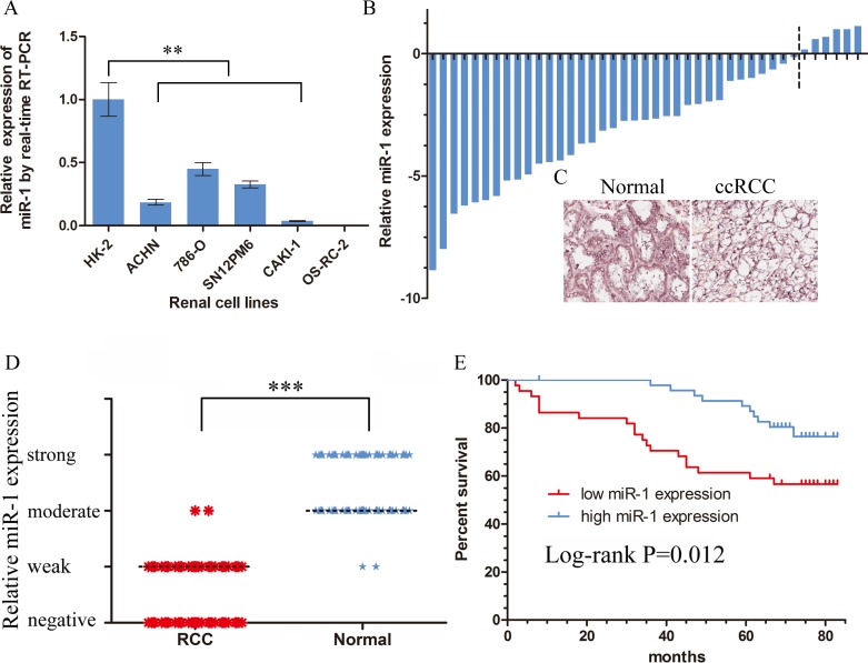
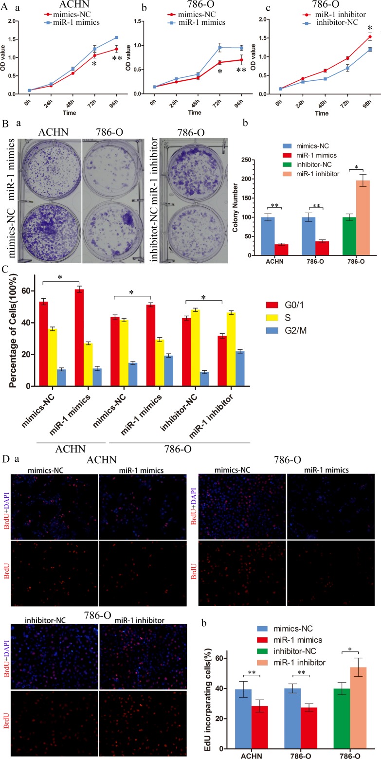
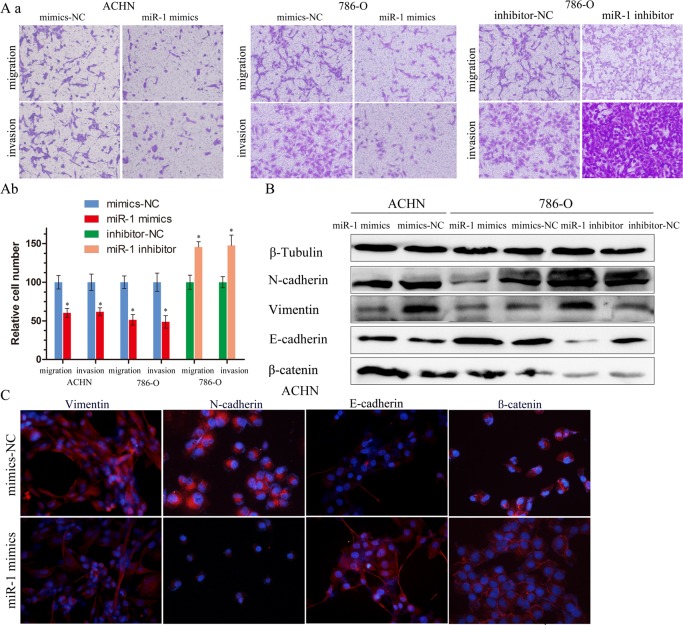
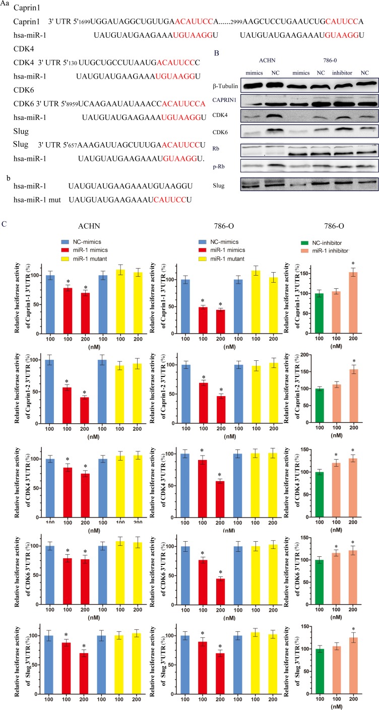
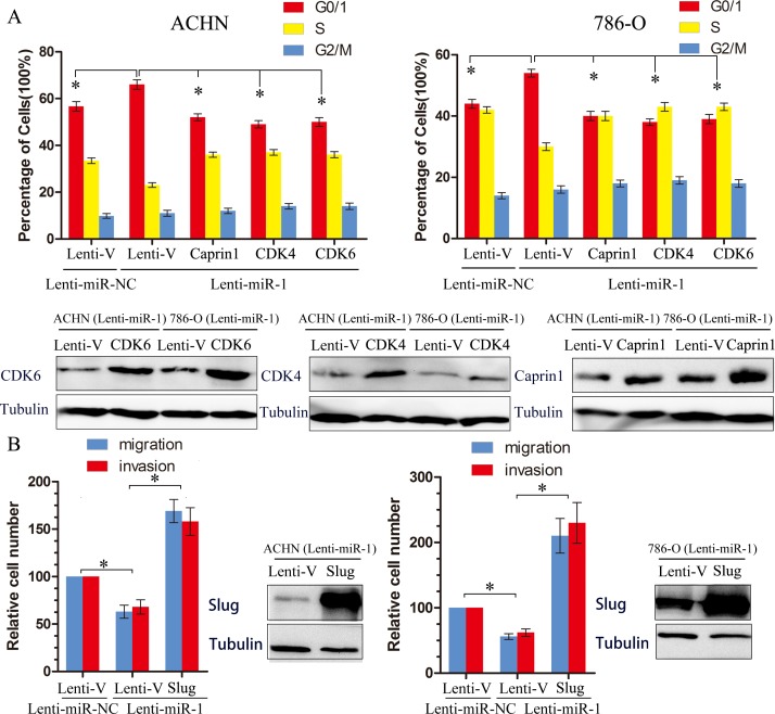
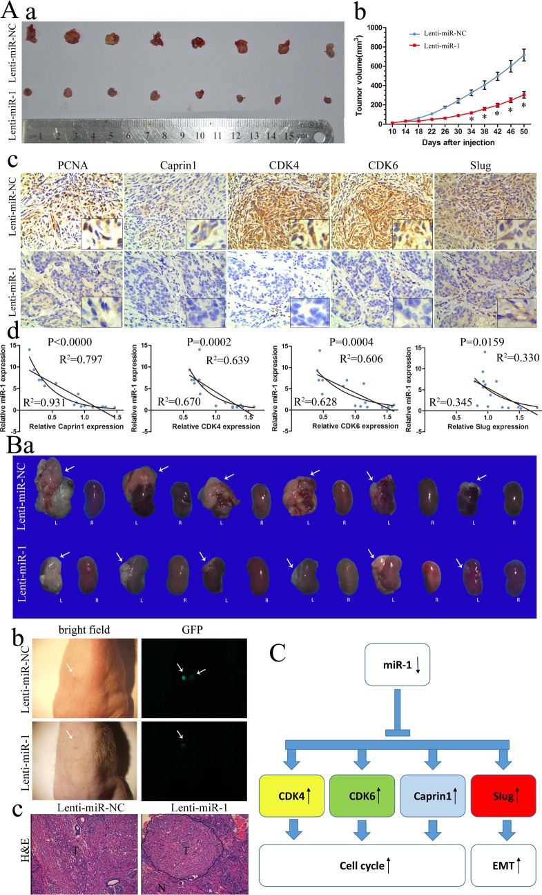
MicroRNAs (miRNA) that are strongly implicated in carcinogenesis have recently reshaped our understanding of the role of noncoding RNAs. Here, we focused on the function and molecular mechanism of miR-1 and its potential clinical application in clear cell renal cell carcinoma (ccRCC). First, miR-1 was significantly downregulated in 87.8% renal cancer samples compared with corresponding noncancerous tissues (NCT), which was significantly associated with clinical stage, T classification and poor overall survival. Functional study demonstrated that enforced overexpression of miR-1 in renal cancer cells inhibited proliferation and metastasis in vitro and in vivo. Conversely, miR-1 inhibitor silencing miR-1 expression promoted cell proliferation and metastasis in ccRCC. CDK4, CDK6, Caprin1 and Slug were each directly targeted for inhibition by miR-1 and restoring their expression reversed miR-1-mediated inhibition of cell cycle progression and metastasis. Taken together, our findings established a tumor suppressive role for miR-1 in the progression of ccRCC by targeting CDK4, CDK6, Caprin1 and Slug and suggested miR-1 can be served as a novel potential therapeutic target for ccRCC.

Keywords: ccRCC; metastasis; miR-1; proliferation.


Conflict of interest statement

CONFLICTS OF INTEREST

The authors declare no conflict of interest.

Figures

Figure 1
Figure 1. Downregulation of miR-1 in ccRCC correlated with poor patient survival
Real-time PCR analysis of miR-1 expression in immortalized human renal tubule epithelial cell line HK-2 and indicated renal carcinoma cell lines. Data were plotted as the mean ± SEM of three independent experiments relative to HK-2 cells. **, P < 0.01. B. Relative expression of miR-1 in 41 pairs of ccRCC tumor tissues and their corresponding adjacent non-cancerous tissues (Δtumor-Δnormal). The average miR-1 expression was normalized by U6 expression. C. Expression of miR-1 in tumor tissues and their corresponding adjacent non-cancerous tissues by in situ hybridization (ISH). D. The expression level of miR-1 was measured by H-score. Negative (−, score: 0), weak (+, score: 1–4), moderate (++, score: 5–8) and strong (+++, score: 9–12). ***, P < 0.001. E. Kaplan-Meier analysis of correlation between the miR-1 level and overall survival of ccRCC patients with high (n = 47) and low (n = 43) miR-1 expression. In the Kaplan-Meier analysis, negative was recognized as low expression, weak and moderate were recognized as high expression.
Figure 2
Figure 2. miR-1 attenuates ccRCC cell proliferation and motility
ccRCC cells were transfected with 100 nM of indicated small RNA molecules. Results were plotted as the mean ± SEM of three independent experiments, with at least three replicates in each independent experiment. *, P < 0.05; **, P < 0.01. A. MTS assays revealed cell growth curves of indicated cells. B. Representative micrographs (left) and relative quantification (right) of crystal violet-stained cell colonies analyzed by clongenic formation. C. Flow cytometric determination of proportion of indicated cells in distinct cell cycle phases. D. Representative micrographs (left) and quantification (right) of EdU incorporated-cells in indicated engineered cell lines.
Figure 3
Figure 3. miR-1 attenuates ccRC cell migration and invasion
A. a. Migration and invasion assay for renal cancer cells. Representative photographs were taken at ×200 magnification; number of migrated cells was quantified in ten random images from each treatment group. b. Results were the mean ± SEM from two independent experiments and plotted as percent (%) migrating cells relative to mimic-NC or inhibitor-NC. *P < 0.05. B. EMT-related proteins were determined by immunoblot analysis. β-Tubulin was used as loading control. C. Representative photographs of immunofluorescence were taken at ×200 magnification. ACHN cells were transfected with 100 nM of indicated small RNA molecules.
Figure 4
Figure 4. miR-1 targeted cell cycle regulators CDK4, CDK6, CAPRIN1 and metastasis related gene Slug
A. a. Schematic miR-1 putative target sites in 3′ UTRs of CDK4, CDK6, Caprin1 and Slug. b. Sequence of miR-1-mut. B. WB analysis of the protein levels of CDK4, CDK6, Caprin1, slug, Rb and p-Rb in response to 100 nM of indicated small RNA molecules. C. Luciferase assay. Luciferase reporters harboring putative target sites in the 3′ UTRs of Caprin1 (Caprin1-1 and Caprin1-2), CDK4, CDK6 and Slug, were co-transfected with 100 and 200 nM of indicated small RNA molecules in ACHN and 786-O cells. Relative luciferase activity was plotted as the mean ± SEM of three independent experiments. *, P < 0.05.
Figure 5
Figure 5. Target genes partly suppresses functions initiated by miR-1
A. The effect of ectopic restoration of Caprin1, CDK4 or CDK6 on proportions of indicated cells in distinct cell cycle phases as determined by flow cytometric analysis (top), the levels of Caprin1, CDK4 or CDK6 protein were measured by Western Blot (bottom). Lenti-V is the negative control for lentiviral-mediated CDK4, CDK6, Caprine1 and Slug ectopic overexpression. Lenti-miR-NC is the negative control for Lenti-miR-1. B. The effect of ectopic restoration of slug on proportions of indicated cells in distinct cell migration and invasion analysis. The levels of slug protein were measured by Western Blot (bottom). *, p < 0.05.
Figure 6
Figure 6. miR-1 inhibited ccRCC tumor growth in subcutaneously and orthotopic
A. a. Photographs of tumors excised 51 days after inoculation of stably transfected cells into nude mice; b. mean tumor volume measured by caliper on the indicated days; c. IHC staining for PCNA, Caprin1, CDK4, CDK6 and Slug in slices of sectioned implanted tumors formed by indicated cells. Original magnification was ×200; d. Correlation of miR-1 with Caprin1, CDK4, CDK6 and Slug in subcutaneous transplantation tumor. B. a. Macroscopic appearance of the tumor xenograft (arrows) in nude mice from the 7th-week. L, left kidney; R, right kidney. b. Representative fluorescence images with primary tumors in the left kidney of nude mice after orthotopic injections of Lenti-miR-1 or Lenti-miR-NC for 7 weeks (arrows). c. H&E staining of the tumor xenograft. N, normal renal tissues; T, primary renal tumors. Original magnification was ×200. C. MiR-1 family modulates G1/S transition by regulating Cdk4/6-cyclin D complexes and Caprin1. Moreover, miR-1 can inhibit metastasis by downregulation of Slug.

Similar articles

Cited by

References

    1. Siegel R, Naishadham D, Jemal A. Cancer statistics, 2013. CA: a cancer journal for clinicians. 2013;63:11–30. - PubMed
    1. Yan BC, Mackinnon AC, Al-Ahmadie HA. Recent developments in the pathology of renal tumors: morphology and molecular characteristics of select entities. Archives of pathology & laboratory medicine. 2009;133:1026–1032. - PubMed
    1. Bartel DP. MicroRNAs: genomics, biogenesis, mechanism, and function. Cell. 2004;116:281–297. - PubMed
    1. Esquela-Kerscher A, Slack FJ. Oncomirs - microRNAs with a role in cancer. Nature reviews Cancer. 2006;6:259–269. - PubMed
    1. Nakada C, Matsuura K, Tsukamoto Y, Tanigawa M, Yoshimoto T, Narimatsu T, Nguyen LT, Hijiya N, Uchida T, Sato F, Mimata H, Seto M, Moriyama M. Genome-wide microRNA expression profiling in renal cell carcinoma: significant down-regulation of miR-141 and miR-200c. The Journal of pathology. 2008;216:418–427. - PubMed

Publication types

Supplementary concepts