. 2015 Aug 6:300:508-17.
doi: 10.1016/j.neuroscience.2015.05.052. Epub 2015 May 30.

Astrocytes regulate α-secretase-cleaved soluble amyloid precursor protein secretion in neuronal cells: Involvement of group IIA secretory phospholipase A2

Affiliations

Astrocytes regulate α-secretase-cleaved soluble amyloid precursor protein secretion in neuronal cells: Involvement of group IIA secretory phospholipase A2

X Yang et al. Neuroscience. .

Abstract

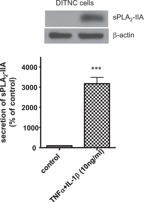
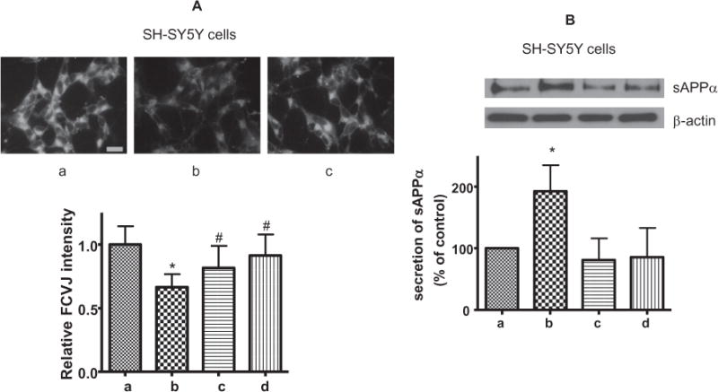
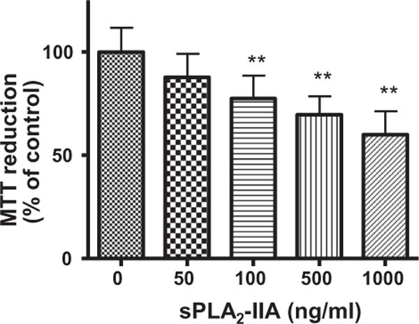
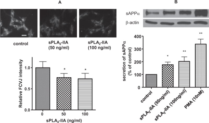
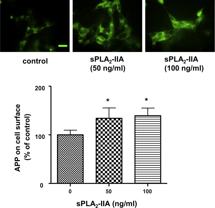
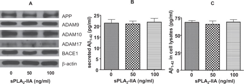
Astrocytes are major supportive cells in brains with important functions including providing nutrients and regulating neuronal activities. In this study, we demonstrated that astrocytes regulate amyloid precursor protein (APP) processing in neuronal cells through secretion of group IIA secretory phospholipase A2 (sPLA2-IIA). When astrocytic cells (DITNC) were mildly stimulated with the pro-inflammatory cytokines, such as TNF α and IL-1β, sPLA2-IIA was secreted into the medium. When conditioned medium containing sPLA2-IIA was applied to human neuroblastoma (SH-SY5Y) cells, there was an increase in both cell membrane fluidity and secretion of α-secretase-cleaved soluble amyloid precursor protein (sAPPα). These changes were abrogated by KH064, a selective inhibitor of sPLA2-IIA. In addition, exposing SH-SY5Y cells to recombinant human sPLA2-IIA also increased membrane fluidity, accumulation of APP at the cell surface, and secretion of sAPPα, but without altering total expressions of APP, α-secretases and β-site APP cleaving enzyme (BACE1). Taken together, our results provide novel information regarding a functional role of sPLA2-IIA in astrocytes for regulating APP processing in neuronal cells.

Keywords: SH-SY5Y; astrocytes; cytokine; membrane fluidity; sAPP(α); sPLA(2)-IIA.


Conflict of interest statement

CONFLICT OF INTEREST DISCLOSURE

The authors have no conflict of interest to declare.

Figures

Fig. 1
Fig. 1
Cytokine induced sPLA2-IIA secretion in DITNC cells. DITNC cells were exposed to cytokines (TNFα and IL-1β, 10 ng/ml) for 8 h, followed by removing cytokines and incubating in serum-free medium for another 40 h. Conditioned medium was used for Western blot analysis of sPLA2-IIA. Data are expressed as percentages of control and mean ± SD from at least three independent experiments (***p < 0.001).
Fig. 2
Fig. 2
SH-SY5Y cells were treated with conditioned medium containing sPLA2-IIA from cytokine-stimulated DITNC cells. (a): The conditioned medium from non-stimulated DITNC cells (control). (b): The conditioned medium from cytokine-stimulated DITNC cells. (c): The conditioned medium from cytokine-stimulated DITNC cells supplemented with the sPLA2-IIA inhibitor (KH064, 1 μM). (d): The conditioned medium from non-stimulated DITNC cells supplemented with KH064. (A, upper) Representative images of SH-SY5Y cells fluorescently labeled with FCVJ with conditioned medium treatment. (Scale bar=20 μm) (A, lower) The relative fluorescent intensity of FCVJ where a lower intensity indicates increased membrane fluidity. (B) Western blot analysis of sAPPα secretion with SH-SY5Y cells treated with the aforementioned DITNC conditioned media treatment groups. Data are expressed as percentages of control and mean ± SD from at least three independent experiments with statistical analysis performed with a one-way ANOVA, followed by Bonferroni’s post hoc tests (*p < 0.05 when compared to the control group (a) and #p < 0.05 when compared to the conditioned media treatment group (b)).
Fig. 3
Fig. 3
Effects of human recombined sPLA2-IIA on the viability of SH-SY5Y cells. MTT assay was applied after treatment of SH-SY5Y cells with different levels of sPLA2-IIA. Data are expressed as percentages of control and mean ± SD from at least three independent experiments (**p < 0.01).
Fig. 4
Fig. 4
(A) Effects of human recombined sPLA2-IIA on SH-SY5Y cell membrane fluidity. Representative images of SH-SY5Y cells fluorescently labeled with FCVJ without (upper, left) and with sPLA2-IIA treatment (upper, middle and right). Scale bar=20 μm. (B) Effects of human recombined sPLA2-IIA on sAPPα secretion in SH-SY5Y cells. After treatment of SH-SY5Y cells with sPLA2-IIA, the culture medium was used for Western blot analysis of sAPPα. Results showed that sPLA2-IIA increased sAPPα secretion to medium from SH-SY5Y cells. PMA treatment known to increase sAPPα secretion in cells was used as a positive control. Data are expressed as percentages of control and mean ± SD from at least three independent experiments (*p < 0.05, **p < 0.01).
Fig. 5
Fig. 5
Immunofluorescence microscopy showing effects of sPLA2-IIA on the presence of APP at the cell surface of SH-SY5Y cells. SH-SY5Y cells were treated with human recombined sPLA2-IIA for 24 h and were labeled with anti-APP antibody and FITC-conjugated secondary antibody without cell permeabilization. Representative images of fluorescently labeled SH-SY5Y cells without (upper, left) and with sPLA2-IIA treatment (upper, middle and right) showed sPLA2-IIA increased the APP accumulation at the cell surface (lower). Scale bar=20 μm. sPLA2-IIA increased the APP accumulation at the cell surface (lower). Data are expressed as percentages of control and mean ± SD from at least three independent experiments (*p < 0.05).
Fig. 6
Fig. 6
(A) sPLA2-IIA on the expressions of APP, α-secretases and BACE1 in SH-SY5Y cells. Western blot analysis showed that sPLA2-IIA did not alter the expressions of APP, different isoforms of α-secretases (ADAM 9, ADAM 10 and ADAM 17) and BACE1. (B) ELISA assay to quantify Aβ1–42 production from SH-SY5Y cells treated with 0, 50 or 100 ng/ml of sPLA2-IIA. sPLA2-IIA did not alter Aβ1–42 secretion from SH-SY5Y cells to culture medium. (C) sPLA2-IIA did not alter the expression of Aβ1–42 within SH-SY5Y cells. Data are expressed as mean ± SD from three independent experiments.

References

    1. Allinson TM, Parkin ET, Turner AJ, Hooper NM. ADAMs family members as amyloid precursor protein alpha-secretases. J Neurosci Res. 2003;74:342–352. - PubMed
    1. Apelt J, Schliebs R. Beta-amyloid-induced glial expression of both pro- and anti-inflammatory cytokines in cerebral cortex of aged transgenic Tg2576 mice with Alzheimer plaque pathology. Brain Res. 2001;894:21–30. - PubMed
    1. Baranowska-Bik A, Bik W, Wolinska-Witort E, Martynska L, Chmielowska M, Barcikowska M, Baranowska B. Plasma beta amyloid and cytokine profile in women with Alzheimer’s disease. Neuro Endocrinol Lett. 2008;29:75–79. - PubMed
    1. Beloosesky Y, Salman H, Bergman M, Bessler H, Djaldetti M. Cytokine levels and phagocytic activity in patients with Alzheimer’s disease. Gerontology. 2002;48:128–132. - PubMed
    1. Cacquevel M, Lebeurrier N, Cheenne S, Vivien D. Cytokines in neuroinflammation and Alzheimer’s disease. Curr Drug Targets. 2004;5:529–534. - PubMed

Publication types

MeSH terms

Substances

LinkOut - more resources