Review
. 2015 Aug;41(4):349-62.
doi: 10.1007/s00068-015-0530-z. Epub 2015 Apr 18.

Fragility fractures of the sacrum: how to identify and when to treat surgically?

Affiliations
Review

Fragility fractures of the sacrum: how to identify and when to treat surgically?

D Wagner et al. Eur J Trauma Emerg Surg. 2015 Aug.

Abstract

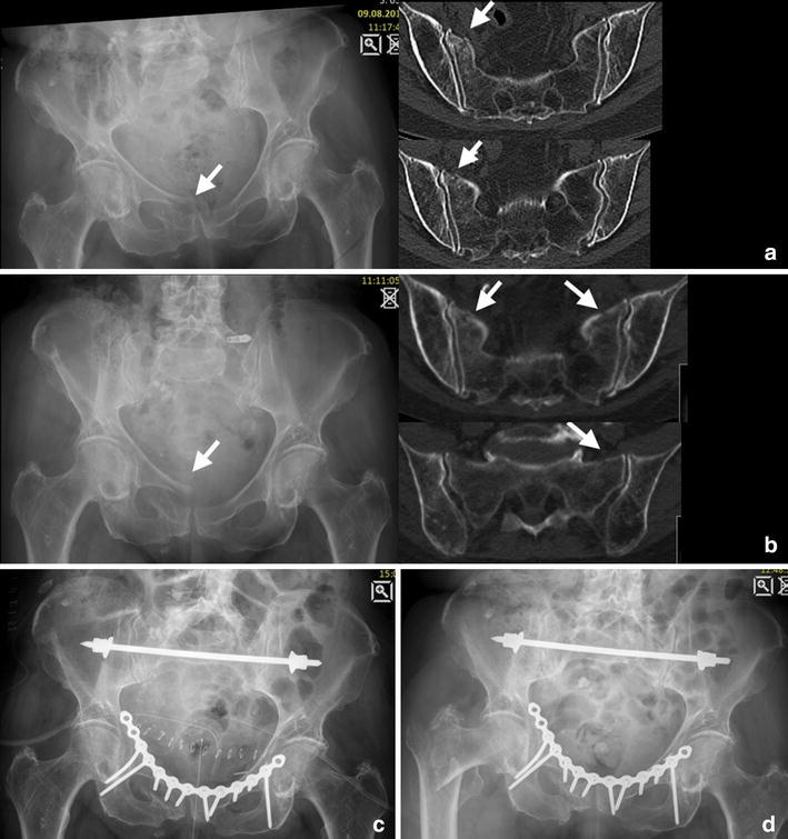
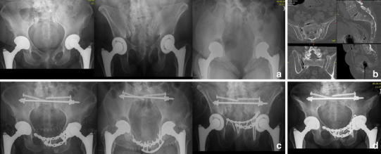
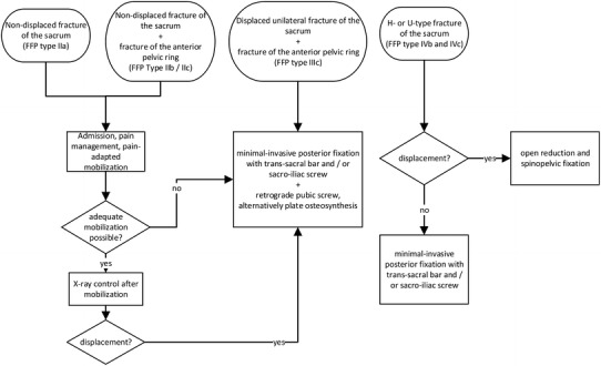
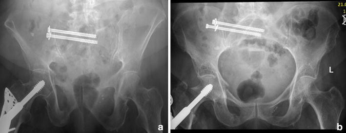
The increasing prevalence of fragility fractures of the sacrum (FFS) occurring predominantly in osteoporotic individuals poses a diagnostic and therapeutic challenge. The clinical presentation varies from longstanding low back pain without the patient remembering a traumatic event to immobilized patients after suffering a low-energy trauma. FFS are often combined with a fracture of the anterior pelvic ring; hence they are classified as a part of fragility fractures of the pelvis (FFP). If not displaced, the patients are treated with weight bearing as tolerated and analgesics; however, we advocate to treat displaced fractures surgically according to the fracture personality and the patient's comorbidities. Surgical options include minimal invasive sacro-iliac screws, trans-sacral bar osteosynthesis, open reduction and internal fixation, or spinopelvic stabilization. In the light of the high complication rate associated with immobilized patients, an operative approach often is indicated to accelerate the patient's mobility.


Figures

Fig. 1
Fig. 1
Diagnostic algorithm
Fig. 2
Fig. 2
FFP type IIa. 84-year-old female with immobilizing lower back pain. Conventional radiograph did not show a bony lesion (a). Also with adequate pain medication mobilization was not possible. The MRI (T1 and STIR sequence in the coronal plane of the sacrum) showed bilateral bone bruise in the sacral ala with a transverse connection on level S2/S3 (b). A CT scan confirmed bilateral sacral involvement without fracture of the anterior pelvic ring (c). The patient was stabilized percutaneously with a trans-sacral bar and bilateral SI-screws (d)
Fig. 3
Fig. 3
FFP type IIb: 81-year-old female with a crush injury of the left sacral ala and a non-displaced fracture of the left anterior pelvic ring (b). With conservative management she went on to consolidation (c radiograph 13 months after trauma)
Fig. 4
Fig. 4
FFP type IIc. 91-year-old female with a unilateral sacral fracture (b) and a slightly displaced anterior pelvic ring fracture (a). Conservative treatment failed because of persisting pain in the dorsal pelvic ring. Minimal-invasive surgery was performed (c): the sacrum was addressed with a trans-sacral bar and a SI-screw on the right side, and the superior pubic ramus was fixed retrogradely with a cannulated screw. Pain at mobilization resided after the operation
Fig. 5
Fig. 5
FFP type IIc. Initial diagnostics showed a unilateral fracture of the sacral ala right-sided and a displaced fracture of the anterior pelvic ring in this 83-year-old female (a). Conservative treatment with mobilization led to a bilateral sacral fracture and progressive displacement anteriorly after 3 weeks (b). She was stabilized subsequently with a trans-sacral bar and an anterior plate osteosynthesis (note the long screws reaching the posterior column) (c). A radiograph taken 5 months later demonstrated no implant failure or displacement; however, the patient sustained a pertrochanteric fracture due to recurrent fall (d)
Fig. 6
Fig. 6
FFP type IVb. This 67-year-old patient presented 10 months after a fall suffering from groin pain and pain projecting in both legs as well as a peroneal lesion on the left side, she was treated conservatively. She had bilateral pseudarthrosis of the sacrum and the left pubic rami with intrusion of the sacrum into the pelvic ring (a, b). Open debridement was performed in all pseudarthrosis with application of iliac bone graft. The posterior instabilities were addressed with a trans-sacral bar and an additional SI-screw on both sides through S1. Anteriorly, symphysiodesis was performed with bone graft and a double-plate osteosynthesis (c). Follow-up at 2 years showed consolidation (d). Mobilization was unlimited and without pain
Fig. 7
Fig. 7
Therapeutic algorithm
Fig. 8
Fig. 8
Backing out of SI-screw one month postoperatively in a 87-year-old female while only the posterior pelvic ring was fixed; however, mobilization was not painful

References

    1. Leidig-Bruckner G, Raue F, Frank-Raue K. Secondary osteoporosis—relevant clinical characteristics in diagnosis and therapy. Dtsch Med Wochenschr. 2012;137:326–332. - PubMed
    1. Consensus Development Conference Prophylaxis and treatment of osteoporosis. Am J Med. 1991;90:107–110. - PubMed
    1. Hernlund E, Svedbom A, Ivergård M, Compston J, Cooper C, Stenmark J, et al. Osteoporosis in the European Union: medical management, epidemiology and economic burden. A report prepared in collaboration with the International Osteoporosis Foundation (IOF) and the European Federation of Pharmaceutical Industry Associations (EFPIA). Arch. Osteoporos. 2013;8:136. - PMC - PubMed
    1. Kanis JA, Johnell O, Oden A, Jonsson B, De Laet C, Dawson A. Risk of hip fracture according to the World Health Organization criteria for osteopenia and osteoporosis. Bone. 2000;27:585–590. - PubMed
    1. Warriner AH, Patkar NM, Curtis JR, Delzell E, Gary L, Kilgore M, et al. Which fractures are most attributable to osteoporosis? J Clin Epidemiol. 2011;64:46–53. - PMC - PubMed

MeSH terms