. 2013 Mar;2(3):e9.
doi: 10.1038/emi.2013.9. Epub 2013 Mar 13.

Pathobiology of hepatitis E: lessons learned from primate models

Affiliations

Pathobiology of hepatitis E: lessons learned from primate models

Robert H Purcell et al. Emerg Microbes Infect. 2013 Mar.

Abstract

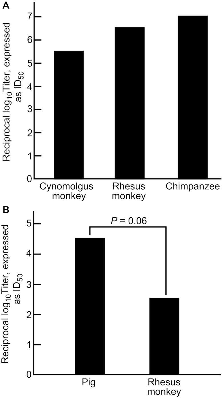
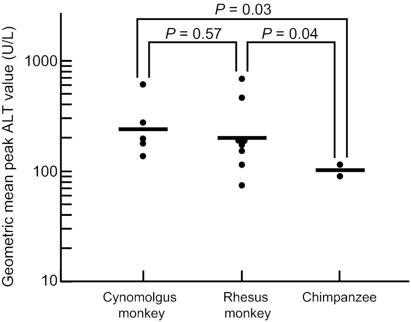
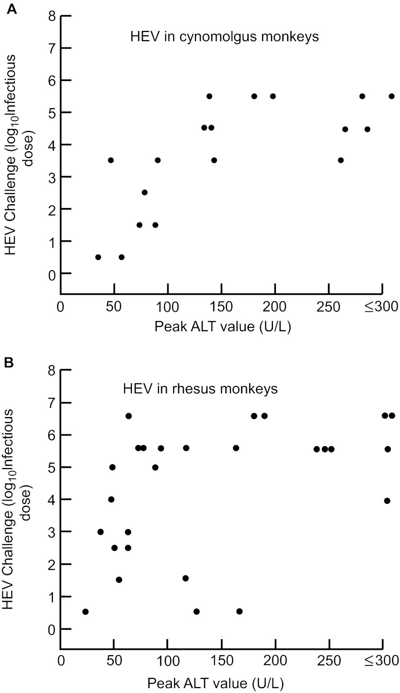
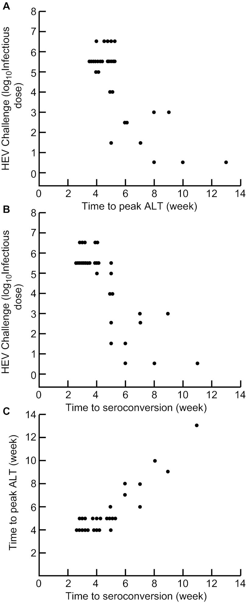
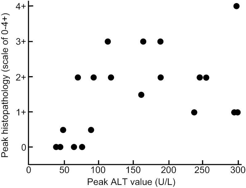
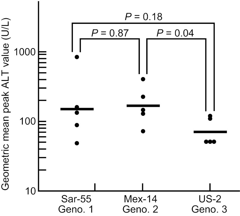
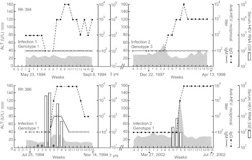
Like the other hepatitis viruses, hepatitis E virus (HEV) has been difficult to study because of limitations in cell culture systems and small animal models. Much of what we know has come from epidemiological studies in developing countries and, more recently, in industrialized countries. However, the epidemiology is very different in these two settings: hepatitis E in developing countries is epidemic as well as sporadic, principally water-borne, most likely to cause disease in older children and young adults and relatively severe, especially in pregnant women; in industrialized countries the disease is sporadic, principally food-borne, most common in the elderly and probably associated with mostly inapparent infections. These differences are believed to be genotypically determined. To examine the biological parameters of hepatitis E, we have studied HEV infections in nonhuman primates, which are surrogates of man. Infections with HEV genotypes 1-3 were compared in rhesus and cynomolgus macaques and chimpanzees. In general, the biological characteristics of the different HEV genotypes mirrored their epidemiological characteristics.

Keywords: HEV; genotypes; host range; nonhuman primates; virulence.


Figures

Figure 1
Figure 1
(A) The infectivity titer of a standard pool of human-derived genotype 1 HEV in three species of nonhuman primate. The data were obtained by reverse titration in order to limit the number of animals used. The differences in titer were not statistically significant. (B) The infectivity titer of a standard pool of pig-derived genotype 3 HEV in rhesus monkeys and pigs. The differences in titer approached statistical significance.
Figure 2
Figure 2
Severity of hepatitis, as measured by peak ALT levels in three species of nonhuman primate inoculated intravenously with the same high dose (105.5 ID50) of a genotype 1 HEV strain. The infection was associated with higher peak ALT levels in the two macaque species than in chimpanzees. The upper limit of normal for these species is 31 U/L.
Figure 3
Figure 3
Relationship between intravenous challenge dose of genotype 1 HEV and the clinical response, as measured by peak ALT levels in two macaque species. The infections were relatively attenuated following challenge doses of 103 or less in both species. (A) cynomolgus monkeys; (B) rhesus monkeys.
Figure 4
Figure 4
Relationship between intravenous challenge dose of genotype 1 HEV and interval to markers of infection in rhesus monkeys. There was an inverse relationship between the challenge dose and interval to (A) peak ALT and (B) seroconversion. (C) Interval to peak ALT paralleled the interval to seroconversion, with the latter occurring, on average, one half week earlier.
Figure 5
Figure 5
Comparison of magnitude of histological versus biochemical evidence of hepatitis in rhesus monkeys inoculated intravenously with a genotype 1 (SAR-55) HEV strain. There was a weak but significant positive correlation (Spearman's correlation coefficient was 0.54; P=0.02).
Figure 6
Figure 6
Comparison of virulence, as measured by mean peak ALT levels of genotype 1, genotype 2 and genotype 3 HEV strains administered intravenously at a high dose (104.0 MID50) to rhesus monkeys. The genotype 3 strain was significantly less virulent than the genotype 2 strain; the difference between the genotype 3 and genotype 1 strains approached significance.
Figure 7
Figure 7
A second HEV infection in rhesus monkeys infected with HEV previously. Both animals were seronegative at the time of the second infection. Rhesus 394 was infected with a genotype 1 HEV and, 3 years late, with a genotype 3 strain. Viremia was not detected during either infection. Rhesus 396 was infected with a genotype 1 strain and, 7 years later, with a different genotype 1 strain. Viremia was detected during both infections (starred sera were not available for testing).

References

    1. Purcell RH, Emerson SU. Hepatitis E: an emerging awareness of an old disease. J Hepatol. 2008;48:494–503. - PubMed
    1. Kamar N, Bendall R, Legrand-Abravanel F, et al. Hepatitis E. Lancet. 2012;379:2477–2488. - PubMed
    1. Hoofnagle JH, Nelson KE, Purcell RH. Hepatitis E. N Engl J Med. 2012;367:1237–1244. - PubMed
    1. Dalton HR, Stableforth W, Thurairajah P, et al. Authtochthonous hepatitis E in Southwest England: natural history, complications and seasonal variation, and hepatitis E virus IgG seroprevalence in blood donors, the elderly and patients with chronic liver disease. Eur J Gastroenterol Hepatol. 2008;20:784–790. - PubMed
    1. Davern TJ, Chalasani N, Fontana RJ, et al. Acute hepatitis E infection accounts for some cases of suspected drug-induced liver injury. Gastroenterology. 2011;141:1665–1672. - PMC - PubMed

LinkOut - more resources