. 2015 Jun;3(6):e12419.
doi: 10.14814/phy2.12419.

Irisin evokes bradycardia by activating cardiac-projecting neurons of nucleus ambiguus

Affiliations

Irisin evokes bradycardia by activating cardiac-projecting neurons of nucleus ambiguus

Eugen Brailoiu et al. Physiol Rep. 2015 Jun.

Abstract

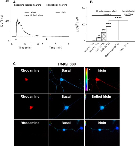
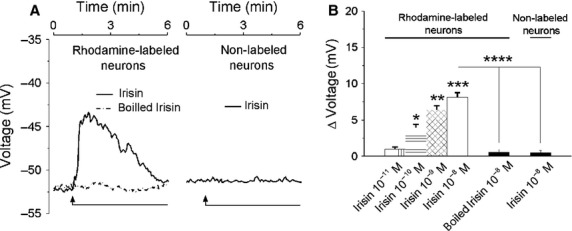
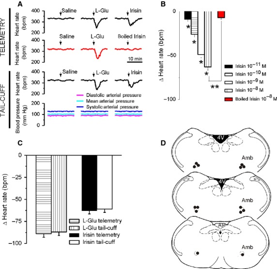
Irisin is a newly identified hormone induced in muscle and adipose tissues by physical activity. This protein and its encoding gene have been identified in the brain; in addition, the precursor for irisin, FNDC5, can cross the blood-brain barrier. The fact that irisin is secreted during exercise together with the lower resting heart rate in athletes prompted us to investigate the effect of irisin on cardiac-projecting vagal neurons of nucleus ambiguus, a key regulatory site of heart rate. In vitro experiments in cultured nucleus ambiguus neurons indicate that irisin activates these neurons, inducing an increase in cytosolic Ca(2+) concentration and neuronal depolarization. In vivo microinjection of irisin into the nucleus ambiguus promotes bradycardia in conscious rats. Our study is the first to report the effects of irisin on the neurons controlling the cardiac vagal tone and to link a myokine to a cardioprotective role, by modulating central cardiovascular regulation.

Keywords: Autonomic cardiovascular regulation; cytosolic Ca2+; membrane potential; vagal tone.


Figures

Figure 1
Figure 1
Irisin increases [Ca2+]i in cardiac vagal neurons of nucleus ambiguus. (A) Representative recordings of the Ca2+ responses produced by irisin (10−8 mol/L), boiled irisin (10−8 mol/L) in rhodamine-labeled neurons, and by irisin (10−8 mol/L) in nonlabeled neurons. (B) Comparison of the mean amplitudes of the Ca2+ responses produced by increasing concentrations of irisin (10−11–10−8 mol/L) and by heat-inactivated irisin (10−8 mol/L) in rhodamine-labeled neurons and by irisin (10−8 mol/L) in nonlabeled neurons; < 0.05 as compared to basal [Ca2+]i, (*), to the increase in [Ca2+]i produced by irisin (10−10 mol/L) (**), irisin 10−9 mol/L (***), and irisin 10−8 mol/L (****). (C) Changes in Fura-2 fluorescence ratio (340 nm/380 nm) of rhodamine-labeled neurons upon administration of 10−8 mol/L irisin (top) or 10−8 mol/L heat-inactivated irisin (middle), and of nonlabeled neurons upon administration of 10−8 mol/L irisin (bottom).
Figure 2
Figure 2
Irisin depolarizes cardiac vagal neurons of nucleus ambiguus. (A) Representative examples indicating changes in neuronal membrane potential produced by irisin (10−8 mol/L) and heat-inactivated (boiled) irisin (10−8 mol/L) in rhodamine-labeled neurons and by irisin (10–8 mol/L) in nonlabeled neurons (B) Concentration-dependent depolarizations produced by irisin (10−11–10−8 mol/L) and lack of effect of boiled irisin (10−8 mol/L) in rhodamine-labeled neurons; irisin (10−8 mol/L) did not affect the membrane potential in nonlabeled neurons; < 0.05 as compared to the resting membrane potential (*), to the response to irisin (10−10 mol/L) (**), irisin 10−9 mol/L (***), and irisin 10−8 mol/L (****).
Figure 3
Figure 3
Microinjection of irisin into the nucleus ambiguus produces bradycardia in conscious rats. (A) Typical recordings of heart rate and blood pressure during microinjection of saline, L-glutamate (L-Glu, 5 mmol/L, 50 nL) and either irisin (10−8 mol/L, 50 nL) or boiled irisin (10−8 mol/L, 50 nL), obtained using the telemetric method (top traces) or the tail-cuff method (bottom traces). (B) Comparison of the bradycardic responses elicited by microinjection of irisin (10−11–10−8 mol/L) and by heat-inactivated irisin (10−8 mol/L); < 0.05 compared to the response to the other concentrations of irisin (*), or to the effect of irisin10−8 mol/L (**). (C) Consistency of heart rate monitoring using invasive (telemetry) or noninvasive (tail-cuff) methods is indicated by the similarity of the responses induced by either L-Glu or irisin in the two paradigms. (D) Illustration of microinjection sites (dark dots) on coronal medullary sections. Abbreviations: AP, area postrema; Amb, nucleus ambiguus; 4V, fourth ventricle.

References

    1. Albrecht E, Norheim F, Thiede B, Holen T, Ohashi T, Schering L, et al. Irisin - a myth rather than an exercise-inducible myokine. Sci. Rep. 2015;5:8889. - PMC - PubMed
    1. Aydin S. Three new players in energy regulation: preptin, adropin and irisin. Peptides. 2014;56:94–110. - PubMed
    1. Aydin S, Kuloglu T, Aydin S, Kalayci M, Yilmaz M, Cakmak T, et al. A comprehensive immunohistochemical examination of the distribution of the fat-burning protein irisin in biological tissues. Peptides. 2014;61:130–136. - PubMed
    1. Azevedo LF, Perlingeiro PS, Hachul DT, Gomes-Santos IL, Brum PC, Allison TG, et al. Sport modality affects bradycardia level and its mechanisms of control in professional athletes. Int. J. Sports Med. 2014;35:954–959. - PubMed
    1. Borresen J. Lambert MI. Autonomic control of heart rate during and after exercise: measurements and implications for monitoring training status. Sports Med. 2008;38:633–646. - PubMed

LinkOut - more resources