. 2014 Oct;3(10):e75.
doi: 10.1038/emi.2014.75. Epub 2014 Oct 22.

Pathobiological features of a novel, highly pathogenic avian influenza A(H5N8) virus

Affiliations

Pathobiological features of a novel, highly pathogenic avian influenza A(H5N8) virus

Young-Il Kim et al. Emerg Microbes Infect. 2014 Oct.

Abstract

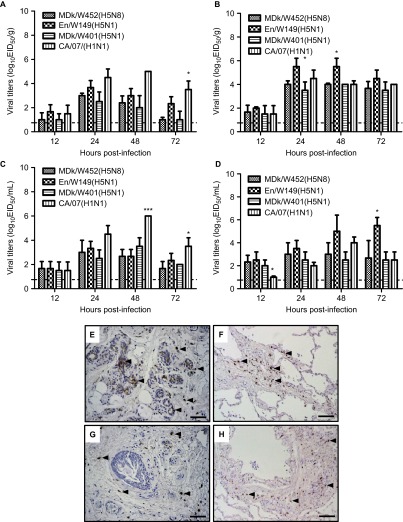
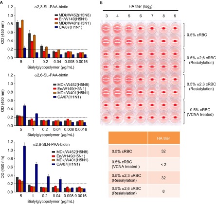
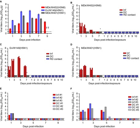
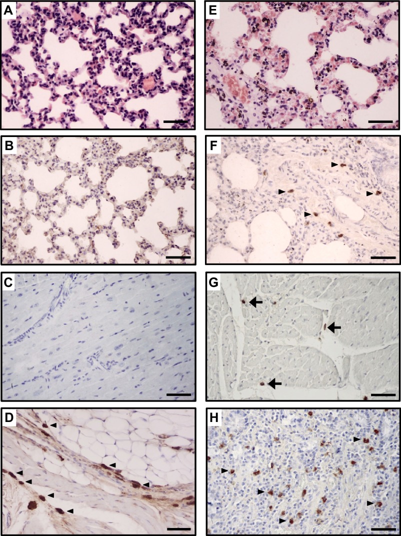
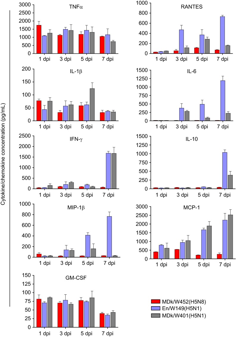
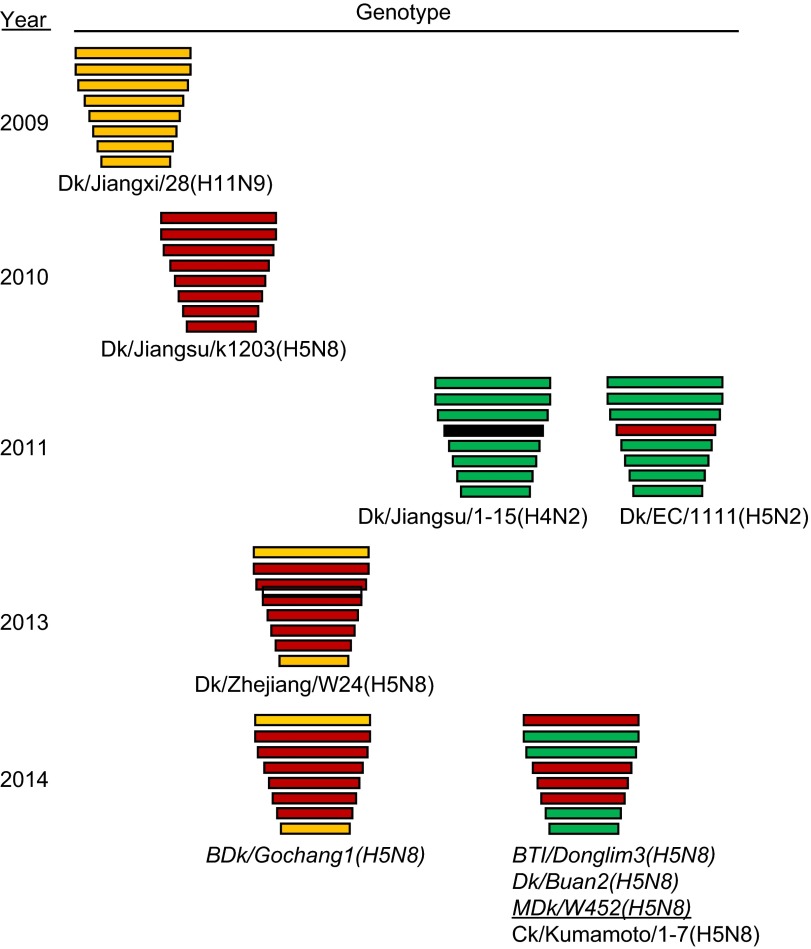
The endemicity of highly pathogenic avian influenza (HPAI) A(H5N1) viruses in Asia has led to the generation of reassortant H5 strains with novel gene constellations. A newly emerged HPAI A(H5N8) virus caused poultry outbreaks in the Republic of Korea in 2014. Because newly emerging high-pathogenicity H5 viruses continue to pose public health risks, it is imperative that their pathobiological properties be examined. Here, we characterized A/mallard duck/Korea/W452/2014 (MDk/W452(H5N8)), a representative virus, and evaluated its pathogenic and pandemic potential in various animal models. We found that MDk/W452(H5N8), which originated from the reassortment of wild bird viruses harbored by migratory waterfowl in eastern China, replicated systemically and was lethal in chickens, but appeared to be attenuated, albeit efficiently transmitted, in ducks. Despite predominant attachment to avian-like virus receptors, MDk/W452(H5N8) also exhibited detectable human virus-like receptor binding and replicated in human respiratory tract tissues. In mice, MDk/W452(H5N8) was moderately pathogenic and had limited tissue tropism relative to previous HPAI A(H5N1) viruses. It also induced moderate nasal wash titers in inoculated ferrets; additionally, it was recovered in extrapulmonary tissues and one of three direct-contact ferrets seroconverted without shedding. Moreover, domesticated cats appeared to be more susceptible than dogs to virus infection. With their potential to become established in ducks, continued circulation of A(H5N8) viruses could alter the genetic evolution of pre-existing avian poultry strains. Overall, detailed virological investigation remains a necessity given the capacity of H5 viruses to evolve to cause human illness with few changes in the viral genome.

Keywords: HPAI A(H5N8); avian influenza virus; genetic evolution; migratory waterfowl; reassortment.


Figures

Figure 1
Figure 1
Growth kinetics and attachment of viruses in human respiratory tissues ex vivo. Replication of the MDk/W452(H5N8) virus was monitored in human nasal respiratory epithelial (A) and lung (B) tissue explants and corresponding culture supernatants (C and D, respectively) starting at 12 hpi and at 24-h intervals thereafter. Growth kinetics were compared to those of the control HPAI A(H5N1) (En/W149 and MDk/W401) and 2009 pandemic CA/07(H1N1) viruses. The titers shown are means±SD from three independently performed experiments. Immunohistochemical detection of viral NP antigen was performed using nasal and lung tissue blocks incubated with CA/07(H1N1) (E and F), the control strain, or MDk/W452(H5N8) (G and H), the test virus. Arrows indicate positive immunostaining. Scale bars=50 µm. *P<0.05, ***P<0.0001 relative to MDk/W452(H5N8).
Figure 2
Figure 2
Virus receptor-binding specificity assays. (A) Binding affinity of inactivated whole viruses to SA α2,3′-SL-PAA-biotin, SA α2,6′-SL-PAA-biotin or SA α2,6′SLN-PAA-biotin glycans. The results shown are means±SD (mean of three replicates). The dashed lines indicate the limit of detection. (B) HA assays using re-sialylated cRBCs. The results obtained with re-sialylated cRBCs were normalized to the results obtained with untreated cRBCs.
Figure 3
Figure 3
Replication of MDk/W452(H5N8) in mammalian animal models. Virus replication was examined in mice, ferrets, dogs and cats that had been experimentally inoculated intranasally with 106 EID50/mL of designated virus. Mouse lung titers shown are means±SD from three animals (A). Individual nasal wash titers are shown for ferrets inoculated with MDk/W452(H5N8) (B), En/W149(H5N1) (C) or MDk/W401(H5N1) (D). To examine transmission, the inoculated animals were individually paired with a DC and an RD-contact animal (1∶1∶1 setup, triplicate). Three infected dogs (E) and cats (F) were also individually cohoused with contact animals in the same cage at 1 dpi and monitored for virus shedding in nasal swabs. The limit of virus detection was 0.7 log10EID50/mL; titers below that limit are shown as 0.5 log10EID50/mL (dashed lines). An asterisk (*) signifies seroconversion as determined by HI analysis at 21 dpi. Inf, directly infected animal.
Figure 4
Figure 4
Histopathology and immunohistochemical staining for influenza virus antigen in tissues of mouse (AD) and ferret (EH) infected with the HPAI A(H5N8) virus. In the lung, the alveolar septum is mildly thickened with increased cellularity (mouse: A and B; ferret: E and F). The influenza viral NP antigen was rarely detected in the lungs and hearts of mice (B and C, respectively) and was infrequently detected in mononuclear phagocytic cells in the lungs and hearts of ferrets (F, arrows; G, arrowheads, respectively). In contrast, in the nasal cavities of mice (D, arrows) and in the intestines of ferrets (H), mononuclear phagocytic cells were commonly immunopositive for influenza virus antigen. Scale bars=50 µm.
Figure 5
Figure 5
Cytokine and chemokine responses in the lungs of infected mice. Concentrations of various cytokines/chemokines in BAL fluids of infected mice at 1, 3, 5 and 7 dpi were measured by protein analysis with the Luminex-based multiplex immunoassay kit (Affymetrix, Santa Clara, CA, USA). The values shown are means±SD (errors bars) from three mouse BAL fluids per time point tested.
Figure 6
Figure 6
Putative generation of the novel HPAI A(H5N8) viruses that caused domestic poultry outbreaks in Korea and Japan in 2014. Genes from top to bottom are PB2, PB1, PA, HA, NP, NA, M and NS. For simplicity, each color represents an individual virus background. Italicized virus names are the Korean strains, and the underlined virus is the isolate we characterized. Virus abbreviations: Dk/Jiangxi/28(H11N9), A/duck/Jiangxi/28/2009(H11N9); Dk/Jiangsu/k1203(H5N8), A/duck/Jiangsu/k1203/2009(H5N8); Dk/Jiangsu/1-15(H4N2), A/duck/Jiangsu/1-15/2011(H4N2); Dk/EC/1111(H4N2), A/duck/Eastern China/1111/2011(H4N2); Dk/Zhejiang/W24(H5N8), A/duck/Zhejiang/W24/2013(H5N8); BDk/Gochang1(H5N8), A/broiler duck/Korea/Gochang1/2014(H5N8; BTl/Donglim3(H5N8), A/Baikal teal/Korea/Donglim3/2014(H5N8); Dk/Buan2(H5N8), A/duck/Korea/Buan2/2014(H5N8); MDk/W452(H5N8), A/mallard duck/W452/2014(H5N8); Ck/Kumamoto/1-7(H5N8), A/chicken/Kumamoto/1-7/2014(H5N8).

References

    1. Eagles D, Siregar ES, Dung DH, Weaver J, Wong F, Daniels P. H5N1 highly pathogenic avian influenza in Southeast Asia. Rev Sci Tech. 2009;28:341–348. - PubMed
    1. World Health Organization/Global Influenza Programme Cumulative number of confirmed human cases for avian influenza A(H5N1) reported to WHO, 2003–2014 Geneva: WHO; 2014. Available at http://www.who.int/influenza/human_animal_interface/EN_GIP_20140124Cumul... (accessed 24 January 2014).
    1. Chen H, Smith GJ, Li KS, et al. Establishment of multiple sublineages of H5N1 influenza virus in Asia: implications for pandemic control. Proc Natl Acad Sci USA. 2006;103:2845–2450. - PMC - PubMed
    1. Gu M, Liu W, Cao Y, et al. Novel reassortant highly pathogenic avian influenza (H5N5) viruses in domestic ducks, China. Emerg Infect Dis. 2011;17:1060–1063. - PMC - PubMed
    1. Gu M, Huang J, Chen Y, et al. Genome sequence of a natural reassortant H5N2 avian influenza virus from domestic mallard ducks in eastern China. J Virol. 2012;86:12463–12464. - PMC - PubMed