Review
. 2014 Mar;3(3):e21.
doi: 10.1038/emi.2014.19. Epub 2014 Mar 26.

Natural reservoirs for homologs of hepatitis C virus

Affiliations
Review

Natural reservoirs for homologs of hepatitis C virus

Stephanie Pfaender et al. Emerg Microbes Infect. 2014 Mar.

Abstract

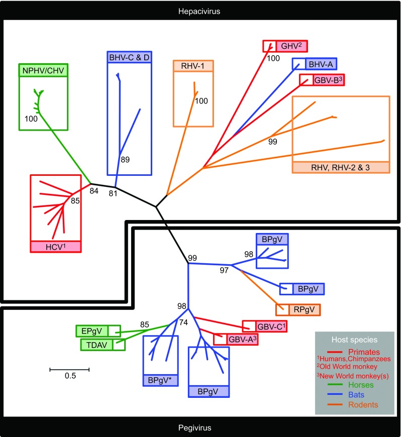
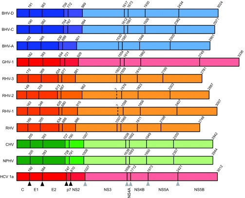
Hepatitis C virus is considered a major public health problem, infecting 2%-3% of the human population. Hepatitis C virus infection causes acute and chronic liver disease, including chronic hepatitis, cirrhosis and hepatocellular carcinoma. In fact, hepatitis C virus infection is the most frequent indication for liver transplantation and a vaccine is not available. Hepatitis C virus displays a narrow host species tropism, naturally infecting only humans, although chimpanzees are also susceptible to experimental infection. To date, there is no evidence for an animal reservoir of viruses closely related to hepatitis C virus which may have crossed the species barrier to cause disease in humans and resulted in the current pandemic. In fact, due to this restricted host range, a robust immunocompetent small animal model is still lacking, hampering mechanistic analysis of virus pathogenesis, immune control and prophylactic vaccine development. Recently, several studies discovered new viruses related to hepatitis C virus, belonging to the hepaci- and pegivirus genera, in small wild mammals (rodents and bats) and domesticated animals which live in close contact with humans (dogs and horses). Genetic and biological characterization of these newly discovered hepatitis C virus-like viruses infecting different mammals will contribute to our understanding of the origins of hepatitis C virus in humans and enhance our ability to study pathogenesis and immune responses using tractable animal models. In this review article, we start with an introduction on the genetic diversity of hepatitis C virus and then focus on the newly discovered viruses closely related to hepatitis C virus. Finally, we discuss possible theories about the origin of this important viral human pathogen.

Keywords: genetic diversity; hepacivirus; hepatitis C virus; homologs of hepatitis C virus; pegivirus.


Figures

Figure 1
Figure 1
Phylogenetic analysis of the NS3 protease domain of diverse members of the hepaciviruses and pegiviruses (Table 2), with susceptible host species incorporated. Radial tree topology highlighting distinct viral clades (boxed) within the hepacivirus (top) and pegivirus (bottom) genera. Viral clades are color-coded according to susceptible host species (primates: red, horses: green, bats: blue and rodents: orange). Phylogeny was generated using 54 nucelotide sequences encoding a 534-bp fragment at the N-terminus of the NS3 gene, corresponding to the virally encoded protease domain (genome coordinates 3420–3954 HCV genotype 1a strain; accession number NC_004102). Sequences were aligned according to overlying amino acids using the Clustal W algorithm implemented in MEGA5, and gap-stripped prior to tree construction. Tree was generated using the maximum likelihood method implemented in MEGA5 under the GTR+I+Γ model of nucleotide substitution. Values assigned to deep internal nodes within the phylogeny represent bootstrap supports for groupings. Values presented are percentages, derived from 1000 replications, with only significant values shown (>70%). The pegivirus grouping and the deep divergences within it are generally well supported, indicating the monophyly of the genus. The grouping of HCV, NPHV/CHV and BHV-C and D within the hepacivirus genus is also well supported. However, the deep divergences apparent between the remaining members of the genus are characterised by extremely long connecting branch-lengths and non-significant bootstrap values. This is indicative of ancient divergence between members, in addition to sparseness of taxon sampling: it is likely that many hepaciviruses in this portion of the tree remain either undiscovered, or have become extinct along with their hosts. Branch lengths are in accordance with the scale bar and are proportional to nucleotide substitutions per site. BPgV* clade containing the virus formerly designated GBV-D. Bat hepacivirus, BHV; bat pegivirus, BPgV; rodent hepacivirus, RHV; rodent pegivirus, RPgV; guereza hepacivirus, GHV; Theiler's disease-associated virus, TDAV; equine pegivirus, EPgV.
Figure 2
Figure 2
Reference cartoons depicting polyprotein organisation and cleavage site coordinates for distinct hepaciviruses. Cartoons of recently described hepaciviral polyproteins relative to an HCV reference (bottom) are shown, depicting locations of homologous protease cleavage sites, with names of represented hepaciviruses located to the left of each polyprotein schematic. Coordinates located above each cartoon represent the first residue of each encoded protein. The 10 cleaved proteins are detailed below the schematic cartoon of the HCV genotype 1a reference polyprotein. Black and grey triangles located below the HCV cartoon indicate host and viral protease cleavage sites, respectively. Peptide cleavage sites have been experimentally validated for HCV, while recently identified hepaciviral homologs have predicted cleavage sites which require further experimental validation. Color coding according to susceptible host species is identical to Figure 1. Structural proteins (core, E1 and E2) associated with infectious virions are shaded dark, non-structural proteins essential for assembly (p7 and NS2) are shaded intermediate and non-structural proteins associated with the replication module (NS3, NS4A, NS4B, NS5A and NS5B) are shaded light. GenBank accession numbers for representative strains are HCV-1a: NC_004102, NPHV: JQ434008, CHV: JF744991, RHV: KC815310, RHV-1: KC411777, RHV-2: KC411784, RHV-3: KC411807, BHV-A: KC796077, BHV-C: KC796090 and BHV-D: KC7960074.

References

    1. Lavanchy D. Evolving epidemiology of hepatitis C virus. Clin Microbiol Infect. 2011;17:107–115. - PubMed
    1. Brown RS. Hepatitis C and liver transplantation. Nature. 2005;436:973–978. - PubMed
    1. Manns MP, von Hahn T. Novel therapies for hepatitis C—one pill fits all. Nat Rev Drug Discov. 2013;12:595–610. - PubMed
    1. Casey LC, Lee WM. Hepatitis C virus therapy update 2013. Curr Opin Gastroenterol. 2013;29:243–249. - PubMed
    1. Moradpour D, Penin F, Rice CM. Replication of hepatitis C virus. Nat Rev Microbiol. 2007;5:453–463. - PubMed