. 2015 Jun 2;112(22):E2891-9.
doi: 10.1073/pnas.1507393112. Epub 2015 May 18.

AMCase is a crucial regulator of type 2 immune responses to inhaled house dust mites

Affiliations

AMCase is a crucial regulator of type 2 immune responses to inhaled house dust mites

Lark Kyun Kim et al. Proc Natl Acad Sci U S A. .

Abstract

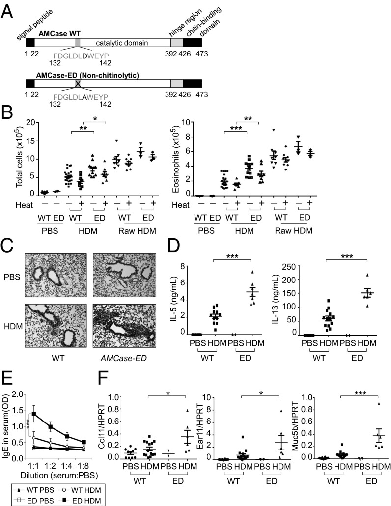
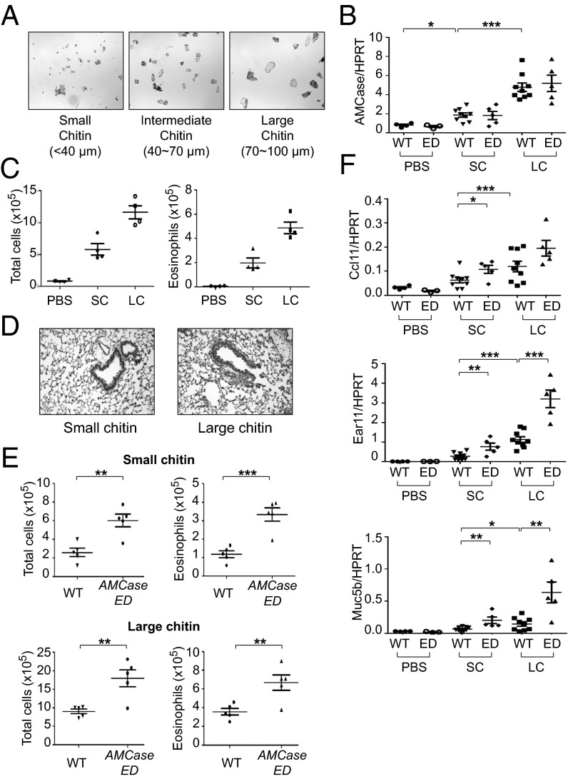
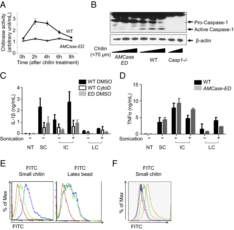
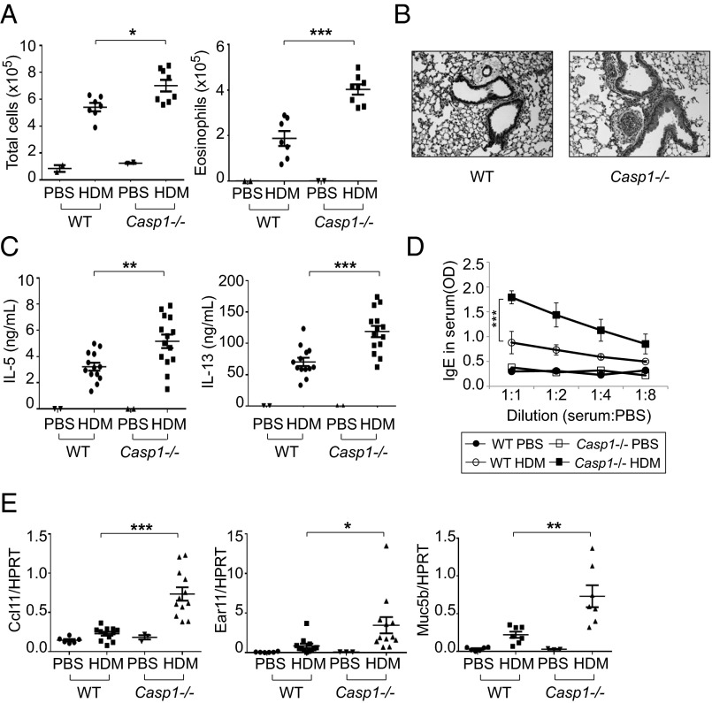
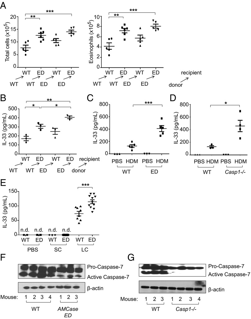
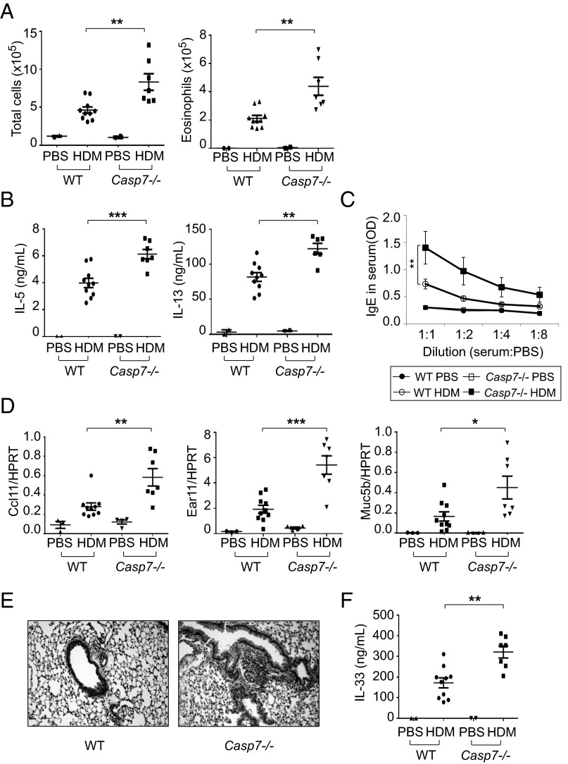
Chitinases are enzymes that cleave chitin, a component of the exoskeleton of many organisms including the house dust mite (HDM). Here we show that knockin mice expressing an enzymatically inactive acidic mammalian chitinase (AMCase), the dominant true chitinase in mouse lung, showed enhanced type 2 immune responses to inhaled HDM. We found that uncleaved chitin promoted the release of IL-33, whereas cleaved chitin could be phagocytosed and could induce the activation of caspase-1 and subsequent activation of caspase-7; this results in the resolution of type 2 immune responses, probably by promoting the inactivation of IL-33. These data suggest that AMCase is a crucial regulator of type 2 immune responses to inhaled chitin-containing aeroallergens.

Keywords: AMCase; IL-33; chitin; house dust mite.


Conflict of interest statement

The authors declare no conflict of interest.

Figures

Fig. 1.
Fig. 1.
Enhanced type 2 immune responses to inhaled HDM in AMCase-ED mice. (A) The schematic domains of AMCase. Aspartic acid is replaced with alanine in active site of catalytic domain of AMCase in AMCase-ED mice. (B) The number of total cells (Left) or eosinophils (Right) in the BAL of WT or AMCase-ED mice after administration of PBS, HDM, or raw HDM with (+) or without (−) pretreatment at 95 °C for 10 min. (C) H&E staining of lung sections of WT or AMCase-ED mice after administration of PBS or HDM (magnification: 20x). (D) The production of IL-5 (Left) and IL-13 (Right). The cells of lung-draining lymph nodes from WT or AMCase-ED mice after administration of PBS or HDM were restimulated in vitro with HDM and mitomycin-C–treated splenocytes for 48 h. Supernatants were collected and analyzed for IL-5 and IL-13 by ELISA. (E) The level of IgE in serum of WT or AMCase-ED mice after administration of PBS or HDM. The serum was diluted with PBS sequentially and analyzed for IgE by ELISA. (F) The expression of ccl11, ear11, and muc5b, normalized to hypoxanthine-guanine phosphoribosyltransferase, in the lung of WT or AMCase-ED mice after administration of PBS or HDM. *P < 0.05, **P < 0.01, and ***P < 0.001, unpaired Student’s t test. Data were combined from at least four independent experiments. Error bars indicate the SEM.
Fig. 2.
Fig. 2.
The enhanced type 2 immune responses to inhaled chitin in AMCase-ED mice. (A) The images of fractionated chitin. (B) The expression of AMCase, normalized to HPRT, in the lung of WT or AMCase-ED mice after administration of PBS, small-chitin, or large-chitin fragments. (C) The number of total cells (Left) or eosinophils (Right) in the BAL of WT mice after administration of PBS, small-chitin, or large-chitin fragments. (D) H&E staining of lung sections of WT mice after administration of small-chitin or large-chitin fragments (magnification: 20x). (E) The number of total cells and eosinophils in the BAL of WT mice after administration of small-chitin (Top) and large-chitin fragments (Bottom). (F) The expression of ccl11, ear11, and muc5b, normalized to HPRT in the lung of WT or AMCase-ED mice after administration of PBS, small-chitin, or large-chitin fragments. *P < 0.05, **P < 0.01, and ***P < 0.001, unpaired Student’s t test. Data were combined from at least three independent experiments. Error bars indicate the SEM.
Fig. 3.
Fig. 3.
Defective caspase-1 activation after administration of chitin in AMCase-ED cells. (A) Chitinase activity in the supernatants of LPS-primed peritoneal macrophages from WT or AMCase-ED mice after chitin mixture (less than 70 μm) treatments. (B) Immunoblot analysis of caspase-1 and β-actin in LPS-primed peritoneal macrophages from WT, AMCase-ED, or Casp1−/− mice. (C) Production of IL-1β in the supernatants of LPS-primed peritoneal macrophages from WT or AMCase-ED mice with pretreatment of DMSO or cytochalasin D after treatment of small, intermediate, or large-chitin fragments with (+) or without (−) sonication. (D) Production of TNFα in the supernatants of LPS-primed peritoneal macrophages from WT or AMCase-ED mice after treatment of small-, intermediate-, or large-chitin fragments with (+) or without (−) sonication. (E) Histograms of fluorescence distribution of the peritoneal macrophages from WT (blue) or AMCase-ED mice (green) after FITC-conjugated small-chitin (Left) or latex bead (Right) treatment. Red represents WT cells without any treatment. (F) Histograms of fluorescence distribution of the mixture of peritoneal macrophages from WT and AMCase-ED mice. Yellow represents 100% of WT cells, green represents the mixture of 66% of WT cells and 33% of AMCase-ED cells, and blue represents the mixture of 33% of WT cells and 66% of AMCase-ED cells after FITC-conjugated small-chitin fragment treatment. Red represents the mixture of 50% of WT cells and 50% of AMCase-ED cells without any treatment. Data are representative of at least three independent experiments. Error bars indicate the SD.
Fig. 4.
Fig. 4.
Enhanced type 2 immune responses to inhaled HDM in Casp1−/− mice. (A) The number of total cells (Left) and eosinophils (Right) in the BAL of WT or Casp1−/− mice after administration of HDM. (B) H&E staining of lung sections of WT or Casp1−/− mice after administration of HDM (magnification: 20x). (C) Production of IL-5 (Left) and IL-13 (Right). The cells of lung-draining lymph nodes from WT or Casp1−/− mice after administration of HDM were restimulated in vitro with HDM and mitomycin-C–treated splenocytes for 48 h. Supernatants were collected and analyzed for IL-5 and IL-13 by ELISA. (D) Levels of IgE in serum of WT or Casp1−/− mice after administration of HDM. The serum was diluted with PBS sequentially and analyzed for IgE by ELISA. (E) Expression of ccl11, ear11, and muc5b, normalized to HPRT, in the lung of WT or Casp1−/− mice after administration of PBS or HDM. *P < 0.05, **P < 0.01, and ***P < 0.001, unpaired Student’s t test. Data were combined from at least three independent experiments. Error bars indicate the SEM.
Fig. 5.
Fig. 5.
Enhanced production of IL-33 in AMCase-ED mice after administration of HDM or chitin. (A) The number of total cells (Left) and eosinophils (Right) in the BAL of WT → WT (where WT bone marrow was injected into irradiated WT recipients), WT → AMCase-ED, AMCase-ED → WT, or AMCase-EDAMCase-ED mice after administration of HDM. (B) Production of IL-33 in the BAL of WT → WT, WT → AMCase-ED, AMCase-ED → WT, or AMCase-EDAMCase-ED mice after administration of HDM. (C) Production of IL-33 in the BAL of WT or AMCase-ED mice after administration of PBS or HDM. (D) Production of IL-33 in the BAL of WT or Casp1−/− mice after administration of PBS or HDM. (E) Production of IL-33 in the BAL of WT or AMCase-ED mice after administration of PBS, small chitin, or large chitin. (F) Immunoblot analysis of caspase-7 and β-actin in lung lysates from WT mice or AMCase-ED mice after administration of PBS or HDM. The number indicates the individual mouse. (G) Immunoblot analysis of caspase-7 and β-actin in lung lysates from WT mice or Casp7−/− mice after administration of PBS or HDM. The number indicates the individual mouse. *P < 0.05, **P < 0.01, and ***P < 0.001, unpaired Student’s t test. Data were combined from at least three independent experiments. Error bars indicate the SEM.
Fig. 6.
Fig. 6.
Enhanced type 2 immune responses to inhaled HDM in Casp7−/− mice. (A) The number of total cells (Left) or eosinophils (Right) in the BAL of WT or Casp7−/− mice after administration of PBS or HDM. (B) Production of IL-5 (Left) and IL-13 (Right). The cells of lung-draining lymph nodes from WT or Casp7−/− mice after administration of PBS or HDM were restimulated in vitro with HDM and mitomycin-C–treated splenocytes for 48 h. Supernatants were collected and analyzed for IL-5 and IL-13 by ELISA. (C) Levels of IgE in serum of WT or Casp7−/− mice after administration of PBS or HDM. The serum was diluted with PBS sequentially and analyzed for IgE by ELISA. (D) Expression of ccl11, ear11, and muc5b, normalized to HPRT, in the lung of WT or Casp7−/− mice after administration of PBS or HDM. (E) H&E staining of lung sections of WT or Casp7−/− mice after administration of PBS or HDM (magnification: 20x). (F) Production of IL-33 in the BAL of WT or Casp7−/− mice after administration of PBS or HDM. *P < 0.05, **P < 0.01, and ***P < 0.001, unpaired Student’s t test. Data were combined from at least three independent experiments. Error bars indicate the SEM.

References

    1. Gregory LG, Lloyd CM. Orchestrating house dust mite-associated allergy in the lung. Trends Immunol. 2011;32(9):402–411. - PMC - PubMed
    1. Lambrecht BN, Hammad H. The airway epithelium in asthma. Nat Med. 2012;18(5):684–692. - PubMed
    1. Hammad H, Lambrecht BN. Dendritic cells and epithelial cells: Linking innate and adaptive immunity in asthma. Nat Rev Immunol. 2008;8(3):193–204. - PubMed
    1. Palm NW, Rosenstein RK, Medzhitov R. Allergic host defences. Nature. 2012;484(7395):465–472. - PMC - PubMed
    1. Pulendran B, Artis D. New paradigms in type 2 immunity. Science. 2012;337(6093):431–435. - PMC - PubMed

Publication types