Review
. 2015 Jun;32(2):182-94.
doi: 10.1055/s-0035-1549377.

Iatrogenic hepatopancreaticobiliary injuries: a review

Affiliations
Review

Iatrogenic hepatopancreaticobiliary injuries: a review

Prasanti G Vachhani et al. Semin Intervent Radiol. 2015 Jun.

Abstract

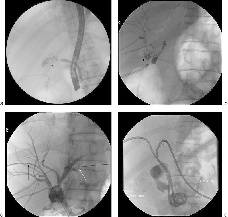
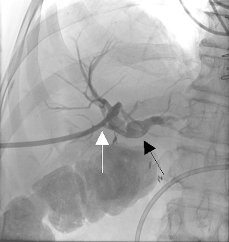
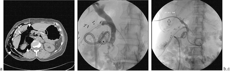
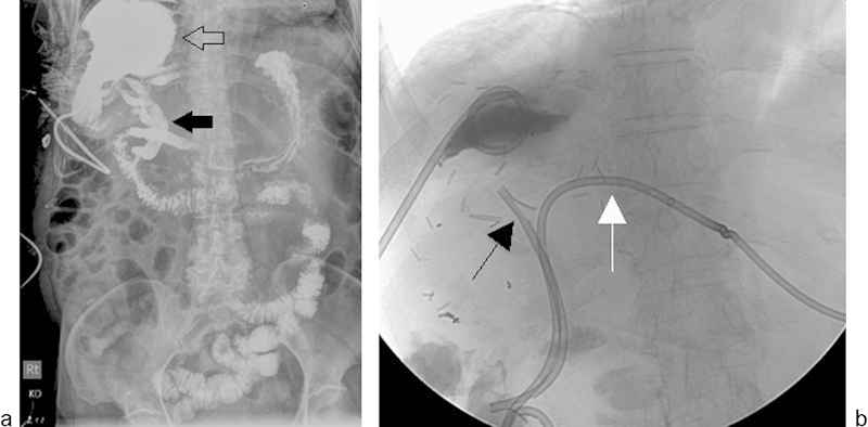
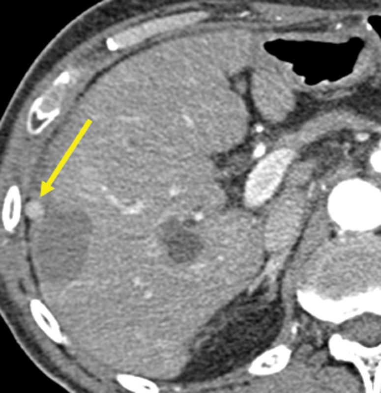
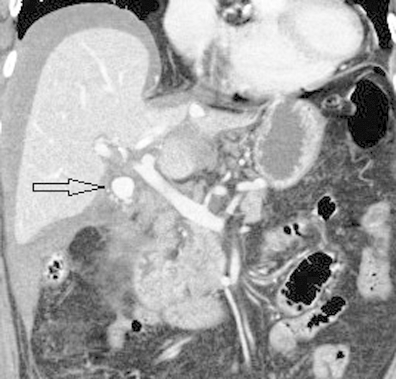
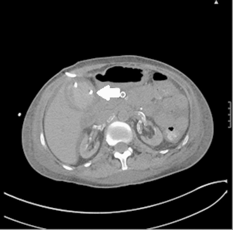
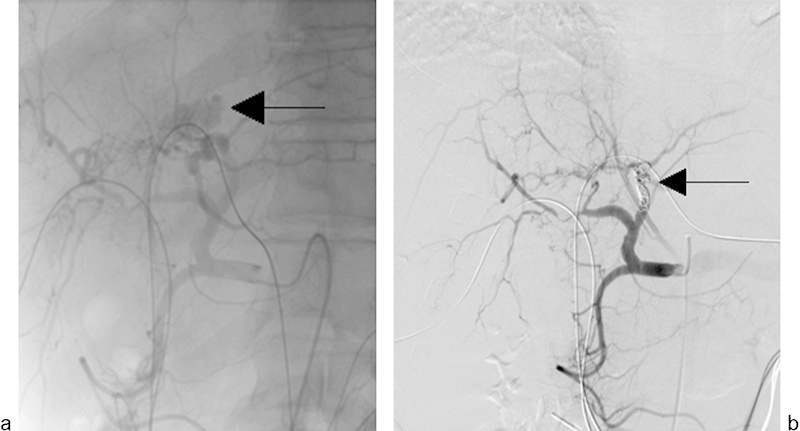
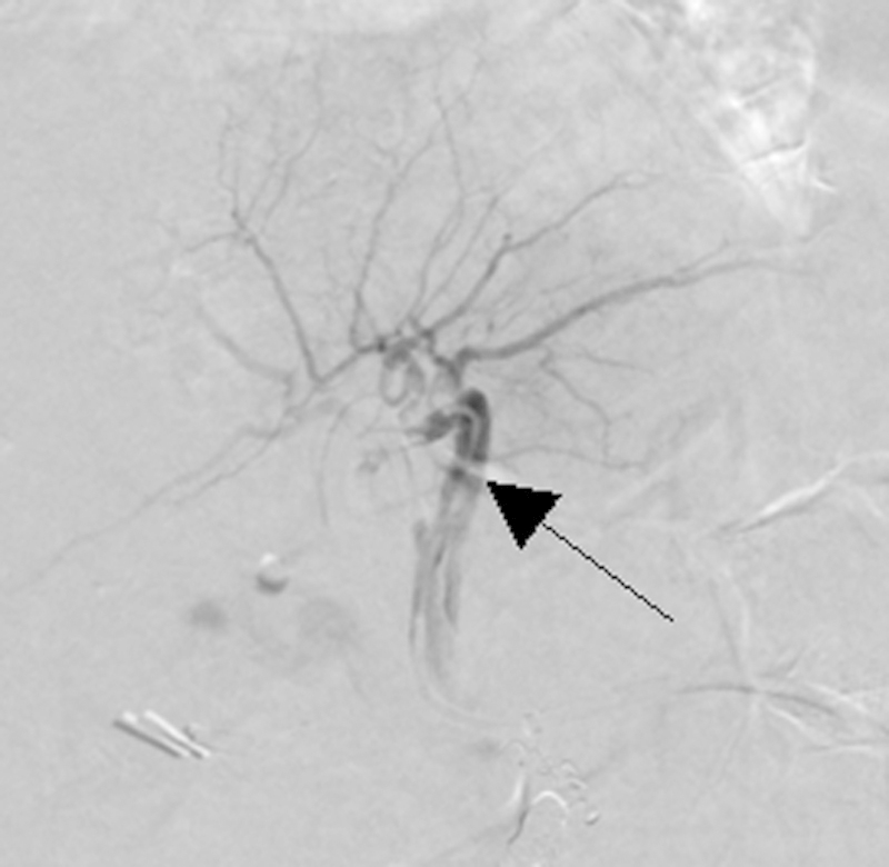
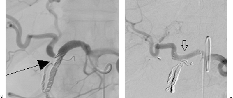
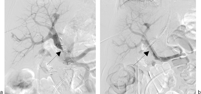
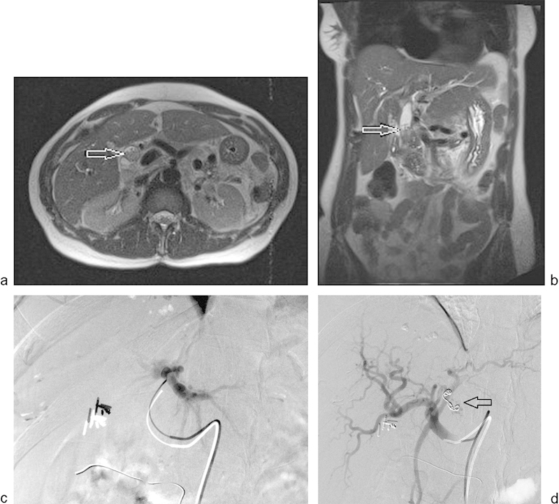
Iatrogenic hepatopancreaticobiliary injuries occur after various types of surgical and nonsurgical procedures. Symptomatically, these injuries may lead to a variety of clinical presentations, including tachycardia and hypotension from hemobilia or hemorrhage. Iatrogenic injuries may be identified during the intervention, immediately afterwards, or have a delayed presentation. These injuries are categorized into nonvascular and vascular injuries. Nonvascular injuries include biliary injuries such as biliary leak or stricture, pancreatic injury, and the development of fluid collections such as abscesses. Vascular injuries include pseudoaneurysms, arteriovenous fistulas, dissection, and perforation. Imaging studies such as ultrasound, computed tomography, magnetic resonance imaging, and digital subtraction angiography are critical for proper diagnosis of these conditions. In this article, we describe the clinical and imaging presentations of these iatrogenic injuries and the armamentarium of minimally invasive procedures (percutaneous drainage catheter placement, balloon dilatation, stenting, and coil embolization) that are useful in their management.

Keywords: bile duct injury; hepatobiliary; hepatopancreaticobiliary; iatrogenic injuries; interventional radiology.


Figures

Fig. 1
Fig. 1
A 52-year-old man underwent laparoscopic cholecystectomy and presented with persistent moderately severe abdominal pain 1 week after the cholecystectomy. Cross-sectional imaging (not shown) revealed a fluid collection in the surgical bed. (a) A fluoroscopic image obtained during ERCP demonstrates contrast extravasation from a blind-ending common bile duct (arrow), consistent with disruption. (b) PTHC was performed for biliary drainage. Cholangiographic image demonstrates disruption of the right hepatic ducts (arrow). There is no opacification of the common bile duct. (c) Fluoroscopic image showing placement of a wire across the disrupted right hepatic duct (black arrow) into the common bile duct. Cannulation of the left hepatic duct (white arrow) also demonstrates left hepatic ductal disruption. (d) Bilateral biliary drainage catheters were placed into the duodenum.
Fig. 2
Fig. 2
A 37-year-old woman presented with progressively increasing jaundice and abdominal pain after she underwent laparoscopic cholecystectomy. A single fluoroscopic image obtained during PTHC shows complete occlusion of the common hepatic duct (black arrow) and drainage by an external biliary drainage catheter (white arrow).
Fig. 3
Fig. 3
A 54-year-old man with jaundice, abdominal pain, and chronic pancreatitis underwent ERCP and pancreatic stent placement. During cannulation of the common bile duct, the patient became hypoxemic and the procedure was terminated, and an emergent CT scan was performed. (a) Axial CT image demonstrates diffuse subcutaneous emphysema, pneumoperitoneum, and pneumoretroperitoneum (white arrows). The patient underwent exploratory laparotomy, which revealed a malpositioned pancreatic stent (white circle) and perforated duodenum. (b) Postoperatively, biliary fluid drained from the incision site. PTHC showed extravasation from the distal common bile duct (black arrow). (c) An internal/external biliary drainage catheter was placed (black arrow), with the pigtail in the duodenum.
Fig. 4
Fig. 4
A 72-year-old woman underwent right hepatectomy and resection of the caudate lobe for hepatocellular carcinoma. On postoperative day 2, a large fluid collection was identified in the surgical bed. The collection was drained by a 12 French drainage catheter placed under CT guidance (not shown). Large volume drainage from the catheter persisted for ∼2 months. (a) Fluoroscopic injection of the large right subphrenic fluid collection cavity (open arrow) shows fistulous communication with the biliary system (solid arrow). (b) Additional drainage by placement of an endoscopic biliary stent (black arrow) and left internal–external biliary drain (white arrow) resulted in complete resolution of communication after 6 weeks. Separate contrast injection of the right upper quadrant abscess drainage catheter and biliary catheter revealed no residual communication between the right perihepatic fluid collection and the bile ducts (not shown).
Fig. 5
Fig. 5
Images from a 50-year-old woman who underwent trisegmentectomy for cholangiocarcinoma. The patient presented with persistent leakage of bile despite PTHC and PTBD (a) Cholangiogram of a segment 3 duct through the sheath demonstrated active leakage of contrast in the perihepatic space (arrow). (b) After glue (n-butyl cyanoacrylate) injection, there was complete obliteration of the fistulous tract (arrow).
Fig. 6
Fig. 6
A 53-year-old man underwent left hepatectomy with Roux-en-Y hepaticojejunostomy for metastatic rectal carcinoma. The patient then underwent PTHC for persistent bile drainage from his surgical drain. (a) PTHC shows leakage of contrast at the hepaticojejunostomy site as a result of partial anastomotic dehiscence (arrow). (b) Fluoroscopic image shows placement of the external/internal biliary drainage catheter. (c) Tube cholangiogram shows resolution of the leak after 8 weeks.
Fig. 7
Fig. 7
A 38-year-old woman with carcinoid tumor metastases to the liver underwent extended left hepatectomy, hepatic artery thrombectomy with interposition graft, and choledochocholedochostomy. The patient had persistent bile leak from the T-tube tract and underwent PTHC. (a) PTHC shows complete loss of integrity of the common bile duct (open arrow), which is replaced by debris. A pigtail catheter is seen in the liver draining a biloma (solid arrow). (b) Digital subtraction angiography of the proper hepatic artery performed via a right common femoral artery approach demonstrates complete occlusion of the hepatic artery (arrow).
Fig. 8
Fig. 8
A 73-year-old woman who underwent pylorus-preserving pancreaticoduodenectomy for pancreatic intraductal mucinous adenoma presented with abdominal pain. (a and b) Axial contrast-enhanced CT images show contrast leak (arrow) at the pancreaticojejunostomy anastomosis with adjacent fluid collection.
Fig. 9
Fig. 9
A 56-year-old man underwent radiofrequency ablation of hepatocellular carcinoma involving the right lobe of the liver. At a 3-month follow-up, contrast-enhanced CT showed a hyperenhancing nodule adjacent to the ablation zone, representing tumor seeding along the needle track (arrow).
Fig. 10
Fig. 10
An 82-year-old woman presented to the emergency room with sudden onset of abdominal pain, tachycardia, and hypotension 3 days after laparoscopic cholecystectomy. Contrast-enhanced coronal CT scan image shows a loculated collection of contrast measuring ∼1.7 × 1.5 cm in the gallbladder fossa (arrow) representing a pseudoaneurysm. There is high-density perihepatic fluid due to intra-abdominal hemorrhage. The pseudoaneurysm was successfully treated by coil embolization of the cystic artery.
Fig. 11
Fig. 11
A 35-year-old woman underwent percutaneous cholecystostomy tube placement for acute cholecystitis due to multiple comorbidities preventing cholecystectomy. Following cholecystostomy tube placement, the patient became mildly tachycardic and complained of severe abdominal pain without a significant change in blood pressure. Axial noncontrast CT image of the abdomen reveals a hyperdense fluid collection within the gallbladder (arrow) after cholecystostomy tube placement, representing acute hemorrhage/hematoma.
Fig. 12
Fig. 12
A 37-year-old man with unresectable cholangiocarcinoma and bilateral internal/external biliary drainage catheters presented with hemodynamic instability and bright red blood from the left internal/external biliary catheter 2.5 months after catheter placement. (a) Emergency hepatic arteriogram revealed active extravasation from a major branch of the left hepatic artery (arrow). (b) The left hepatic artery was successfully embolized using 0.018 microcoils (arrow).
Fig. 13
Fig. 13
Image from a 62-year-old woman with unresectable hepatocellular carcinoma. Digital subtraction angiography performed during embolotherapy of the hepatic artery shows a non–flow-limiting intimal dissection of the left hepatic artery due to catheter manipulation (arrow).
Fig. 14
Fig. 14
A 64-year-old woman with hepatocellular carcinoma underwent gastroduodenal artery embolization before hepatic radioembolization. (a) A common hepatic angiogram shows displacement of the last coil (arrow) into the common hepatic artery, resulting in spasm and mechanical obstruction. (b) A digital subtraction angiogram shows successful exclusion of the displaced coil by a 5-mm-diameter covered endograft (arrow).
Fig. 15
Fig. 15
A 58-year-old woman underwent pancreaticoduodenectomy and partial portal vein resection repaired by a Gore-Tex patch for pancreatic adenocarcinoma. Follow-up ultrasound revealed a severe stenosis at the site of reconstruction. (a) Transhepatic portogram shows severe stenosis at the site of portal vein reconstruction (arrow). (b) A 16-mm-diameter Wallstent (Boston Scientific Inc., Marlborough, MA) was deployed across the stenosis using a transhepatic approach. Portogram after wall stent placement shows a patent portal vein with mild residual stenosis at the site of stenosis (arrow).
Fig. 16
Fig. 16
A 41-year-old woman underwent percutaneous biopsy of the left lobe of the liver and presented to the emergency room with upper gastrointestinal hemorrhage and decreasing hemoglobin level. Upper gastrointestinal endoscopy revealed blood oozing from the papilla. (a and b) T2-weighted MRI images/MRCP revealed dilated common bile duct and amorphous material in the common bile duct representing acute hemorrhage (arrow). No imaging abnormality was seen at the biopsy site. (c and d) Selective left hepatic angiogram performed via a right common femoral approach revealed an arterioportal fistula that was successfully embolized using two 0.0184-mm hydrocoils (arrow). On follow-up, no further evidence of clinical gastrointestinal hemorrhage was reported.

References

    1. Lau W Y, Lai E C, Lau S H. Management of bile duct injury after laparoscopic cholecystectomy: a review. ANZ J Surg. 2010;80(1–2):75–81. - PubMed
    1. Nuzzo G, Giuliante F, Giovannini I. et al.Advantages of multidisciplinary management of bile duct injuries occurring during cholecystectomy. Am J Surg. 2008;195(6):763–769. - PubMed
    1. Flum D R, Cheadle A, Prela C, Dellinger E P, Chan L. Bile duct injury during cholecystectomy and survival in Medicare beneficiaries. JAMA. 2003;290(16):2168–2173. - PubMed
    1. Saad N, Darcy M. Iatrogenic bile duct injury during laparoscopic cholecystectomy. Tech Vasc Interv Radiol. 2008;11(2):102–110. - PubMed
    1. Roslyn J J, Binns G S, Hughes E F, Saunders-Kirkwood K, Zinner M J, Cates J A. Open cholecystectomy. A contemporary analysis of 42,474 patients. Ann Surg. 1993;218(2):129–137. - PMC - PubMed