Review
. 2015 Jun;32(2):195-208.
doi: 10.1055/s-0035-1549378.

Iatrogenic urinary tract injuries: etiology, diagnosis, and management

Affiliations
Review

Iatrogenic urinary tract injuries: etiology, diagnosis, and management

Anthony M Esparaz et al. Semin Intervent Radiol. 2015 Jun.

Abstract

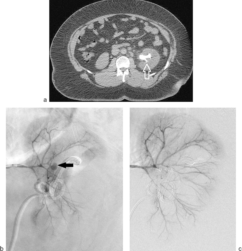
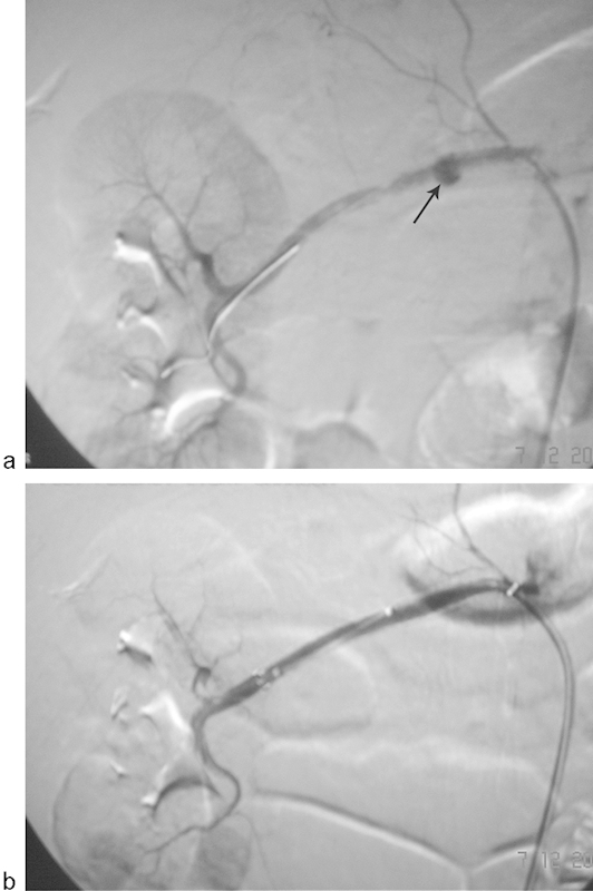
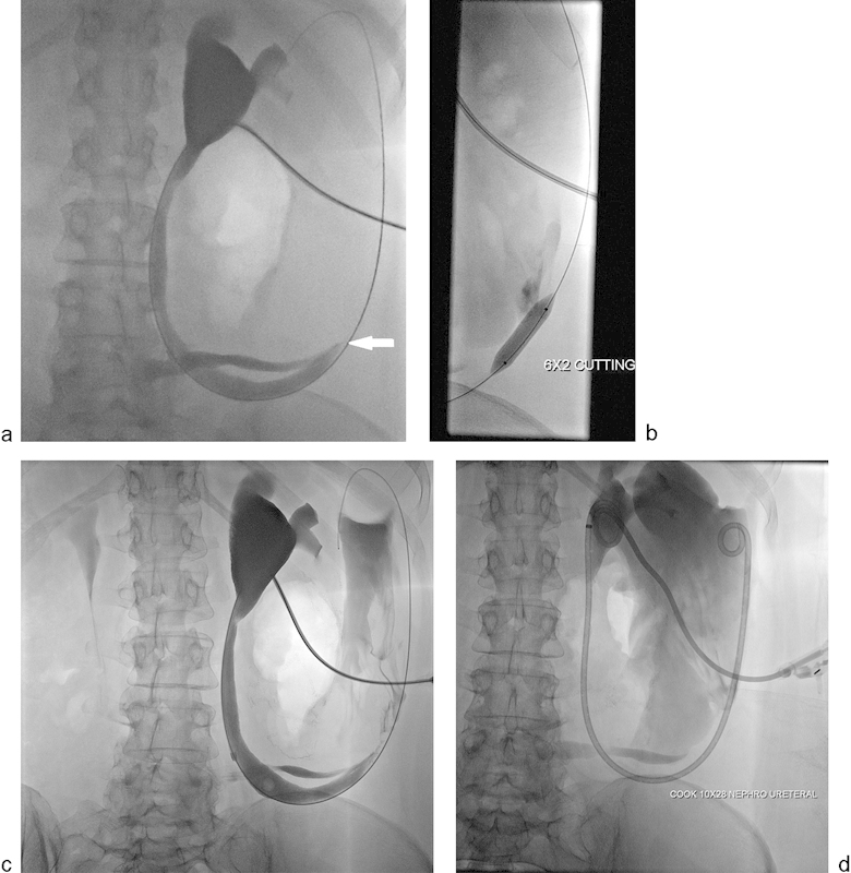
Iatrogenic injury to the urinary tract, including the kidneys, ureters, bladder, and urethra, is a potential complication of surgical procedures performed in or around the retroperitoneal abdominal space or pelvis. While both diagnostic and interventional radiologists often play a central and decisive role in the identification and initial management of a variety of iatrogenic injuries, discussions of these injuries are often directed toward specialists such as urologists, obstetricians, gynecologists, and general surgeons whose procedures are most often implicated in iatrogenic urinary tract injuries. Interventional radiologic procedures can also be a source of an iatrogenic urinary tract injury. This review describes the clinical presentation, risk factors, imaging findings, and management of iatrogenic renal vascular and urinary tract injuries, as well as the radiologist's role in the diagnosis, treatment, and cause of these injuries.

Keywords: bladder; iatrogenic injury; interventional radiology; renal vascular; ureter.


Conflict of interest statement

Disclosures None.

Figures

Fig. 1
Fig. 1
Axial (a) and coronal (b) noncontrast CT images of a 59-year-old man who presented to the emergency room in hemodynamic shock on postoperative day 9, following open partial nephrectomies for renal cell carcinoma. Noncontrast CT scan of the abdomen shows high attenuation fluid in the perinephric area and layering of high attenuation material, consistent with acute hemorrhage (arrow). Note mass effect on the kidney displacing the kidney anteriorly.
Fig. 2
Fig. 2
(a) An 82-year-old man presented with persistent hematuria following left partial nephrectomy for renal cell carcinoma. Contrast-enhanced CT scan shows a large pseudoaneurysm (arrow) and simultaneous opacification of the renal artery and renal vein representing arteriovenous fistula in the surgical bed. (b) Digital subtraction angiogram of the left renal artery confirms the pseudoaneurysm (arrow) and high flow arteriovenous fistula. (c) Digital subtraction angiogram of the left renal artery showing successful occlusion of the left renal artery pseudoaneurysm and arteriovenous fistula using combination of microcoils and thrombin. During embolization of the pseudoaneurysm and arteriovenous fistula, venous outflow was occluded using a balloon catheter placed in the left renal vein via right common femoral approach to prevent the possibility of nontarget embolization.
Fig. 3
Fig. 3
(a) Noncontrast CT scan of a 33-year-old man showing a staghorn calculus (arrow) within the left renal collecting system. The patient underwent nephrolithotomy for the stone and developed gross hematuria. (b) Left renal angiogram demonstrating an intrarenal pseudoaneurysm arising from a third-order branch of the left renal artery in the interpolar region (arrow). (c) Renal angiogram following embolization of the arterial supply using three 3-mm microcoils demonstrates complete occlusion of the pseudoaneurysm. Clinically, there was complete cessation of hematuria.
Fig. 4
Fig. 4
Digital subtraction angiography images of a 62-year-old woman following right renal artery angioplasty using 5 mm × 40 mm balloon catheter for renal artery stenosis. (a) Renal angiogram demonstrates development of a focal collection of contrast (arrow), consistent with a pseudoaneurysm. (b) Renal angiogram following deployment of 6 mm × 60 mm covered Fluency (Bard Peripheral Vascular, Inc. Tempe, AZ) stent. The pseudoaneurysm has been successfully excluded and good flow is noted in the artery.
Fig. 5
Fig. 5
A 76-year-old woman with history of rectal carcinoma underwent limited anterior resection and neoadjuvant chemoradiation therapy. Following chemoradiation and anterior resection, the patient developed bilateral hydronephrosis that was managed with bilateral ureteral stents. Six weeks after the stent placement, the patient presented with hematuria. (a) Contrast-enhanced CT scan demonstrates a pseudoaneurysm arising from the left external iliac artery (arrow), presumed to be due to erosion of the left distal ureter and external iliac artery by the ureteral stent. (b) Digital subtraction angiography of the left common iliac artery performed via a right common femoral artery approach confirms the above-described pseudoaneurysm (arrow) arising from the left external iliac artery. (c) Digital subtraction angiogram of the left common iliac artery following placement of an 8 mm × 50 mm Viabahn covered stent (Gore Inc., Flagstaff, AZ) across the above-described pseudoaneurysm demonstrating complete occlusion of the pseudoaneurysm. Clinically, following left common iliac artery endograft placement, there was complete cessation of hematuria.
Fig. 6
Fig. 6
A single fluoroscopic image of a 62-year-old woman demonstrating complete right ureteral obstruction (arrow) after a laparoscopic partial nephrectomy for renal cell carcinoma. This was successfully drained using an 8 French nephrostomy tube, and 2 months later it was successfully treated with pyeloplasty.
Fig. 7
Fig. 7
A 57-year-old woman with a history of interstitial cystitis underwent cystectomy and Indiana pouch urinary diversion. (a) Antegrade nephrostogram showing severe short segment, almost occlusive, stricture involving ureteroileal anastomosis (arrow). (b) Fluoroscopic image showing balloon dilation of the above-described stricture using 6 mm × 20 mm cutting balloon after failed regular balloon dilation. (c and d) Antegrade nephrostogram showing mild improvement in the appearance of the stricture to allow placement of a 10 mm × 28 cm nephroureteral stent placement.
Fig. 8
Fig. 8
A 70-year-old man underwent sigmoid colectomy for diverticulitis that was complicated by colorectal anastomotic stricture. During surgical repair of the colorectal stricture, the patient sustained a bladder laceration and right ureteral transection. (a) AP and (b) RAO cystography images showing extravasation of contrast adjacent to the bladder on right lateral and posterior aspects (arrows).
Fig. 9
Fig. 9
A 52-year-old woman with urine leak following decortication of a renal cyst. (a, b) Select axial and coronal images from a CT of the abdomen and pelvis performed in the excretory phase show layering of extravasated contrast (arrow) in an otherwise low attenuation fluid collection in the decortication bed of the right kidney. The patient was initially managed conservatively but subsequently developed fever; percutaneous catheter drainage was placed into the urinoma.

References

    1. Selzman A A, Spirnak J P. Iatrogenic ureteral injuries: a 20-year experience in treating 165 injuries. J Urol. 1996;155(3):878–881. - PubMed
    1. Hairston J C, Ghoniem G M. Urinary tract injuries: recognition and management. Clin Colon Rectal Surg. 2003;16:27–38. - PMC - PubMed
    1. Delacroix S E Jr, Winters J C. Urinary tract injuries: recognition and management. Clin Colon Rectal Surg. 2010;23(3):104–112. - PMC - PubMed
    1. Summerton D J Kitrey N D Lumen N Serafetinidis E Djakovic N; European Association of Urology. EAU guidelines on iatrogenic trauma Eur Urol 2012624628–639. - PubMed
    1. Brandes S, Coburn M, Armenakas N, McAninch J. Diagnosis and management of ureteric injury: an evidence-based analysis. BJU Int. 2004;94(3):277–289. - PubMed