. 2015 Jun 3;10(6):e0128680.
doi: 10.1371/journal.pone.0128680. eCollection 2015.

A Multifaceted Study of Scedosporium boydii Cell Wall Changes during Germination and Identification of GPI-Anchored Proteins

Affiliations

A Multifaceted Study of Scedosporium boydii Cell Wall Changes during Germination and Identification of GPI-Anchored Proteins

Sarah Ghamrawi et al. PLoS One. .

Abstract

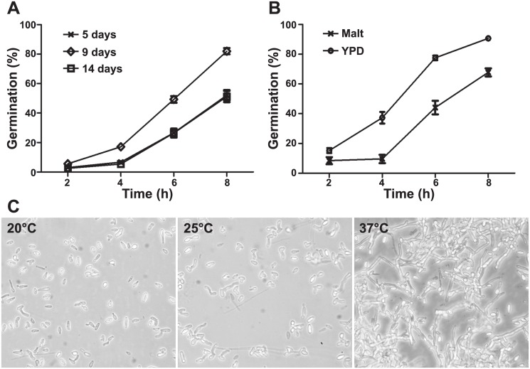
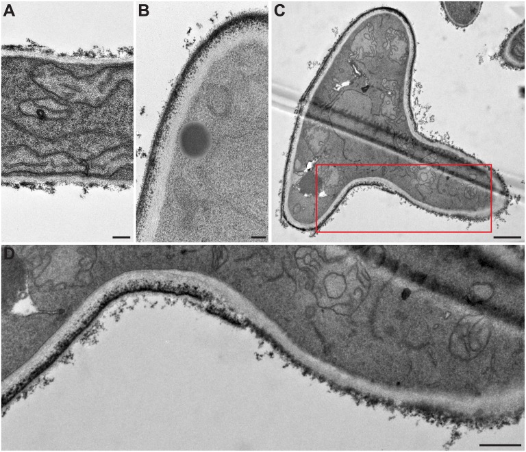
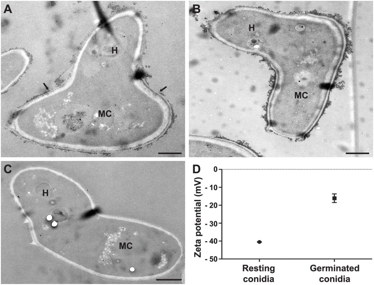
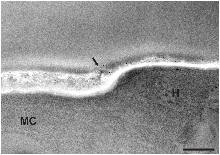
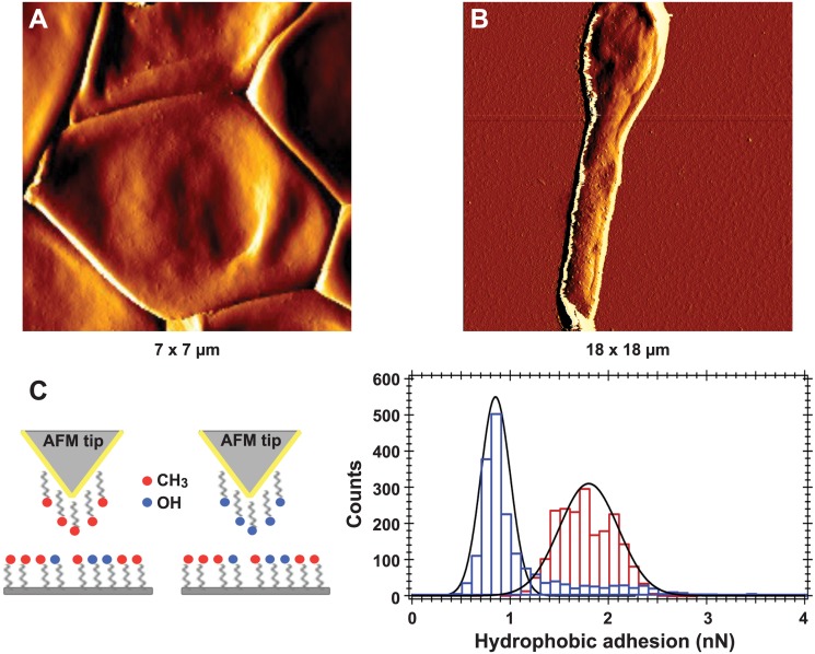
Scedosporium boydii is a pathogenic filamentous fungus that causes a wide range of human infections, notably respiratory infections in patients with cystic fibrosis. The development of new therapeutic strategies targeting S. boydii necessitates a better understanding of the physiology of this fungus and the identification of new molecular targets. In this work, we studied the conidium-to-germ tube transition using a variety of techniques including scanning and transmission electron microscopy, atomic force microscopy, two-phase partitioning, microelectrophoresis and cationized ferritin labeling, chemical force spectroscopy, lectin labeling, and nanoLC-MS/MS for cell wall GPI-anchored protein analysis. We demonstrated that the cell wall undergoes structural changes with germination accompanied with a lower hydrophobicity, electrostatic charge and binding capacity to cationized ferritin. Changes during germination also included a higher accessibility of some cell wall polysaccharides to lectins and less CH3/CH3 interactions (hydrophobic adhesion forces mainly due to glycoproteins). We also extracted and identified 20 GPI-anchored proteins from the cell wall of S. boydii, among which one was detected only in the conidial wall extract and 12 only in the mycelial wall extract. The identified sequences belonged to protein families involved in virulence in other fungi like Gelp/Gasp, Crhp, Bglp/Bgtp families and a superoxide dismutase. These results highlighted the cell wall remodeling during germination in S. boydii with the identification of a substantial number of cell wall GPI-anchored conidial or hyphal specific proteins, which provides a basis to investigate the role of these molecules in the host-pathogen interaction and fungal virulence.


Conflict of interest statement

Competing Interests: The authors have declared that no competing interests exist.

Figures

Fig 1
Fig 1. Kinetics of germination of S. boydii in various conditions.
(A) Conidia isolated from 5-, 9- and 14-day-old cultures on yeast peptone dextrose (YPD) agar were incubated in YPD liquid medium over 8 h at 37°C. (B) Conidia isolated from 9-day-old cultures on Malt or YPD agar were incubated in Malt or YPD liquid media over 8 h at 37°C. (C) Conidia isolated from 9-day-old cultures on YPD agar were incubated in YPD liquid medium for 16 h at 20°C, 25°C or 37°C (200X).
Fig 2
Fig 2. The life cycle of S. boydii under scanning electron microscopy.
After release, conidia (A) germinate (B) and the hyphal part of germ tubes elongates (C) until a first branch emerges near the mother cell (D). Both hyphae grow and more branching sites appear on filaments at the subapical region of the articles (E) until the mother cell can no more be distinguished (F). Arrows indicate sites of first branching, and later branching are indicated by arrowheads. All cultures were performed in YPD broth with incubation at 37°C for 6h (B), 8h (C), 10 h (D and E) or 24 h (F). Bars: 1 μm in A, B and C; 0.5 μm in D; and 5 μm in E and F.
Fig 3
Fig 3. Cell wall modifications during germination of S. boydii under transmission electron microscopy.
(A) hyphal cell wall; (B) conidial cell wall; and (C) cell wall of a germinating conidium. (D) Enlarged part of (C) highlighting the cell wall structural modifications during germination, particularly the electron dense outer layer being less continuous at the surface of the hyphal part of germ tubes. Bars: 0.2 μm in A and B; 1 μm in C; and 0.5 μm in D.
Fig 4
Fig 4. Surface charge modifications during germination of S. boydii by ferritin labeling and zeta potential measurements.
TEM images of germ tubes labeled with cationized ferritin (A), germ tubes treated with neuraminidase prior cationized ferritin labeling (B) or germ tubes incubated with native ferritin (C). (D) Comparison of the surface electrostatic charge of resting and germinated conidia calculated from the electrophoretic mobility of 10 000 cells using Zetasizer Nano ZS (P = 0.0005). H: hyphal part of germ tube; MC: mother cell of germ tube. Bars: 1 μm.
Fig 5
Fig 5. Fluorescence labeling of S. boydii surface carbohydrates with FITC-conjugated lectins.
Germ tubes after labeling with concanavalin A (A and C) or wheat germ agglutinin (B and D) lectins. The same fields are presented under fluorescence (A and B) and phase contrast microscopy (C and D) respectively. Arrows indicate mother cells.
Fig 6
Fig 6. Gold labeling of cell wall mannan groups in S. boydii germ tubes.
Germ tubes labeled with gold-conjugated concanavalin A (Con A; 5-nm gold particles) showing higher affinity of gold particles to the hyphal part (H) of germ tubes compared to the mother cell (MC) under transmission electron microscopy. Arrow indicates the limit of the outer cell wall layer of the mother cell. Bar: 0.5 μm.
Fig 7
Fig 7. High resolution imaging and chemical force spectroscopy analysis of S. boydii resting conidia and germ tubes.
AFM amplitude images of a resting (A) or germinated (B) S. boydii conidium. (C) Left, scheme for chemical functionalization of AFM tips. Gold-coated tips were modified with CH3-terminated alkanethiols or OH-terminated alkanethiols. (C) Right, histograms of hydrophobic adhesion forces measured on the surface of a resting conidium (1.8 ± 0.3 nN, in red) and the hyphal part of a germinated conidium (0.85 ± 0.15 nN, in blue).

References

    1. Guarro J, Kantarcioglu AS, Horré R, Luis Rodriguez-Tudela J, Cuenca Estrella M, Berenguer J, et al. 2006. Scedosporium apiospermum: changing clinical spectrum of a therapy-refractory opportunist. Med Mycol 44:295–327. - PubMed
    1. Kaltseis J, Rainer J, De Hoog GS. 2009. Ecology of Pseudallescheria and Scedosporium species in human-dominated and natural environments and their distribution in clinical samples. Med Mycol 47:398–405. 10.1080/13693780802585317 - DOI - PubMed
    1. Harun A, Gilgado F, Chen SC, Meyer W. 2010. Abundance of Pseudallescheria/Scedosporium species in the Australian urban environment suggests a possible source for scedosporiosis including the colonization of airways in cystic fibrosis. Med Mycol 48 (Suppl 1):S70–S76. 10.3109/13693786.2010.515254 - DOI - PubMed
    1. Rougeron A, Schuliar G, Leto J, Sitterlé E, Landry D, Bougnoux M-E, et al. 2015. Human-impacted areas of France are environmental reservoirs of the Pseudallescheria boydii/Scedosporium apiospermum species complex. Environ Microbiol 17:1039–1048. 10.1111/1462-2920.12472 - DOI - PubMed
    1. Beguin H, Nolard N. 1994. Mould biodiversity in homes I. Air and surface analysis of 130 dwellings. Aerobiologia 10: 157–166.

Publication types

MeSH terms

LinkOut - more resources