Review
. 2015 Jun;21(3 Behavioral Neurology and Neuropsychiatry):646-59.
doi: 10.1212/01.CON.0000466658.05156.54.

Executive dysfunction

Review

Executive dysfunction

Gil D Rabinovici et al. Continuum (Minneap Minn). 2015 Jun.

Abstract

Purpose of review: Executive functions represent a constellation of cognitive abilities that drive goal-oriented behavior and are critical to the ability to adapt to an ever-changing world. This article provides a clinically oriented approach to classifying, localizing, diagnosing, and treating disorders of executive function, which are pervasive in clinical practice.

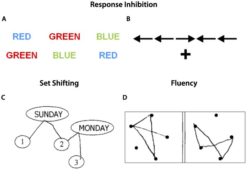
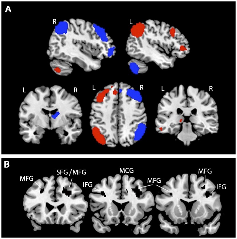
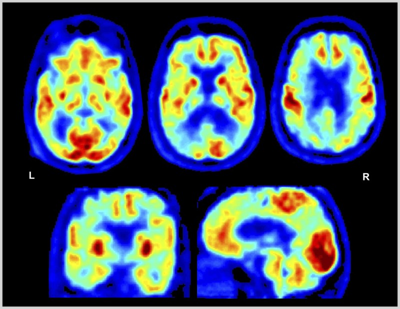
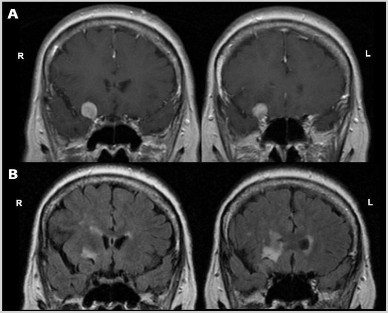
Recent findings: Executive functions can be split into four distinct components: working memory, inhibition, set shifting, and fluency. These components may be differentially affected in individual patients and act together to guide higher-order cognitive constructs such as planning and organization. Specific bedside and neuropsychological tests can be applied to evaluate components of executive function. While dysexecutive syndromes were first described in patients with frontal lesions, intact executive functioning relies on distributed neural networks that include not only the prefrontal cortex, but also the parietal cortex, basal ganglia, thalamus, and cerebellum. Executive dysfunction arises from injury to any of these regions, their white matter connections, or neurotransmitter systems. Dysexecutive symptoms therefore occur in most neurodegenerative diseases and in many other neurologic, psychiatric, and systemic illnesses. Management approaches are patient specific and should focus on treatment of the underlying cause in parallel with maximizing patient function and safety via occupational therapy and rehabilitation.

Summary: Executive dysfunction is extremely common in patients with neurologic disorders. Diagnosis and treatment hinge on familiarity with the clinical components and neuroanatomic correlates of these complex, high-order cognitive processes.


Figures

Figure 4-1
Figure 4-1
Example tasks to assess response inhibition, set shifting, and fluency. A, For the Stroop test, patients are asked to first read the words ignoring the colors (color naming), then name the colors ignoring the word (interference). B, For the Flanker task, patients are asked to identify the direction of the central arrow, with the adjacent arrows pointing either in the same direction (congruent) or opposite direction (incongruent). Stroop interference and incongruent trials on the Flanker task are considered tests of response inhibition. C, Trail making is a classic set shifting task in which patients write lines alternating in order between numbers and days of the week. D, Design fluency is an example of a nonverbal fluency task, in which patients are asked to draw as many unique designs as possible connecting the dots with four lines in 1 minute.
Figure 4-2
Figure 4-2
Neuroimaging correlates of executive functions. A, Left (red) and right (blue) hemisphere functional networks that are activated on functional MRI during executive control tasks are shown on a template brain in neurologic orientation. B, Gray matter correlates of a composite executive function score derived from the Executive Abilities: Measures and Instruments for Neurobehavioral Evaluation and Research (EXAMINER) battery identified via voxel-based morphometry. Black clusters represent brain regions in which, across subjects, higher gray matter volumes were correlated with higher composite executive function scores. All results are thresholded at P<.001 and corrected for multiple comparisons using permutation analysis at P<.05. R = right; L = left; MFG = middle frontal gyrus; SFG = superior frontal gyrus; IFG = inferior frontal gyrus; MCG = middle cingulate gyrus. Panel A network templates courtesy of Michael Greicius, MD, and the Functional Imaging in Neuropsychiatric Disorders Laboratory, Stanford University. Panel B is reprinted with permission from Possin KL, et al, J Int Neuropsychol Soc. © 2013 The International Neuropsychological Society. journals.cambridge.org/action/displayAbstract?fromPage=online&aid=9135793&fileId=S1355617713000611.
Figure 4-3
Figure 4-3
Fluorodeoxyglucose positron emission tomography (FDG-PET) of the patient in Case 4-2. Images displayed in the National Institutes of Health color scale in neurologic orientation. Hypometabolism is noted in bilateral temporoparietal and dorsal prefrontal cortex. Temporoparietal hypometabolism is the FDG pattern associated with Alzheimer disease, while additional involvement of the prefrontal cortex may explain the prominent executive dysfunction experienced by the patient. L = left; R = right.
Figure 4-4
Figure 4-4
Brain MRI of the patient in Case 4-3. Coronal postcontrast T1-weighted (A) and noncontrast fluid-attenuated inversion recovery (FLAIR) (B) images show a dural-based enhancing extraaxial mass along the right anterior clinoid process, suggestive of a meningioma, with displacement of the right inferior frontal cortex, and surrounding vasogenic edema. R = right; L = left.

References

    1. Jurado MB, Rosselli M. The elusive nature of executive functions: a review of our current understanding. Neuropsychol Rev 2007; 17 (3): 213– 233. doi:10.1007/s11065-007-9040-z. - PubMed
    1. Cahn-Weiner DA, Boyle PA, Malloy PF. Tests of executive function predict instrumental activities of daily living in community-dwelling older individuals. Appl Neuropsychol 2002; 9 (3): 187– 191. doi:10.1207/S15324826AN0903_8. - PubMed
    1. Baddeley AD, Hitch GJ. Working memory. In: Bower G, ed. Recent advances in learning and motivation. London: Academic Press, 1974: 47– 90.
    1. Stuss DT, Benson DF. The frontal lobes. New York: Raven Press, 1986.
    1. Anderson VA, Anderson P, Northam E, et al. Development of executive functions through late childhood and adolescence in an Australian sample. Dev Neuropsychol 2001; 20 (1): 385– 406. doi:10.1207/S15326942DN2001_5. - PubMed