

Conditional steroidogenic cell-targeted deletion of TSPO unveils a crucial role in viability and hormone-dependent steroid formation
- PMID: 26039990
- PMCID: PMC4466704
- DOI: 10.1073/pnas.1502670112
Conditional steroidogenic cell-targeted deletion of TSPO unveils a crucial role in viability and hormone-dependent steroid formation
Abstract
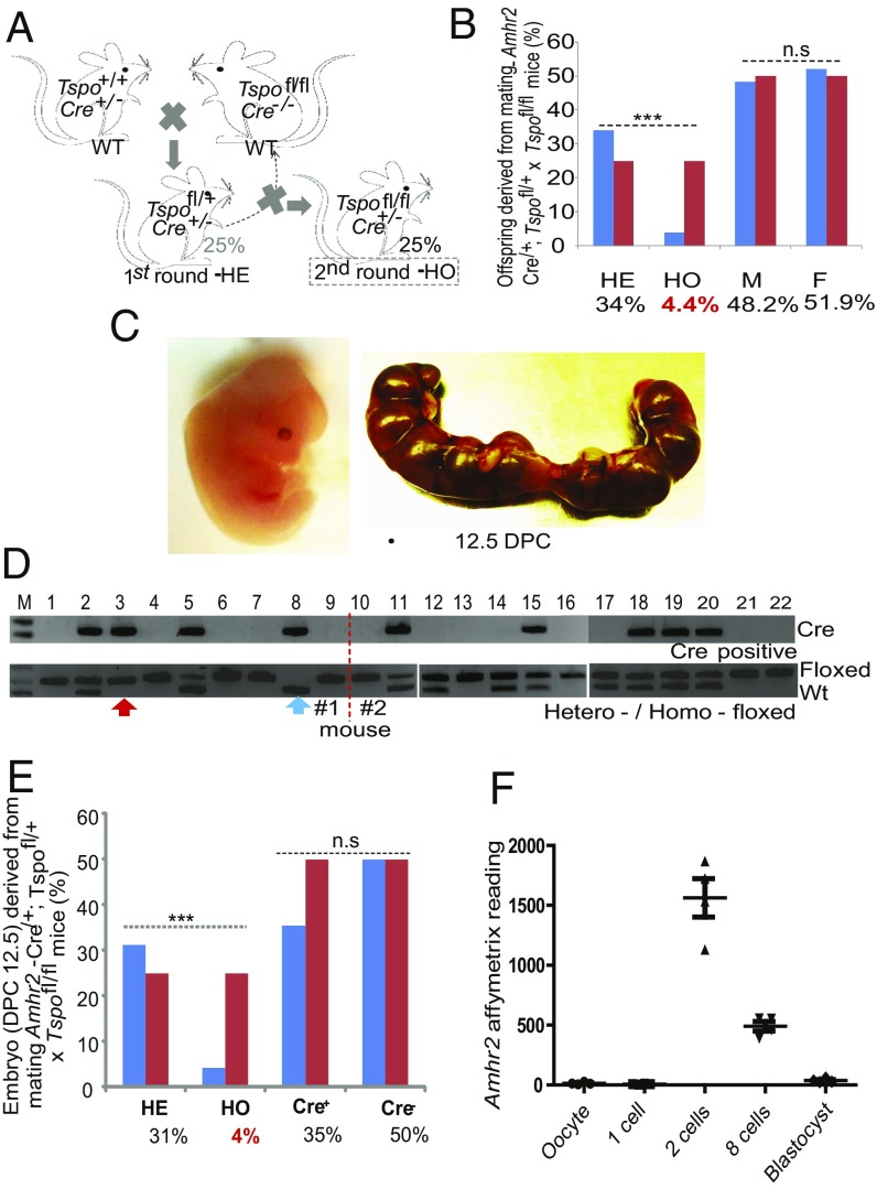
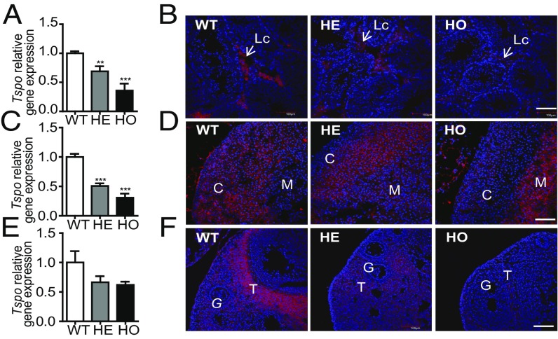
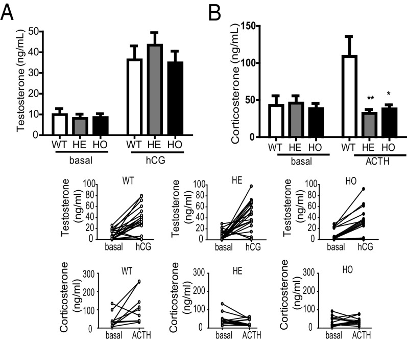
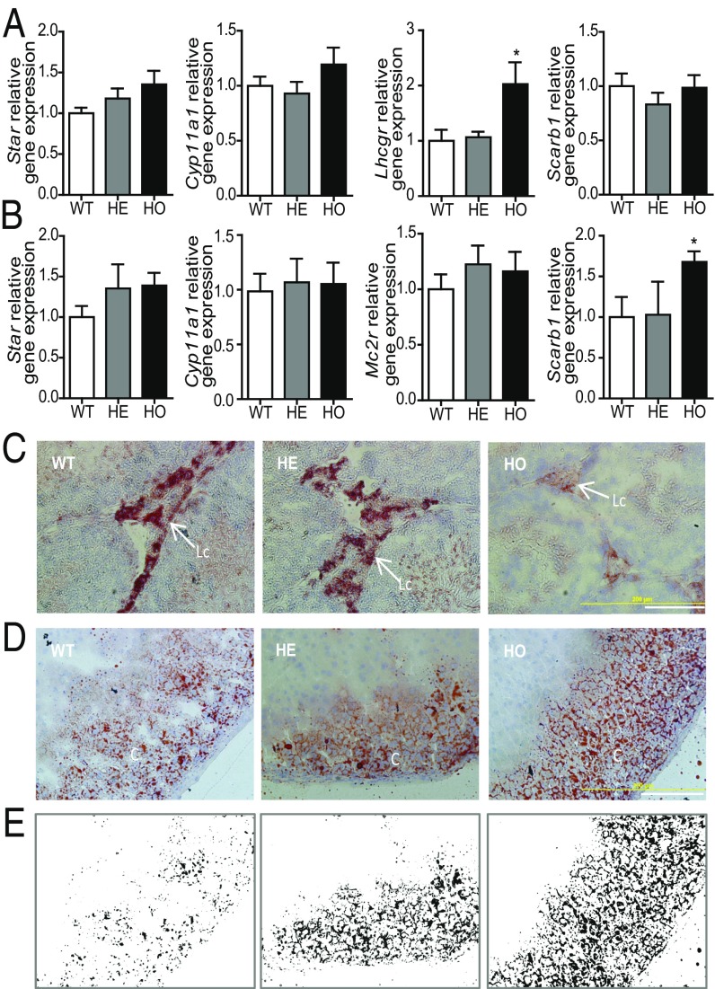
Translocator protein (TSPO) is a key member of the mitochondrial cholesterol transport complex in steroidogenic tissues. To assess the function of TSPO, we generated two lines of Cre-mediated Tspo conditional knockout (cKO) mice. First, gonadal somatic cell-targeting Amhr2-Cre mice were crossed with Tspo-floxed mice to obtain F1 Tspo Amhr2 cKO mice (Tspo(fl/fl);Amhr2-Cre(/+)). The unexpected Mendelian ratio of 4.4% cKO mice was confirmed by genotyping of 12.5-day-postcoitum (dpc) embryos. As Amhr2-Cre is expressed in gonads at 12.5 dpc, these findings suggest preimplantation selection of embryos. Analysis of expression databases revealed elevated levels of Amhr2 in two- and eight-cell zygotes, suggesting ectopic Tspo silencing before the morula stage and demonstrating elevated embryonic lethality and involvement of TSPO in embryonic development. To circumvent this issue, steroidogenic cell-targeting Nr5a1-Cre mice were crossed with Tspo-floxed mice. The resulting Tspo(fl/fl);Nr5a1-Cre(/+) mice were born at a normal Mendelian ratio. Nr5a1-driven Tspo cKO mice exhibited highly reduced Tspo levels in adrenal cortex and gonads. Treatment of mice with human chorionic gonadotropin (hCG) resulted in increased circulating testosterone levels despite extensive lipid droplet depletion. In contrast, Nr5a1-driven Tspo cKO mice lost their ability to form corticosterone in response to adrenocorticotropic hormone (ACTH). Important for ACTH-dependent steroidogenesis, Mc2r, Stard1, and Cypa11a1 levels were unaffected, whereas Scarb1 levels were increased and accumulation of lipid droplets was observed, indicative of a blockade of cholesterol utilization for steroidogenesis. TSPO expression in the adrenal medulla and increased epinephrine production were also observed. In conclusion, TSPO was found necessary for preimplantation embryo development and ACTH-stimulated steroid biosynthesis.
Keywords: anti-Mullerian hormone receptor type II; knockout mice; nuclear receptor subfamily 5 group A member 1; steroidogenesis; translocator protein.
Conflict of interest statement
The authors declare no conflict of interest.
Figures
Comment in
-
Crucial Role Reported for TSPO in Viability and Steroidogenesis is a Misconception. Commentary: Conditional Steroidogenic Cell-Targeted Deletion of TSPO Unveils a Crucial Role in Viability and Hormone-Dependent Steroid Formation.Front Endocrinol (Lausanne). 2016 Jul 18;7:91. doi: 10.3389/fendo.2016.00091. eCollection 2016. Front Endocrinol (Lausanne). 2016. PMID: 27489176 Free PMC article. No abstract available.
References
-
- Papadopoulos V, et al. Translocator protein (18kDa): New nomenclature for the peripheral-type benzodiazepine receptor based on its structure and molecular function. Trends Pharmacol Sci. 2006;27(8):402–409. - PubMed
-
- Rupprecht R, et al. Translocator protein (18 kDa) (TSPO) as a therapeutic target for neurological and psychiatric disorders. Nat Rev Drug Discov. 2010;9(12):971–988. - PubMed
-
- Lacapère JJ, Papadopoulos V. Peripheral-type benzodiazepine receptor: Structure and function of a cholesterol-binding protein in steroid and bile acid biosynthesis. Steroids. 2003;68(7-8):569–585. - PubMed
Publication types
MeSH terms
Substances
Grants and funding
LinkOut - more resources
Full Text Sources
Other Literature Sources
Molecular Biology Databases
Research Materials
