. 2015 Aug 15;309(4):C205-14.
doi: 10.1152/ajpcell.00347.2014. Epub 2015 Jun 3.

Role of nitric oxide in murine conventional outflow physiology

Affiliations

Role of nitric oxide in murine conventional outflow physiology

Jason Y H Chang et al. Am J Physiol Cell Physiol. .

Abstract

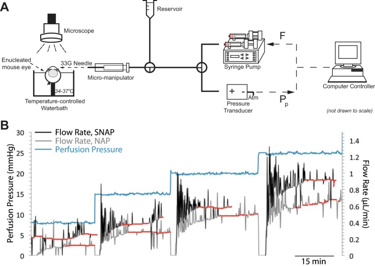
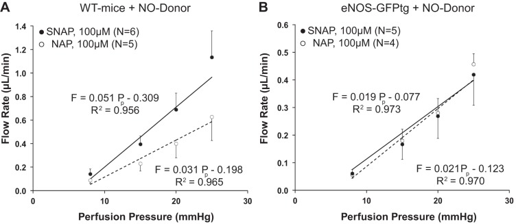
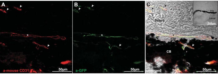
Elevated intraocular pressure (IOP) is the main risk factor for glaucoma. Exogenous nitric oxide (NO) decreases IOP by increasing outflow facility, but whether endogenous NO production contributes to the physiological regulation of outflow facility is unclear. Outflow facility was measured by pressure-controlled perfusion in ex vivo eyes from C57BL/6 wild-type (WT) or transgenic mice expressing human endothelial NO synthase (eNOS) fused to green fluorescent protein (GFP) superimposed on the endogenously expressed murine eNOS (eNOS-GFPtg). In WT mice, exogenous NO delivered by 100 μM S-nitroso-N-acetylpenicillamine (SNAP) increased outflow facility by 62 ± 28% (SD) relative to control eyes perfused with the inactive SNAP analog N-acetyl-d-penicillamine (NAP; n = 5, P = 0.016). In contrast, in eyes from eNOS-GFPtg mice, SNAP had no effect on outflow facility relative to NAP (-9 ± 4%, P = 0.40). In WT mice, the nonselective NOS inhibitor N(G)-nitro-l-arginine methyl ester (l-NAME, 10 μM) decreased outflow facility by 36 ± 13% (n = 5 each, P = 0.012), but 100 μM l-NAME had no detectable effect on outflow facility (-16 ± 5%, P = 0.22). An eNOS-selective inhibitor (cavtratin, 50 μM) decreased outflow facility by 19 ± 12% in WT (P = 0.011) and 39 ± 25% in eNOS-GFPtg (P = 0.014) mice. In the conventional outflow pathway of eNOS-GFPtg mice, eNOS-GFP expression was localized to endothelial cells lining Schlemm's canal and the downstream vessels, with no apparent expression in the trabecular meshwork. These results suggest that endogenous NO production by eNOS within endothelial cells of Schlemm's canal or downstream vessels contributes to the physiological regulation of aqueous humor outflow facility in mice, representing a viable strategy to more successfully lower IOP in glaucoma.

Keywords: aqueous humor outflow; glaucoma; intraocular pressure; mouse model; nitric oxide.


Figures

Fig. 1.
Fig. 1.
Perfusion system to measure outflow facility in enucleated ex vivo mouse eyes. A: computer-controlled syringe pump controls perfusion flow rate (F) into the mouse eye to maintain a user-defined perfusion pressure (Pp), which is measured by a pressure transducer (see Eq. 1). Mouse eye is held within an isotonic bath at 34–37°C and cannulated by a 33-gauge needle that is positioned in the anterior chamber using a micromanipulator while the preparation is viewed under a stereomicroscope. Perfusion is open to a reservoir set to 8 mmHg above the eye during needle insertion and during the initial pressurization period, but the reservoir is closed during perfusion. B: representative perfusion traces showing perfusion pressure and flow rate data from paired eyes perfused with 100 μM N-acetyl-d-penicillamine (NAP) or 100 μM S-nitroso-N-acetylpenicillamine (SNAP). Red-highlighted regions represent data used to calculate average flow rate at each corresponding pressure step. Spikes represent rapid increases in flow rate to maintain user-defined perfusion pressure. Only 1 perfusion pressure trace is shown, but pressure traces are generally similar for both eyes.
Fig. 2.
Fig. 2.
Characterization of nitric oxide (NO) concentration following release by SNAP or NAP under varying lighting conditions. A: NO release following initial high-intensity light (700–1,000 lumens/m2) exposure of stock solution (113 mM) for 10 min followed by dilution to the working solution [1 mM in Dulbecco's PBS including divalent cations and 5.5 mM d-glucose (DBG)] and either low-intensity (100–200 lumens/m2) or prolonged high-intensity (700–1,000 lumens/m2) light. Data are from 1 experiment but are representative of 3 individual experiments. Time 0 corresponds to the end of the initial 10-min period, during which SNAP stock solution was exposed to high-intensity light and after which NO release was measured in the working solution by the probe. B: NO release from 100 μM SNAP or 100 μM NAP in DBG (n = 3 each; bars are SD). Each stock solution was treated with an initial 10-min exposure to high-intensity light (700–1,000 lumens/m2) followed by low-intensity light (100–200 lumens/m2) starting at time 0. NO was measured using an NO-sensitive probe (ISO-NOS II). Data in B are representative of NO release during 100 μM SNAP/NAP perfusions.
Fig. 3.
Fig. 3.
Effects of NO donor on conventional outflow facility in eyes from wild-type (WT) and eNOS-GFPtg mice. A: perfusion flow rate measured as a function of pressure in enucleated eyes from WT mice perfused with 100 μM SNAP (NO donor) or 100 μM NAP (inactive analog). Outflow facility was estimated on the basis of the slope on the linear regression through the data points, as described by Eq. 1. B: perfusion flow rate measured as a function of pressure in enucleated eyes from eNOS-GFPtg mice perfused with 100 μM SNAP or 100 μM NAP. Bars represent SD. Note different vertical axis scales in A and B.
Fig. 4.
Fig. 4.
Effect of NOS inhibitors on conventional outflow facility in enucleated eyes from WT and eNOS-GFPtg mice. A and B: perfusion flow rate measured as a function of pressure in WT eyes perfused with l-NAME (nonselective NOS inhibitor) at 10 μM or 100 μM in DBG vs. DBG vehicle. C and D: perfusion flow rate measured as a function of pressure in eyes perfused with 50 μM cavtratin (eNOS-selective inhibitor) vs. vehicle (DBG + 0.6% DMSO) from WT or eNOS-GFPtg mice. Outflow facility is estimated on the basis of the slope on the linear regression through the data points, as described by Eq. 1. Error bars represent SD.
Fig. 5.
Fig. 5.
Localization of transgene expression in the conventional outflow pathway of eNOS-GFPtg mice. A: sagittal section through the trabecular meshwork (TM) and Schlemm's canal (SC; ∗) labeled with the endothelial marker CD31, showing labeling of SC endothelium as well as blood vessels (arrowheads) in the ciliary body (CB) and sclera. B: indirect immunofluorescence against GFP from the same section shown in A, revealing colocalization of the eNOS-GFP transgene along the endothelium of SC and along blood vessels in the CB. C: merged color image showing labeling for CD31 and GFP superimposed on the bright-field image to show location of SC and TM with respect to CB, iris, and sclera. Inset: lower magnification of bright-field sagittal section showing location of the outflow pathway with respect to the iris and lens. Data are representative images from 2 eNOS-GFPtg mouse eyes.

Comment in

References

    1. Alvarado JA, Katz LJ, Trivedi S, Shifera AS. Monocyte modulation of aqueous outflow and recruitment to the trabecular meshwork following selective laser trabeculoplasty. Arch Ophthalmol 128: 731–737, 2010. - PubMed
    1. Ashpole NE, Overby DR, Ethier CR, Stamer WD. Shear stress-triggered nitric oxide release from Schlemm's canal cells. Invest Ophthalmol Vis Sci 55: 8067–8076, 2014. - PMC - PubMed
    1. Aspelund A, Tammela T, Antila S, Nurmi H, Leppänen VM, Zarkada G, Stanczuk L, Francois M, Mäkinen T, Saharinen P, Immonen I, Alitalo K. The Schlemm's canal is a VEGF-C/VEGFR-3-responsive lymphatic-like vessel. J Clin Invest 124: 3975–3986, 2014. - PMC - PubMed
    1. Bachmann B, Birke M, Kook D, Eichhorn M, Lütjen-Drecoll E. Ultrastructural and biochemical evaluation of the porcine anterior chamber perfusion model. Invest Ophthalmol Vis Sci 47: 2011–2020, 2006. - PubMed
    1. Barany E. The mode of action of pilocarpine on outflow resistance in the eye of a primate (Cercopithecus ethiops). Invest Ophthalmol 1: 712–727, 1962. - PubMed

Publication types