. 2015 Aug;89(16):8406-15.
doi: 10.1128/JVI.00799-15.

Poxvirus Protein MC132 from Molluscum Contagiosum Virus Inhibits NF-B Activation by Targeting p65 for Degradation

Poxvirus Protein MC132 from Molluscum Contagiosum Virus Inhibits NF-B Activation by Targeting p65 for Degradation

Gareth Brady et al. J Virol. 2015 Aug.

Abstract

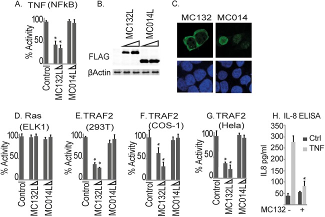
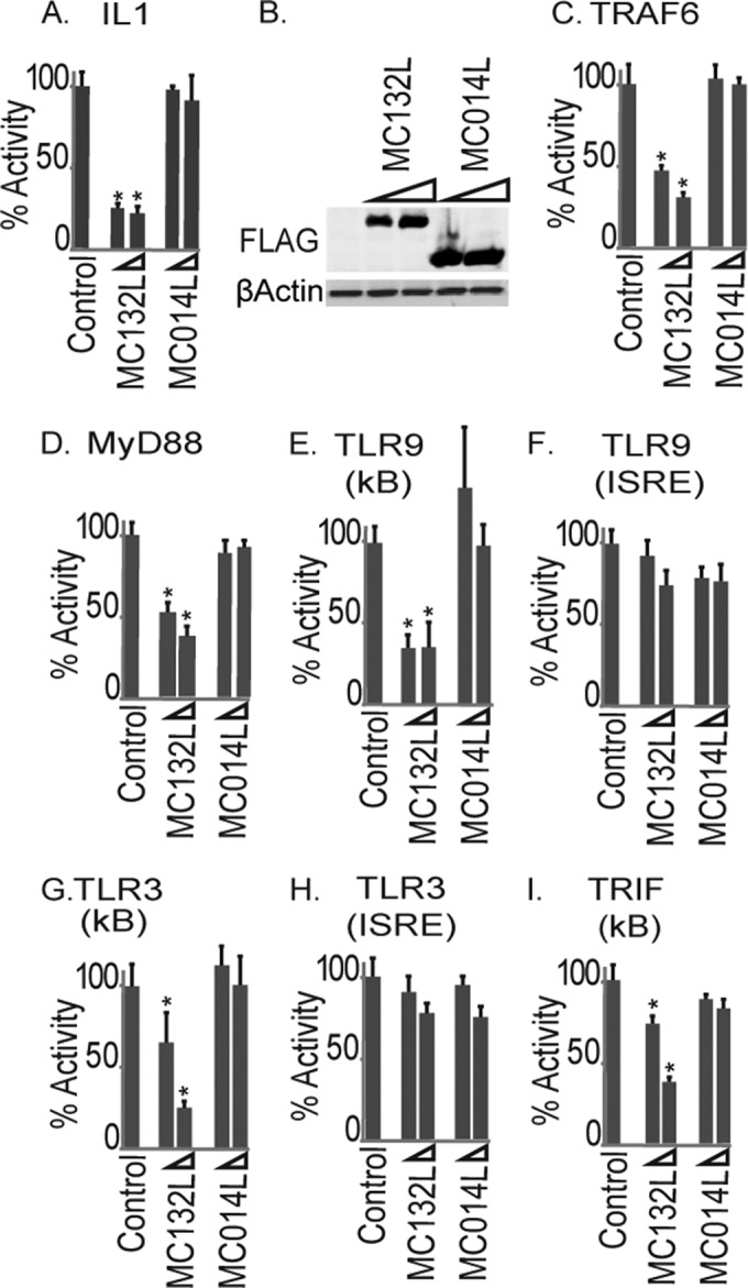
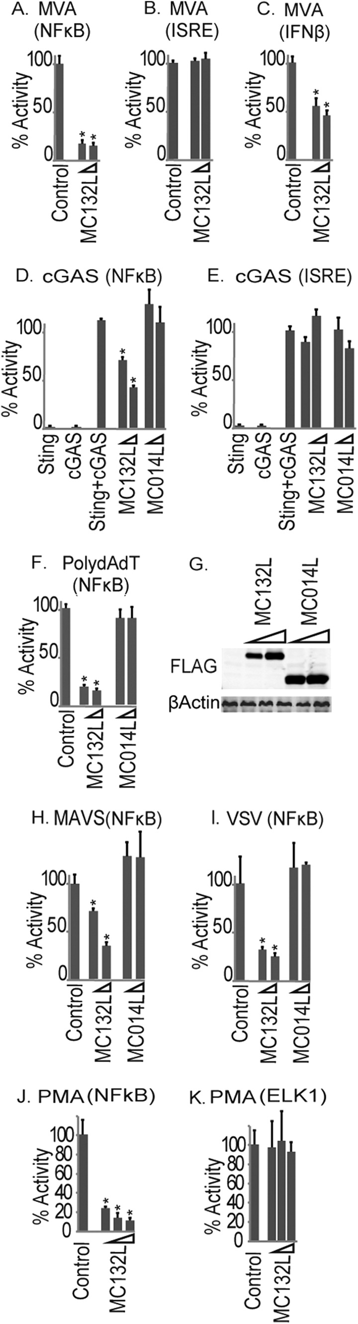
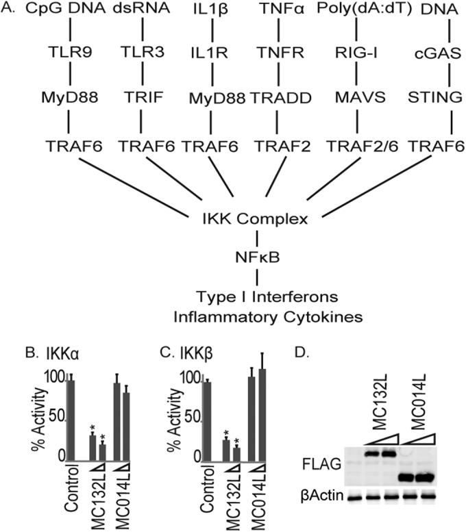
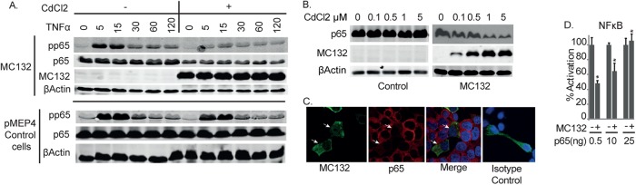
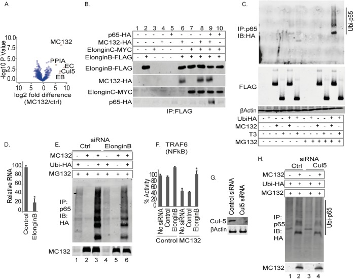
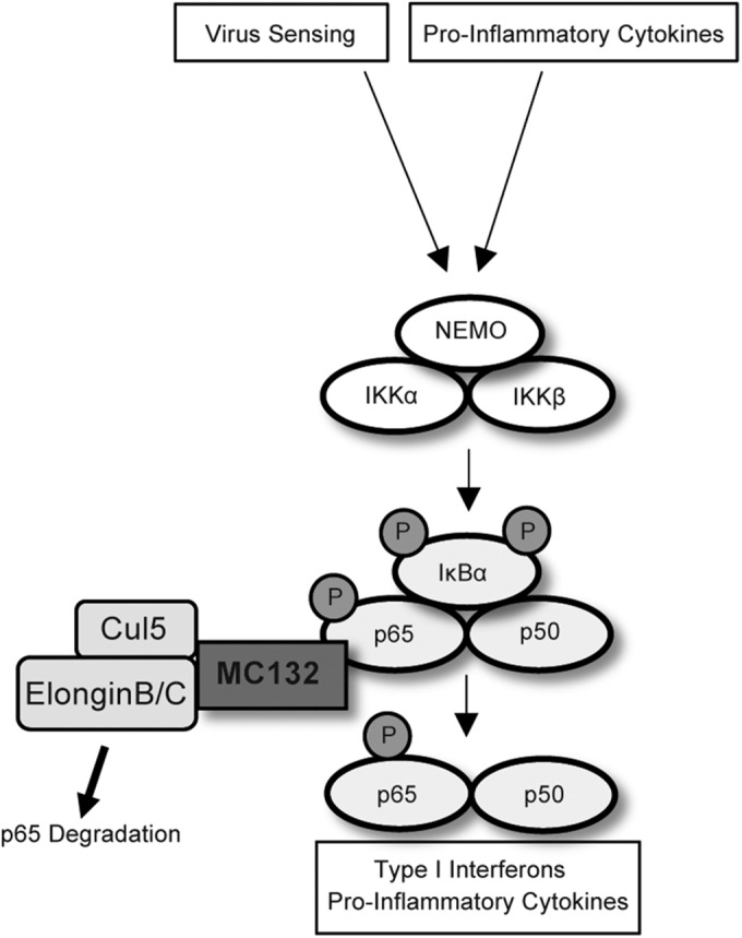
Molluscum contagiosum virus (MCV) is unique in being the only known extant, human-adapted poxvirus, yet to date, it is very poorly characterized in terms of host-pathogen interactions. MCV causes persistent skin lesions filled with live virus, but these are generally immunologically silent, suggesting the presence of potent inhibitors of human antiviral immunity and inflammation. Fewer than five MCV immunomodulatory genes have been characterized in detail, but it is likely that many more remain to be discovered given the density of such sequences in all well-characterized poxviruses. Following virus infection, NF-B activation occurs in response to both pattern recognition receptor (PRR) signaling and cellular activation by virus-elicited proinflammatory cytokines, such as tumor necrosis factor (TNF). As such, NF-B activation is required for virus detection, antiviral signaling, inflammation, and clearance of viral infection. Hence, we screened a library of MCV genes for effects on TNF-stimulated NF-B activation. This revealed MC132, a unique protein with no orthologs in other poxviral genomes, as a novel inhibitor of NF-B. Interestingly, MC132 also inhibited PRR- and virus-activated NF-B, since MC132 interacted with the NF-B subunit p65 and caused p65 degradation. Unbiased affinity purification to identify host targets of MC132 revealed that MC132 acted by targeting NF-B p65 for ubiquitin-dependent proteasomal degradation by recruiting p65 to a host Cullin-5/Elongin B/Elongin C complex. These data reveal a novel mechanism for poxviral inhibition of human innate immunity and further clarify how the human-adapted poxvirus MCV can so effectively evade antiviral immunity to persist in skin lesions.


Figures

FIG 1
FIG 1
Identification of MC132 as an inhibitor of TNF-α-stimulated NF-κB activation. (A) HEK293T cells were seeded at 2 × 10⁵ cells per ml, transfected with 80 ng NF-κB reporter gene, 40 ng TK renilla reporter gene, and 25 or 50 ng (indicated by wedge) empty vector (control) or pCEP4 plasmids expressing the indicated MCV ORFs, stimulated with 50 ng/ml TNF-α for 6 h, and then harvested and assayed for NF-κB reporter gene activity. Data are percentages of the stimulation activity for control cells and are means ± standard deviations (SD) for triplicate samples from a representative experiment (n = 3). (B) Extracts from the samples used for panel A were probed for expression of Flag-tagged viral proteins. (C) Localization of MC132 and MC014 in HEK293T cells. Cells were transfected with 3 μg pCEP4-Flag vector containing the indicated MCV ORFs. Cells were fixed 24 h later and stained with DAPI (blue) or for MCV protein expression (green). Representative images are shown (n = 4). (D) Elk1 activation by Ras in cells transfected with empty vector (control) or pCEP4 plasmids expressing MCV ORFs was measured by a reporter gene assay. Data are percentages of the stimulation activity for control cells for a representative experiment performed in triplicate (n = 3). (E to G) The same as panel A, except that HEK293T (E), COS-1 (F), or HeLa (G) cells were transfected with 50 ng TRAF2-Flag instead of TNF-α stimulation. (H) Cells stably expressing pMEP4-MC132 were seeded at 2 × 10⁵ cells per ml, cultured with (+) or without (−) 1 μM CdCl2 to induce MC132 expression, and then, 24 h later, stimulated with 50 ng/ml TNF-α for 24 h. IL-8 production was then assayed by ELISA. Data are means ± SD for triplicate samples from a representative experiment (n = 3). *, P < 0.001 compared to the control.
FIG 2
FIG 2
Inhibition of IL-1 and TLR activation of NF-κB by MC132. (A) HEK293T cells were seeded at 2 × 10⁵ cells per ml, transfected with 80 ng NF-κB reporter gene, 40 ng TK renilla reporter gene, and 25 or 50 ng (indicated by wedge) empty vector (control) or pCEP4 plasmids expressing the indicated MCV ORFs, stimulated with 50 ng/ml IL-1β for 6 h, and then harvested and assayed for NF-κB reporter gene activity. Data are percentages of the stimulation activity for control cells and are means ± SD for triplicate samples from a representative experiment (n = 4). (B) Extracts from the samples for panel A were probed for expression of Flag-tagged viral proteins. (C to I) The same as panel A, except that instead of IL-1 stimulation, cells were transfected with 50 ng plasmid expressing TRAF6 (C), MyD88 (D), CD4-TLR9 (E and F), CD4-TLR3 (G and H), or TRIF (I) for 24 h. For panels F and H, cells were transfected with an ISRE-luciferase reporter in place of the NF-κB reporter. *, P < 0.001 compared to the control.
FIG 3
FIG 3
Inhibition of cytosolic DNA-sensing-pathway- and virus-stimulated NF-κB activation by MC132. HEK293T cells were seeded at 2 × 10⁵ cells per ml and transfected with the indicated reporter genes and empty vector (control) or pCEP4 plasmids expressing MC132. Cells were then infected with MVA for 16 h, and NF-κB reporter activity (A), ISRE reporter activity (B), and IFN-β promoter reporter gene activity (C) were measured. Data are percentages of the stimulation activity for control cells and are means ± SD for triplicate samples from a representative experiment (n = 4). (D and E) The same as panel A, except that cells were transfected with cGAS- and STING-expressing plasmids (25 ng each), and 24 h later, NF-κB (D) or ISRE (E) reporter gene activity was measured. (F, H, and I) The same as panel A, except that cells were transfected with 500 ng/ml poly(dA:dT) (F) or 50 ng MAVS-expressing plasmid (H) or infected with VSV for 16 h (I). (G) Extracts from the samples used for panel F were probed for expression of Flag-tagged viral proteins. (J) The same as panel A, except that cells were transfected with 10 ng, 25 ng, and 50 ng of pCEP4-MC132flag and stimulated with 10 nM PMA for 6 h. (K) The same as panel J, except that Elk1 reporter gene activity was measured. *, P < 0.001 compared to the control.
FIG 4
FIG 4
MC132 inhibits NF-κB activation by IKKs, a common convergence point in the network of signal transduction pathways activated during viral infection. (A) Schematic showing multiple signal transduction pathways to NF-κB expected to be activated during a poxviral infection, all of which were shown to be sensitive to MC132 inhibition (Fig. 1 to 3). (B and C) HEK293T cells were seeded at 2 × 10⁵ cells per ml, transfected with 25 or 50 ng (indicated by wedge) empty vector (control) or pCEP4 plasmids expressing the indicated MCV ORFs, together with 50 ng IKKα (B) or IKKβ (C), and then harvested and assayed for NF-κB reporter gene activity 24 h later. Data are percentages of the stimulation activity for control cells and are means ± SD for triplicate samples from a representative experiment (n = 3). *, P < 0.001 compared to the control. (D) Extracts from the samples used for panel C were probed for expression of Flag-tagged viral proteins.
FIG 5
FIG 5
MC132 expression causes a depletion of p65 protein expression. (A) Effect of inducible MC132 expression on TNF-α-stimulated NF-κB activation. HEK293T cells stably transfected with pMEP4 and pMEP4-MC132 were seeded at 6 × 10⁵ cells per well in 6-well dishes and treated with (+) or without (−) 1 μM CdCl2 to induce MCV protein expression. Twenty-four hours later, cells were stimulated with 50 ng/ml TNF-α for the indicated times, and cell lysates were immunoblotted with the indicated antibodies. Representative blots are shown (n = 3). (B) HEK293T cells stably containing pMEP4-MC132 or control cells containing pMEP4 were seeded at 6 × 10⁵ cells per well in 6-well dishes for 24 h with the indicated concentrations of CdCl2 to induce MC132 expression, and cell lysates were immunoblotted for p65 and MC132. Representative blots are shown (n = 3). (C) HEK293T cells were seeded at 6 × 10⁵ cells per well in 6-well dishes, transfected with 3 μg pCEP4-MC132, fixed 24 h later, and stained with DAPI (blue), for MC132-Flag (green) and endogenous p65 (red), and with an anti-rabbit isotype control. Representative images are shown (n = 3). MC132-positive cells are indicated with white arrows. (D) HEK293T cells were seeded at 2 × 10⁵ cells per ml and transfected with the indicated amounts of p65 expression vector, with or without 50 ng of pCEP4-MC132. Cells were harvested 24 h later and assayed for NF-κB reporter gene activity. Data are presented as percentages of reporter activation by increasing amounts of p65 and display the effect of 50 ng of MC132 on this activation, with data given as means ± SD for triplicate samples from a representative experiment (n = 3). *, P < 0.001 compared to the control.
FIG 6
FIG 6
MC132 interacts with p65 via the RHD of p65. (A) MC132 and p65 interact. HEK293T cells were seeded at 4 × 10⁶ cells per 10-cm plate and transfected with 8 μg pCEP4-MC132 or a plasmid expressing control p38-Flag, and 24 h later, the cells were lysed and immunoprecipitated (IP) with anti-Flag beads. Immunoblots were probed with the indicated antibodies. Representative blots are shown (n = 4). (B) MC132 interacts with the p65 RHD. The schematic shows full-length p65(1–551) and the different truncations tested. HEK293T cells were seeded at 4 × 10⁶ cells per 10-cm plate and transfected with 4 μg pCEP4-MC132 together with 4 μg vector encoding full-length p65-HA or the indicated p65 truncation mutants. Cells were lysed the following day, immunoprecipitated with anti-Flag beads, and probed for Flag-tagged MC132 or p65-HA. Representative blots are shown (n = 4). (C) Truncation of MC132 abolishes interaction with p65 and inhibition of NF-κB. HEK293T cells were seeded at 4 × 10⁶ cells per ml and transfected the following day with 8 μg pCEP4-MC132 or the MC132 truncation mutants indicated in the schematic. Cells were lysed the following day, immunoprecipitated with anti-Flag beads, and probed along with lysate controls for Flag-tagged proteins and endogenous p65. Representative blots are shown (n = 4). (D) The effect of MC132 truncations on TRAF2-stimulated NF-κB activation was measured by a reporter gene assay as described in the legend to Fig. 1. Data are percentages of the stimulation activity for control cells and are means ± SD for triplicate samples from a representative experiment (n = 4). *, P < 0.001 compared to the control.
FIG 7
FIG 7
MC132 recruits p65 to a Cullin-5/Elongin B/Elongin C complex to target p65 for ubiquitin-mediated degradation. (A) Volcano plot of MC132-interacting proteins identified from MC132-expressing HEK293T cells by AP-MS. (B) MC132 targets p65 to the Elongin B/C complex. HEK293T cells were seeded at 4 × 10⁶ cells per 10-cm plate and transfected with 2 μg expression vector(s) for MC132-HA, Elongin B-Flag, and/or Elongin c-Myc, as indicated. Twenty-four hours later, cells were lysed, immunoprecipitated with anti-Flag beads, and then immunoblotted with the indicated antibodies. Representative blots are shown (n = 3). (C) MC132 induces polyubiquitination of p65. HEK293T cells were seeded at 4 × 10⁶ cells per 10-cm plate, transfected with 4 μg expression vector(s) for ubiquitin-HA, MC132-Flag, and/or T3-Flag, as indicated, and treated with 20 μM MG132 for 4 h prior to harvest. Twenty-four hours later, cells were lysed, immunoprecipitated with anti-Flag beads, and then immunoblotted with the indicated antibodies. Ubi-p65, polyubiquitinated p65. A representative blot is shown (n = 3). (D) HEK293T cells were seeded at 2 × 10⁵ cells per ml in 24-well dishes and transfected with 2.5 nM Elongin B siRNA at 24 and 48 h, and then cDNAs from cells were assayed for levels of Elongin B normalized to β-actin by real-time PCR. (E) HEK293T cells were seeded at 4 × 10⁶ cells per 10-cm plate, transfected with 2.5 nM Elongin B or control siRNA for 24 h and again at 48 h, and transfected with the indicated constructs prior to MG132 treatment for 4 h and harvest. Total p65 was immunoprecipitated, and immunoblots were probed with the indicated antibodies. (F) The effect of Elongin B siRNA on TRAF6-stimulated NF-κB activation and MC132 inhibition was measured by a reporter gene assay as described in the legend to Fig. 1. Data are percentages of the stimulation activity for control cells and are means ± SD for triplicate samples from a representative experiment (n = 4). *, P < 0.001 compared to the control. (G) HEK293T cells were seeded and treated as described for panel E, but they were transfected with 25 μM Cullin-5 or control siRNA for 24 h and then immunoblotted for Cullin-5 expression. (H) Cullin-5 siRNA reduces MC132-stimulated p65 ubiquitination. Cells were treated as described for panel C, except that Cullin-5 or control siRNA was transfected prior to MG132 treatment and harvest. Representative blots are shown (n = 4).
FIG 8
FIG 8
Model for MC132-mediated inhibition of human NF-κB signaling. Innate virus-sensing and cytokine signaling pathways converge on the IKK complex, leading to IKK-dependent phosphorylation of p65 and IκBα. MC132 recruits a Cullin-5/Elongin B/Elongin C complex to p65, causing ubiquitin-mediated degradation of p65 and subsequent suppression of NF-κB activity.

References

    1. Gurtler C, Bowie AG. 2013. Innate immune detection of microbial nucleic acids. Trends Microbiol 21:413–420. doi:10.1016/j.tim.2013.04.004. - DOI - PMC - PubMed
    1. Bowie AG, Unterholzner L. 2008. Viral evasion and subversion of pattern-recognition receptor signalling. Nat Rev Immunol 8:911–922. doi:10.1038/nri2436. - DOI - PMC - PubMed
    1. Brady G, Bowie AG. 2014. Innate immune activation of NFkappaB and its antagonism by poxviruses. Cytokine Growth Factor Rev 25:611–620. doi:10.1016/j.cytogfr.2014.07.004. - DOI - PubMed
    1. Senkevich TG, Koonin EV, Bugert JJ, Darai G, Moss B. 1997. The genome of molluscum contagiosum virus: analysis and comparison with other poxviruses. Virology 233:19–42. doi:10.1006/viro.1997.8607. - DOI - PubMed
    1. Struzik J, Szulc-Dabrowska L, Niemialtowski M. 2014. Modulation of NF-κB transcription factor activation by Molluscum contagiosum virus proteins. Postepy Hig Med Dosw 68:129–136. doi:10.5604/17322693.1088053. - DOI - PubMed

Publication types

MeSH terms