. 2015 Aug;89(16):8334-45.
doi: 10.1128/JVI.00908-15.

Broadly Neutralizing Human Immunodeficiency Virus Type 1 Antibody Gene Transfer Protects Nonhuman Primates from Mucosal Simian-Human Immunodeficiency Virus Infection

Broadly Neutralizing Human Immunodeficiency Virus Type 1 Antibody Gene Transfer Protects Nonhuman Primates from Mucosal Simian-Human Immunodeficiency Virus Infection

Kevin O Saunders et al. J Virol. 2015 Aug.

Abstract

Broadly neutralizing antibodies (bnAbs) can prevent lentiviral infection in nonhuman primates and may slow the spread of human immunodeficiency virus type 1 (HIV-1). Although protection by passive transfer of human bnAbs has been demonstrated in monkeys, durable expression is essential for its broader use in humans. Gene-based expression of bnAbs provides a potential solution to this problem, although immune responses to the viral vector or to the antibody may limit its durability and efficacy. Here, we delivered an adeno-associated viral vector encoding a simianized form of a CD4bs bnAb, VRC07, and evaluated its immunogenicity and protective efficacy. The expressed antibody circulated in macaques for 16 weeks at levels up to 66 g/ml, although immune suppression with cyclosporine (CsA) was needed to sustain expression. Gene-delivered simian VRC07 protected against simian-human immunodeficiency virus (SHIV) infection in monkeys 5.5 weeks after treatment. Gene transfer of an anti-HIV antibody can therefore protect against infection by viruses that cause AIDS in primates when the host immune responses are controlled.


Figures

FIG 1
FIG 1
Design of simian VRC07 IgG. (A) Amino acid alignment of huVRC07 and simVRC07 heavy and light chain variable regions. Residues that are identical to the inferred germ line gene (Mac gl), shown on the top line, are indicated by periods. The complementarity-determining region (CDR) residues (red) and grafted framework region (FWR) residues (green) are shown. HuVRC07 CDR3 is shown below the germ line sequence, since the original sequences after recombination could not be predicted with a high degree of certainty. Somatic mutations in the FWRs that were not grafted to make simVRC07 are highlighted in blue. The percent similarity of each segment of simVRC07 compared to the macaque germ line is denoted above the sequences. The number of grafted residues is indicated in parentheses above each segment. (B) Tertiary sequence location of amino acids selected for engraftment onto the macaque germ line gene. Somatic mutations in the FWRs of the heavy chain (HC) and light chain (LC) of huVRC07 are shown as spheres on the crystal structure (14). The CDRs (red) and somatic mutations in the FWRs (blue or green) are colored to correspond to the alignment in panel A and are depicted in proximity to HIV-1 gp120 (gray). (C) Comparison of HIV-1 neutralization titers of simVRC07 and huVRC07 by a TZM-bl cell assay. Neutralization titers (IC50s) (in micrograms per milliliter) of the indicated antibodies are shown and are color-coded as follows: red, <0.1; orange, 0.1 to 0.99; yellow, 1 to 4.9; green, 5 to 50; white >50. NA indicates that the fold change could not be calculated due to a lack of neutralization by both antibodies.
FIG 2
FIG 2
AAV8 delivery of simVRC07 genes confers systemic and mucosal expression in immunodeficient mice and immunocompetent rhesus macaques. (A) Vector transgene design for single-stranded AAV8 vectors. ITR, inverted terminal repeat; pA, polyadenylation sequence. (B) Comparison of huVRC07 and simVRC07 serum concentrations in SCID mice after administration of 2.5 × 10¹⁰ vg of AAV8 vectors. Values represent means ± standard errors of the means of results from three or six mice. (C) Plasma simVRC07 concentration in rhesus macaques administered 1 × 10¹³ vg of AAV8-simVRC07. Values indicate means ± standard errors of the means of data from triplicate independent measurements. (D) Immunoperoxidase staining for simVRC07 IgG in cross sections of macaque rectal tissue by HIV envelope reverse immunohistochemistry. Rectal biopsy specimens were collected before and 3 weeks after AAV8-simVRC07 administration and stained with a labeled RSC3 protein. Original magnification, ×40. Images are representative of staining performed on tissues from macaques DBM8 and AZ36.
FIG 3
FIG 3
Characterization of anti-simVRC07 humoral immunity in macaques. (A) Time course of anti-simVRC07 antibody levels measured by biolayer interferometry in macaques administered 1 × 10¹³ vg of AAV8-simVRC07 (Fig. 2C). Plasma immunoglobulin binding to simVRC07 (solid lines) or control macaque IgG (dashed lines) is shown for each macaque. Values shown are means ± standard errors of the means from triplicate independent measurements. (B) Spearman correlation between the plasma simVRC07 concentration and the anti-simVRC07 plasma antibody response, quantified as described above for panel A (Spearman r = −0.7819). Symbols are coded to correspond to each rhesus macaque in panel A. Anti-simVRC07 responses were inversely correlated with detectable plasma simVRC07 concentrations. (C) Blocking of simVRC07, huVRC07, or VRC03 binding to HIV-1 gp120. Macaque plasma obtained before AAV8-simVRC07 vector administration (week 0) or 9 weeks after vector administration (week 9) was tested for blocking of the monoclonal antibodies. Binding in the presence of no plasma served as the baseline binding control. The means from triplicate measurements are shown.
FIG 4
FIG 4
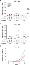
Cellular and humoral immune responses against the F2A peptide in macaques administered 1 × 10¹³ vg of AAV8-simVRC07. (A and B) Intracellular cytokine staining of CD8+ T cells (A) and CD4+ T cells (B) stimulated with a pool of overlapping F2A peptides. The dotted line represents the previously determined threshold for positivity for each cytokine (37). Due to PBMC availability, T cell responses were analyzed at 12 and 25 weeks after vector administration for monkeys AZ36 and DBM8 and at 5 and 7 weeks after vector administration for monkeys AZ66 and 2F7. (C) ELISA of direct plasma IgG binding to pooled F2A peptides. The binding titer is represented as the log10 area under the curve (AUC) for the four macaques listed in panels A and B over time.
FIG 5
FIG 5
Transient immune suppression during gene transfer increases peak simVRC07 concentrations and duration of expression. (A) SimVRC07 plasma concentrations in five rhesus macaques treated with cyclosporine (CsA), as indicated on the x axis. AAV8-simVRC07 was administered at week 0 (solid vertical line). The time point for SHIV-BaLP4 challenge is shown as a dashed vertical line. Values represent the means ± standard errors of data from triplicate independent measurements. (B) Comparison of peak plasma simVRC07 concentrations after AAV8-simVRC07 injection in untreated macaques (Fig. 2C) or the macaques treated with CsA listed in panel A. The mean peak concentration ± standard error of the mean for each group of animals is shown by the horizontal bars. Significantly higher peak plasma concentrations were observed for macaques that received CsA than for untreated macaques (P = 0.0095 by Mann-Whitney test). (C) Biolayer interferometry binding of plasma immunoglobulin from the macaques listed in panel A to macaque IgG (left) and simVRC07 IgG (right). The duration of CsA treatment is indicated on the x axis. Mean values ± standard errors of the means of data from triplicate independent measurements are shown.
FIG 6
FIG 6
SimVRC07 gene transfer protects against mucosal SHIV challenge. (A) Plasma neutralization titers (ID50s) against the SHIV-BaLP4 challenge stock on the day of challenge. The neutralization titer in the plasma for monkey A11E035 was not determined (ND), since it did not have detectable simVRC07 in its plasma at this time point. The ID50 is shown as the reciprocal plasma dilution and is color-coded as follows: red, >500; orange, 300 to 500; yellow, 100 to 299; green, 10 to 99; white, <10. Murine leukemia virus (SVA-MLV) was analyzed as a negative-control virus. (B) Plasma viral loads in rhesus macaques administered AAV8 vectors encoding control IgG (left) or simVRC07 (right) and challenged with SHIV-BaLP4 5.5 weeks later. Prior to SHIV-BaLP4 challenge, both groups of animals received the same course of CsA treatment beginning 1 week before AAV8 administration and ending at week 4. Significantly more macaques were infected in the control group than in the simVRC07 group (treatment group, n = 6; control group, n = 5 [P = 0.0455 by Fisher's exact test]).

References

    1. Doria-Rose NA, Klein RM, Manion MM, O'Dell S, Phogat A, Chakrabarti B, Hallahan CW, Migueles SA, Wrammert J, Ahmed R, Nason M, Wyatt RT, Mascola JR, Connors M. 2009. Frequency and phenotype of human immunodeficiency virus envelope-specific B cells from patients with broadly cross-neutralizing antibodies. J Virol 83:188–199. doi:10.1128/JVI.01583-08. - DOI - PMC - PubMed
    1. Li Y, Migueles SA, Welcher B, Svehla K, Phogat A, Louder MK, Wu X, Shaw GM, Connors M, Wyatt RT, Mascola JR. 2007. Broad HIV-1 neutralization mediated by CD4-binding site antibodies. Nat Med 13:1032–1034. doi:10.1038/nm1624. - DOI - PMC - PubMed
    1. Hraber P, Seaman MS, Bailer RT, Mascola JR, Montefiori DC, Korber BT. 2014. Prevalence of broadly neutralizing antibody responses during chronic HIV-1 infection. AIDS 28:163–169. doi:10.1097/QAD.0000000000000106. - DOI - PMC - PubMed
    1. Ruprecht RM. 2009. Passive immunization with human neutralizing monoclonal antibodies against HIV-1 in macaque models: experimental approaches. Methods Mol Biol 525:559–566. doi:10.1007/978-1-59745-554-1_31. - DOI - PubMed
    1. Mascola JR, Montefiori DC. 2010. The role of antibodies in HIV vaccines. Annu Rev Immunol 28:413–444. doi:10.1146/annurev-immunol-030409-101256. - DOI - PubMed

Publication types

MeSH terms