. 2015 Jul 15;195(2):683-94.
doi: 10.4049/jimmunol.1402983. Epub 2015 Jun 3.

CARMA3 Is Critical for the Initiation of Allergic Airway Inflammation

Affiliations

CARMA3 Is Critical for the Initiation of Allergic Airway Inflammation

Benjamin Causton et al. J Immunol. .

Abstract

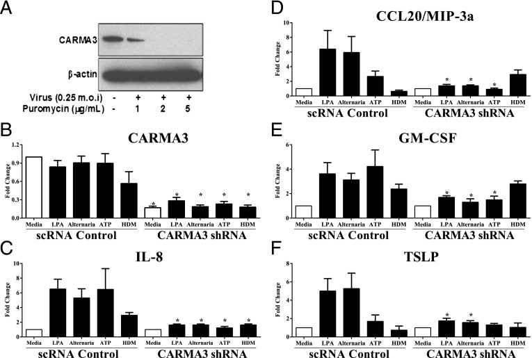
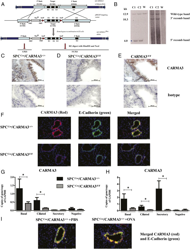
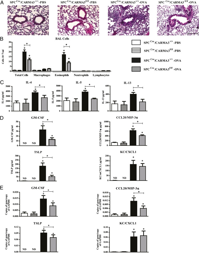
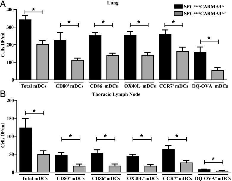
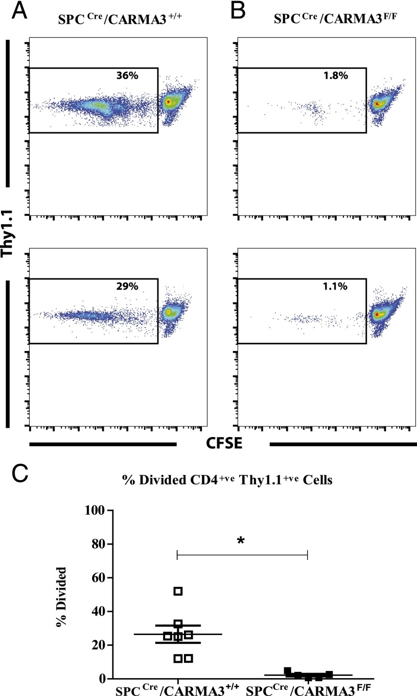
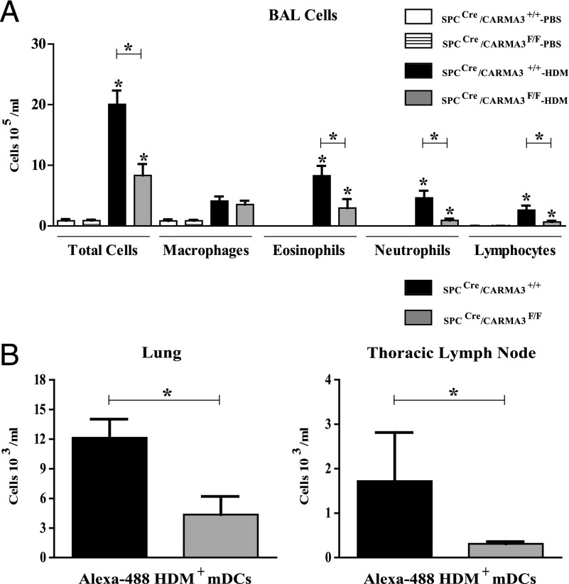
Innate immune responses to allergens by airway epithelial cells (AECs) help initiate and propagate the adaptive immune response associated with allergic airway inflammation in asthma. Activation of the transcription factor NF-κB in AECs by allergens or secondary mediators via G protein-coupled receptors (GPCRs) is an important component of this multifaceted inflammatory cascade. Members of the caspase recruitment domain family of proteins display tissue-specific expression and help mediate NF-κB activity in response to numerous stimuli. We have previously shown that caspase recruitment domain-containing membrane-associated guanylate kinase protein (CARMA)3 is specifically expressed in AECs and mediates NF-κB activation in these cells in response to stimulation with the GPCR agonist lysophosphatidic acid. In this study, we demonstrate that reduced levels of CARMA3 in normal human bronchial epithelial cells decreases the production of proasthmatic mediators in response to a panel of asthma-relevant GPCR ligands such as lysophosphatidic acid, adenosine triphosphate, and allergens that activate GPCRs such as Alternaria alternata and house dust mite. We then show that genetically modified mice with CARMA3-deficient AECs have reduced airway eosinophilia and proinflammatory cytokine production in a murine model of allergic airway inflammation. Additionally, we demonstrate that these mice have impaired dendritic cell maturation in the lung and that dendritic cells from mice with CARMA3-deficient AECs have impaired Ag processing. In conclusion, we show that AEC CARMA3 helps mediate allergic airway inflammation, and that CARMA3 is a critical signaling molecule bridging the innate and adaptive immune responses in the lung.


Figures

FIGURE 1.
FIGURE 1.
Asthma-relevant GPCR profile of mouse tracheal epithelial cells. RNA was isolated from naive, unstimulated mouse tracheal epithelial cells, and the expression profile of a panel of 380 GPCRs was measured using a real-time qPCR mini-array. Shown is an asthma-relevant subset from 67 medium- and high-abundance GPCRs detected. GAPDH was used to normalize the values of GPCR genes tested.
FIGURE 2.
FIGURE 2.
CARMA3 mediates proinflammatory cytokine release from normal human bronchial epithelial cells in response to GPCR stimulation. (A) A lentivirus containing a shRNA against CARMA3 and a puromycin resistance cassette was used to infect NHBE cells (multiplicity of infection of 0.25). The infected cells were selected for puromycin resistance for 5 d and then Western immunoblotting was used to detect CARMA3. (BF) NHBEs were grown on an ALI postinfection with lentivirus containing a nontargeting shRNA (scRNA) or a CARMA3-targeting shRNA. These cells were then stimulated with either 10 μM LPA, 100 μg/ml Alternaria, 100 μM ATP, or 100 μg/ml HDM for 6 h. RNA was then isolated from the cells and the levels of (B) CARMA3, (C) CXCL8/IL-8, (D) GM-CSF, (E) CCL20/MIP-3α, and (F) TSLP were determined. GAPDH levels were used to normalize the values of genes tested. Data are expressed as fold change from media. Values are the mean of three to six samples ± SEM. This experiment was repeated twice. *p < 0.05 versus scRNA cells by unpaired t test. ND, not detected.
FIGURE 3.
FIGURE 3.
Generation of SPCCre/CARMA3F/F mice. (A) Generation of CARMA3 targeting construct. (B) Southern blot analysis to identify correctly targeted ES cell clones obtained from ES cells electroporated with the targeting construct. Two ES cell clones (C1 and C2) were identified in which there was the expected 5′ and 3′ recombinations. W, wild-type mouse. Immunohistochemistry of lungs from naive (C) SPCCre/CARMA3+/+, (D) SPCCre/CARMA3F/F, and (E) CARMA3F/F mice stained with an Ab against CARMA3 (top panels) or an isotype control Ab (bottom panels). Scale bars, 200 μm. (F) Immunofluorescence of lungs from naive SPCCre/CARMA3+/+ and SPCCre/CARMA3F/F mice stained with an Abs against CARMA3 and E-cadherin. All images were taken using the same exposure. Scale bars, 20 μm. Basal, ciliated, and secretory cells sorted from the lungs of SPCCre/CARMA3+/+ and SPCCre/CARMA3F/F mice that were either (G) naive or (H) received one OVA/alum immunization and one OVA challenge. RNA levels of CARMA3 were determined by qPCR. Data are means ± SEMs of six mice per group. *p < 0.05 (SPCCre/CARMA3+/+ compared with SPCCre/CARMA3F/F). (I) Immunofluorescence of lungs from PBS- and OVA-immunized and challenged SPCCre/CARMA3+/+ mice stained for CARMA3. Scale bars, 20 μm.
FIGURE 4.
FIGURE 4.
Attenuation of airway inflammation in OVA-immunized and -challenged SPCCre/CARMA3F/F mice. (A) Histopathologic analysis of lung sections stained with H&E from SPCCre/CARMA3+/+-PBS–, SPCCre/CARMA3F/F-PBS–, SPCCre/CARMA3+/+-OVA–, and SPCCre/CARMA3F/F-OVA–treated mice. Scale bar, 50 μm. (B) Total cells, macrophages, eosinophils, neutrophils, and lymphocytes were enumerated in BAL fluid. (C) Protein levels of IL-4, IL-5, and IL-13 in the lung quantified by ELISA. (D) Protein levels of GM-CSF, CCL20/MIP-3α, TSLP, and CXCL1/KC in the BAL quantified by ELISA. (E) RNA levels of GM-CSF, CCL20/MIP-3α, TSLP, and CXCL1/KC in the lung quantified by qPCR. Data are means ± SEM of eight mice per group from two experiments. *p < 0.05 (OVA treatment compared with same genotype that was treated with PBS or SPCCre/CARMA3+/+ OVA compared with SPCCre/CARMA3F/F OVA). ND, not detected.
FIGURE 5.
FIGURE 5.
DCs isolated from SPCCre/CARMA3F/F mice show impaired maturation, migration, and Ag processing. SPCCre/CARMA3+/+ and SPCCre/CARMA3F/F mice received one OVA/alum immunization on day 0, followed by 40 μg DQ-OVA i.t. on day 7. Twenty-four hours after DQ-OVA administration the (A) lungs and (B) thoracic lymph nodes were isolated and the number of mDCs, CD80+ mDCs, CD86+ mDCs, OX40L+ mDCs, CCR7+ mDCs, and DQ-OVA+ mDCs were determined by flow cytometry. Data are means ± SEM of eight mice per group from two experiments. *p < 0.05.
FIGURE 6.
FIGURE 6.
Impaired T cell proliferation in SPCCre/CARMA3F/F mice. SPCCre/CARMA3+/+ and SPCCre/CARMA3F/F mice received two OVA/alum immunizations on days 0 and 7 followed by two aerosolized OVA challenges on days 14 and 15. On day 16 the thoracic lymph nodes were isolated and single-cell suspensions were incubated with CFSE-labeled Thy1.1+ OT-II CD4+ T cells for 3 d. Representative flow cytometry plots from (A) SPCCre/CARMA3+/+ and (B) SPCCre/CARMA3F/F cultures are shown. (C) The percentage of divided OT-II cells (Thy1.1+/CFSElow) was measured. Data are means ± SEM of five to seven mice per group from two experiments. *p < 0.05.
FIGURE 7.
FIGURE 7.
Attenuation of airway inflammation in HDM-treated SPCCre/CARMA3F/F mice. (A) Total cells, macrophages, eosinophils, neutrophils, and lymphocytes were enumerated in BAL fluid. Data are means ± SEM of six to eight mice per group from two experiments. *p < 0.05 (HDM treatment compared with same genotype that was treated with PBS or SPCCre/CARMA3+/+ HDM compared with SPCCre/CARMA3F/F HDM). (B) SPCCre/CARMA3+/+ and SPCCre/CARMA3F/F mice received three doses of HDM followed by 25 μg Alexa Fluor 488–labeled HDM intranasally. Twenty-four hours after Alexa Fluor 488–labeled HDM administration, the lungs and TLNs were isolated and the number of Alexa Fluor 488–labeled HDM+ mDCs were determined by flow cytometry. Data are means ± SEM of six to seven mice per group from one experiment. *p < 0.05.

References

    1. Busse W. W., Lemanske R. F., Jr. 2001. Asthma. N. Engl. J. Med. 344: 350–362. - PubMed
    1. Mellman I., Steinman R. M. 2001. Dendritic cells: specialized and regulated antigen processing machines. Cell 106: 255–258. - PubMed
    1. Huh J. C., Strickland D. H., Jahnsen F. L., Turner D. J., Thomas J. A., Napoli S., Tobagus I., Stumbles P. A., Sly P. D., Holt P. G. 2003. Bidirectional interactions between antigen-bearing respiratory tract dendritic cells (DCs) and T cells precede the late phase reaction in experimental asthma: DC activation occurs in the airway mucosa but not in the lung parenchyma. J. Exp. Med. 198: 19–30. - PMC - PubMed
    1. Holt P. G., Schon-Hegrad M. A., Oliver J., Holt B. J., McMenamin P. G. 1990. A contiguous network of dendritic antigen-presenting cells within the respiratory epithelium. Int. Arch. Allergy Appl. Immunol. 91: 155–159. - PubMed
    1. de Jong E. C., Smits H. H., Kapsenberg M. L. 2005. Dendritic cell-mediated T cell polarization. Springer Semin. Immunopathol. 26: 289–307. - PubMed

Publication types

MeSH terms