

LeishVet update and recommendations on feline leishmaniosis
- PMID: 26041555
- PMCID: PMC4462189
- DOI: 10.1186/s13071-015-0909-z
LeishVet update and recommendations on feline leishmaniosis
Abstract
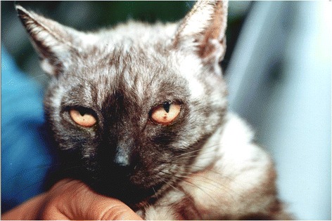
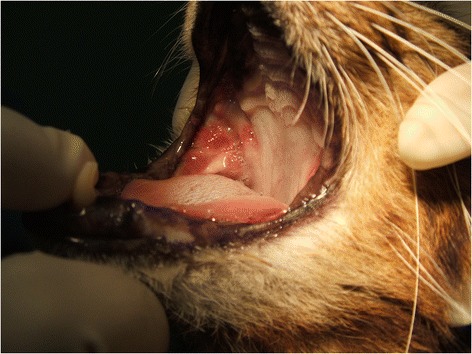
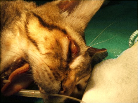
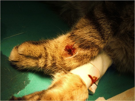
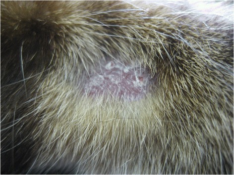
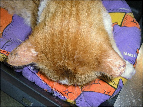
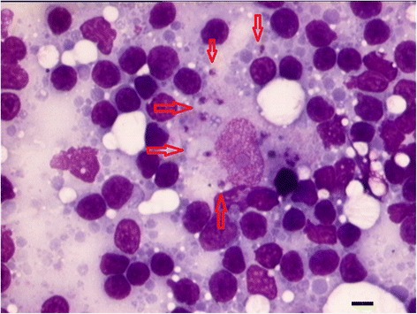
Limited data is available on feline leishmaniosis (FeL) caused by Leishmania infantum worldwide. The LeishVet group presents in this report a review of the current knowledge on FeL, the epidemiological role of the cat in L. infantum infection, clinical manifestations, and recommendations on diagnosis, treatment and monitoring, prognosis and prevention of infection, in order to standardize the management of this disease in cats. The consensus of opinions and recommendations was formulated by combining a comprehensive review of evidence-based studies and case reports, clinical experience and critical consensus discussions. While subclinical feline infections are common in areas endemic for canine leishmaniosis, clinical illness due to L. infantum in cats is rare. The prevalence rates of feline infection with L. infantum in serological or molecular-based surveys range from 0% to more than 60%. Cats are able to infect sand flies and, therefore, they may act as a secondary reservoir, with dogs being the primary natural reservoir. The most common clinical signs and clinicopathological abnormalities compatible with FeL include lymph node enlargement and skin lesions such as ulcerative, exfoliative, crusting or nodular dermatitis (mainly on the head or distal limbs), ocular lesions (mainly uveitis), feline chronic gingivostomatitis syndrome, mucocutaneous ulcerative or nodular lesions, hypergammaglobulinaemia and mild normocytic normochromic anaemia. Clinical illness is frequently associated with impaired immunocompetence, as in case of retroviral coinfections or immunosuppressive therapy. Diagnosis is based on serology, polymerase chain reaction (PCR), cytology, histology, immunohistochemistry (IHC) or culture. If serological testing is negative or low positive in a cat with clinical signs compatible with FeL, the diagnosis of leishmaniosis should not be excluded and additional diagnostic methods (cytology, histology with IHC, PCR, culture) should be employed. The most common treatment used is allopurinol. Meglumine antimoniate has been administered in very few reported cases. Both drugs are administered alone and most cats recover clinically after therapy. Follow-up of treated cats with routine laboratory tests, serology and PCR is essential for prevention of clinical relapses. Specific preventative measures for this infection in cats are currently not available.
Figures
References
-
- Solano-Gallego L, Koutinas A, Miró G, Cardoso L, Pennisi MG, Ferrer L, et al. Directions for the diagnosis, clinical staging, treatment and prevention of canine leishmaniosis. Vet Parasitol. 2009;165:1–18. - PubMed
-
- Sergent E, Sergent E, Lombard J, Quilichini M. La leishmaniose à Alger. Infection simultanée d’un enfant, d’un chien et d’un chat dans la même habitation. Bull Soc Pathol Exot. 1912;5:93–8.
-
- Morsy TA, el Seoud SM A. Natural infection in two pet cats in a house of a zoonotic cutaneous leishmaniasis patient in Imbaba area, Giza Governorate, Egypt. J Egypt Soc Parasitol. 1994;24:199–204. - PubMed
-
- Giordano A. Le chat dans la transmission de la leishmaniose viscérale de la méditérranée. Bull Sez Ital Soc Internaz Microbiol. 1933;5:330–2.
Publication types
MeSH terms
LinkOut - more resources
Full Text Sources
Other Literature Sources
Medical
Miscellaneous
