. 2015 Jul 30;126(5):629-39.
doi: 10.1182/blood-2014-11-612903. Epub 2015 Jun 3.

Eradication of B-ALL using chimeric antigen receptor-expressing T cells targeting the TSLPR oncoprotein

Affiliations

Eradication of B-ALL using chimeric antigen receptor-expressing T cells targeting the TSLPR oncoprotein

Haiying Qin et al. Blood. .

Erratum in

Abstract

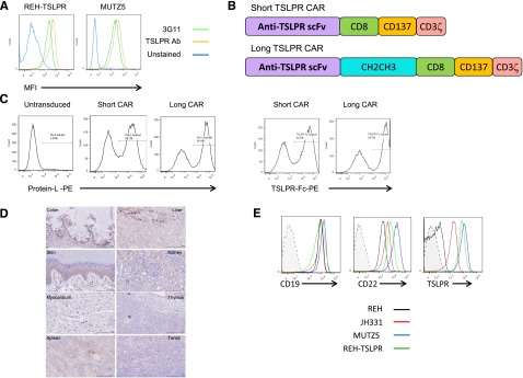
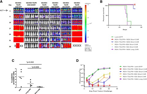
Adoptive transfer of T cells genetically modified to express chimeric antigen receptors (CARs) targeting the CD19 B cell-associated protein have demonstrated potent activity against relapsed/refractory B-lineage acute lymphoblastic leukemia (B-ALL). Not all patients respond, and CD19-negative relapses have been observed. Overexpression of the thymic stromal lymphopoietin receptor (TSLPR; encoded by CRLF2) occurs in a subset of adults and children with B-ALL and confers a high risk of relapse. Recent data suggest the TSLPR signaling axis is functionally important, suggesting that TSLPR would be an ideal immunotherapeutic target. We constructed short and long CARs targeting TSLPR and tested efficacy against CRLF2-overexpressing B-ALL. Both CARs demonstrated activity in vitro, but only short TSLPR CAR T cells mediated leukemia regression. In vivo activity of the short CAR was also associated with long-term persistence of CAR-expressing T cells. Short TSLPR CAR treatment of mice engrafted with a TSLPR-expressing ALL cell line induced leukemia cytotoxicity with efficacy comparable with that of CD19 CAR T cells. Short TSLPR CAR T cells also eradicated leukemia in 4 xenograft models of human CRLF2-overexpressing ALL. Finally, TSLPR has limited surface expression on normal tissues. TSLPR-targeted CAR T cells thus represent a potent oncoprotein-targeted immunotherapy for high-risk ALL.


Figures

Figure 1
Figure 1
Construction of short and long anti-TSLPR CAR constructs and lentiviral transduction of T cells. (A) Anti-TSLPR hybridoma (3G11) supernatant binds to the surface of TSLPR-overexpressing ALL. Binding was detected using phycoerytherin-conjugated goat-anti-mouse antibody. Binding of a commercially available, directly conjugated anti-TSLPR antibody (Ab) is shown for comparison. (B) Schematic representation of anti-TSLPR chimeric antigen receptor constructs. Both constructs contain an anti-TSPLR single-chain fragment variable sequence from the 3G11 hybridoma, a CD8 transmembrane domain, a CD137 (41BB) costimulatory domain, and a CD3 ζ-signaling domain. The long version of the CAR also contains a spacer region derived from an immunoglobulin CH2CH3 domain. (C) Transduction efficiency of activated CD3/CD28 bead-expanded human T cells with lentiviral-based vectors expressing short and long anti-TSLPR constructs. The left panels show detection of CAR using Protein L. The right panels show detection using a TSLPR protein Fc construct. (D) TSLPR expression on normal pediatric tissues, representative of 48 healthy donors. Colon: Weak cytoplasmic granular staining in crypt base columnar cells and weak stromal tissue positivity can be seen. Liver: Mild-moderate diffuse cytoplasmic granular staining in liver sinusoid. Kupffer cells around the lining of liver sinusoid shows stronger granular staining compared with the surrounding parenchyma. Heart: Absent or negative staining on myocardial tissue. Thymus: Weak and diffuse granular staining over thymus tissue. Tonsil: Some, but not all, lymphoid tissue in the tonsil shows weak granular cytoplasmic staining. Spleen: Weak cytoplasmic staining over splenic cords. There is also hemosiderin-laden macrophages and lipofusin (yellow droplets). Kidney: Proximal tubules, distal tubules, and collecting ducts show mild-to-moderate cytoplasmic reaction, but the glomeruli are negative. Skin: Lower level of stratum spinosum and stratum basale (basal layer) show granular cytoplasmic staining. There is no staining of keratinized squamous cell layer and collagen tissue. (E) Expression level of TSLPR on B-ALL relative to expression of CD19 and CD22. JH331 is a patient-derived xenograft model of CRLF2-rearranged ALL with endogenous TSLPR overexpression. Shaded histograms represent isotype control.
Figure 2
Figure 2
Both short and long demonstrate activity in vitro but only the short CAR is active in vivo. (A) Production of IFNγ and TNFα by short and long anti-TSLPR CAR T cells as measured by enzyme-linked immunosorbent assay of supernatant after coincubation with TSLPRhi ALL (REH TSLPR or MUTZ5). (B) Lysis of REH-TSLPR and MUTZ5 TSLPRhi ALL by both short and long CAR T cells measured by 51Cr release after 4-hour coculture. Results show specific lysis calculated as described in Methods. (C) Short TSLPR CAR T cells (15 × 106) administered IV on day 4 after injection of luciferase-expressing REH-TSLPR demonstrate potent activity as measured by bioluminescent imaging, whereas long TSLPR CAR given at the same dose fail to alter leukemia progression. Control mice received the same dose of expanded GFP-transduced T cells. A single animal in the short TSLPR CAR group was sacrificed as the result of a wasting syndrome consistent with xenogeneic graft-versus-host disease. ACT, adoptive cell transfer. (D) In a separate experiment, mice were treated as in Figure 2C, and peripheral blood was analyzed on days 16 and 27 after injection of 15 × 106 short or long TSLPR CAR T cells or GFP-transduced control T cells. Representative dot plots showing increased persistence of short CD8+ TSLPR CAR T cells compared with long CAR T cells on days 16 and 27 after injection. (E) Significantly increased absolute number of short TSLPR CAR T cells at day 27 after injection compared with long CAR T cells as measured using a bead calibration as described in “Methods.”
Figure 3
Figure 3
Potent activity of short TSLPR CAR T cells is associated with a relative expansion of CD8+ CAR T cells in vivo. (A) Mice were injected with high-dose REH-TSLPR (5 × 10⁶) followed by late injection of short TSLPR CAR T cells (10 × 106) on day 16. ACT, adoptive cell transfer. (B) Slightly increased relative number of CD4+ CAR T cells after CD3/CD28 bead-mediated expansion. This converts to a predominance CD8+/TSLPR CAR+ (measured by TSLPR Fc) at day 50 after injection. (C) Representative dot plots showing phenotype of short TSLPR CAR T cells before injection at day 20 after injection. (D) Relative percentages of naïve, central memory, and effector memory CAR T cells at day 20 after injection.
Figure 4
Figure 4
Short TSLPR CAR T cells demonstrate activity over a broad dose range and mediate durable eradication of TSLPRhi ALL at high leukemic burden. (A) Dose titration of short TSLPR CAR T cells given on day 3 after high-dose REH TSLPR ALL injection (5 × 10⁶ cells/mouse). ACT, adoptive cell transfer. (B) Survival plot of mice treated in Figure 4A. (C) Flow cytometry of peripheral blood for the presence of CD45+ leukemia cells at day 12 after injection. (D) Quantitation of bioluminescence in mice treated in Figure 4A.
Figure 5
Figure 5
Leukemia eradication in human CRLF2-rearranged Ph-like ALL patient-derived xenograft models by short TSLPR CAR T cells. (A) Flow cytometric surface expression of CD19 and TSLPR in human ALL cells from TSLPRhi PDX models JH352, NH362, and JH331. Shaded histograms denoted by dotted lines represent unstained controls. (B) Complete eradication of leukemia in a TSLPRhi luciferase–expressing PDX model (JH331) by short TSLPR CAR T cells. ACT, adoptive cell transfer. (C-D) Analysis of human CD45+ CD19+ ALL cells in peripheral blood (C) and bone marrow (D) of TSLPRhi PDX models JH352 and NH362 injected with 1 × 10⁶ ALL cells per mouse, then treated 22 days later with 15 × 106 short TSLPR CAR T cells. (E-G) Tissue analyses of an aggressive relapsed TSLPRhi ALL PDX model (ALL4364) treated with TSLPR CAR T cells. One million ALL cells per mouse were injected IV on day 1, then animals were treated with 1.2 million of TSLPR CAR+ T cells IV on day 14. (E) Spleens from short TSLPR CAR-treated and untreated mice on days 35 and 42 after leukemia injection (2 and 3 weeks after CAR injection). (F) Representative dot plot of bone marrow, spleen, and peripheral blood on day 35 after leukemia injection. (G) Scatter plots of organ and blood leukemia infiltration at day 35 after TSLPRhi ALL injection comparing short TSLPR CAR treated and untreated mice. (H) Separate experiment showing survival analysis demonstrating prolonged survival of TSLPR CAR T cell–treated ALL4364 PDX animals (n = 5/group). ACT, adoptive cell therapy. (I) Expression of TSLPR on human CD45+/CD19+ leukemia cells in the bone marrow of a mouse with relapse after short TSLPR CAR treatment.
Figure 6
Figure 6
TSLPR CAR demonstrates activity comparable with CD19 CAR. (A) Schematic diagram of second-generation CD19 and the CD22 CAR constructs. Both CARs have identical transmembrane and signaling domains to the TSLPR CAR. (B) Flow cytometric surface expression of protein L on TSLPR, CD19, and CD22 CAR T cells at 5 days postviral transduction, demonstrating efficacy of CAR transduction. (C) TSLPR, CD19, and CD22 CAR-redirected T cells were injected IV into NSG mice (3 × 10⁶/mouse) previously engrafted with the patient xenograft cell line JH331-LUC 29 days earlier and followed by bioluminescent imaging. T cells transduced with GFP were used as a negative control. ACT, adoptive cell transfer; D, day. (D) JH331-LUC–bearing NSG mice were treated as in Figure 6C but including a group receiving a log lower dose of CAR T cell. Control mice received 3 × 10⁶ GD2-targeted CAR T cells.

Comment in

References

    1. Pui CH, Evans WE. A 50-year journey to cure childhood acute lymphoblastic leukemia. Semin Hematol. 2013;50(3):185–196. - PMC - PubMed
    1. Hunger SP, Loh ML, Whitlock JA, et al. COG Acute Lymphoblastic Leukemia Committee. Children’s Oncology Group’s 2013 blueprint for research: acute lymphoblastic leukemia. Pediatr Blood Cancer. 2013;60(6):957–963. - PMC - PubMed
    1. Inaba H, Greaves M, Mullighan CG. Acute lymphoblastic leukaemia. Lancet. 2013;381(9881):1943–1955. - PMC - PubMed
    1. Bhojwani D, Pui CH. Relapsed childhood acute lymphoblastic leukaemia. Lancet Oncol. 2013;14(6):e205–e217. - PubMed
    1. Raetz EA, Bhatla T. Where do we stand in the treatment of relapsed acute lymphoblastic leukemia? Hematol Am Soc Hematol Educ Program. 2012;2012:129–136. - PubMed

Publication types

MeSH terms