. 2015 Sep 1;24(17):4817-28.
doi: 10.1093/hmg/ddv206. Epub 2015 Jun 3.

DICER/AGO-dependent epigenetic silencing of D4Z4 repeats enhanced by exogenous siRNA suggests mechanisms and therapies for FSHD

Affiliations

DICER/AGO-dependent epigenetic silencing of D4Z4 repeats enhanced by exogenous siRNA suggests mechanisms and therapies for FSHD

Jong-Won Lim et al. Hum Mol Genet. .

Abstract

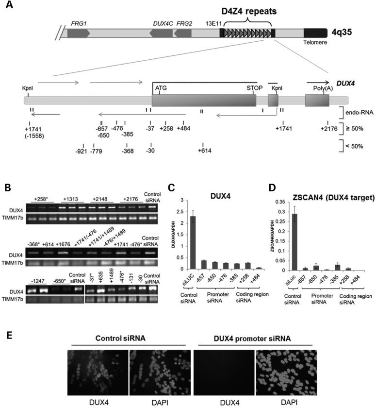
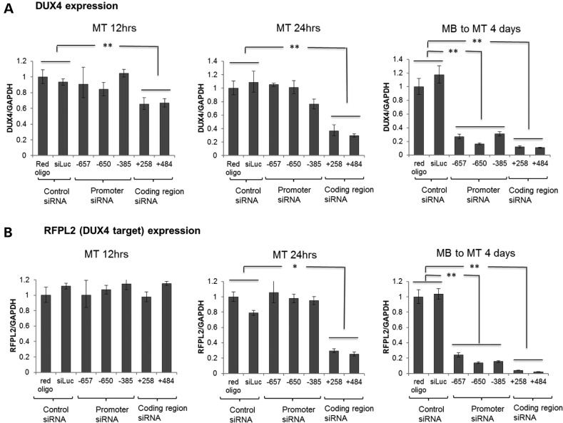
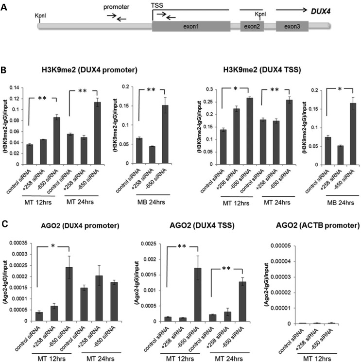
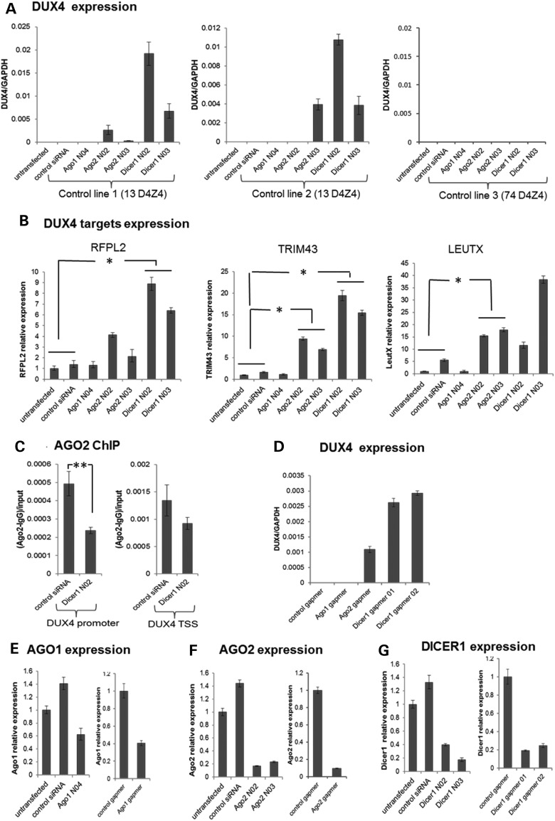
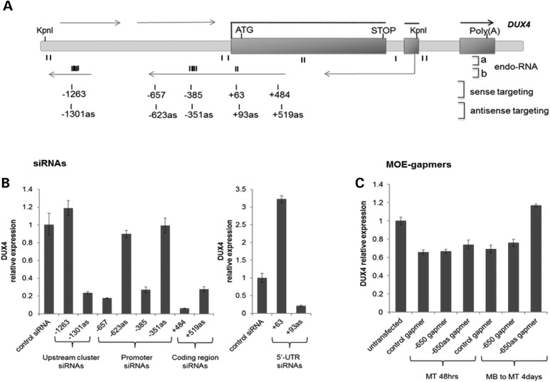
Facioscapulohumeral muscular dystrophy (FSHD) is caused by the aberrant expression of the DUX4 transcription factor in skeletal muscle. The DUX4 retrogene is encoded in the D4Z4 macrosatellite repeat array, and smaller array size or a mutation in the SMCHD1 gene results in inefficient epigenetic repression of DUX4 in skeletal muscle, causing FSHD1 and FSHD2, respectively. Previously we showed that the entire D4Z4 repeat is bi-directionally transcribed with the generation of small si- or miRNA-like fragments and suggested that these might suppress DUX4 expression through the endogenous RNAi pathway. Here we show that exogenous siRNA targeting the region upstream of the DUX4 transcription start site suppressed DUX4 mRNA expression and increased both H3K9 methylation and AGO2 recruitment. In contrast, similarly targeted MOE-gapmer antisense oligonucleotides that degrade RNA but do not engage the RNAi pathway did not repress DUX4 expression. In addition, knockdown of DICER or AGO2 using either siRNA or MOE-gapmer chemistries resulted in the induction of DUX4 expression in control muscle cells that normally do not express DUX4, indicating that the endogenous RNAi pathway is necessary to maintain repression of DUX4 in control muscle cells. Together these data demonstrate a role of the endogenous RNAi pathway in repeat-mediated epigenetic repression of the D4Z4 macrosatellite repeat, and show that enhancing the activity of this pathway by supplying exogenous siRNA oligonucleotides represents a potential therapeutic approach to silencing DUX4 in FSHD.


Figures

Figure 1.
Figure 1.
Inhibition of DUX4 expression in FSHD cells by siRNAs targeting D4Z4 regions. (A) Schematic diagram of the distal tip of 4q35 showing the D4Z4 repeat region and enlargement of the distal D4Z4 unit flanked by KpnI sites and the adjacent pLAM sequence with the imbedded DUX4 gene. Exons 1–3 of DUX4 are indicated by rectangles. Thick horizontal lines correspond to the DUX4 mRNA, whereas thin horizontal lines depict multiple sense and antisense D4Z4 transcripts. Direction of transcription is indicated with arrows. Previously identified D4Z4-derived endogenous small RNAs (6) are shown by short vertical lines and indicated as endo-RNA. Numbered vertical marks show the location of exogenous duplex siRNAs targeting the D4Z4 region. siRNA numbering indicates location of the 5-prime nucleotide of the targeted sequences relative to the most promoter proximal DUX4 transcription start site (TSS = 1) (6) with all siRNAs in this figure designed to target the sense sequence relative to the DUX4 transcript (see Supplementary Material, Table S1 for each targeted sequence). Top lane shows siRNAs that efficiently reduce DUX4 mRNA levels (≥50%), and bottom lane shows less-efficient siRNAs (<50%) as determined by RT- and qRT–PCR analysis of DUX4 and DUX4 target gene expression (B and C). (B) Representative sample of RT–PCR analysis of DUX4 mRNA abundance following transient transfection of FSHD1 muscle cells with duplex siRNAs targeting D4Z4 regions as depicted in (A). TIMM17b RT–PCR on each sample serves as internal control. Biological replicates are indicated by horizontal lines above lanes; otherwise, single representative PCR results from multiple experiments are shown. Asterisk mark indicates siRNAs that were verified by qRT–PCR and selected for further investigation. (C and D) DUX4 and DUX4-target mRNA levels in FSHD2 muscle cells following transfection with the siRNAs, targeting coding and non-coding DUX4 sequences, were quantified by real-time qRT–PCR analysis (see Supplementary Material, Fig. S1 for details). DUX4 and DUX4-target qRT–PCR data were analyzed by standard curve, normalized to GAPDH and presented as mean ± SEM based on triplicate PCR reactions for each sample. (E) Immunofluorescence analysis of FSHD2 muscle cells transfected with control (siLuc) and promoter (−650) targeting siRNAs. Cells were stained with DUX4 antibody and DAPI, as indicated. Single representative images of FSHD muscle cells transfected with the control and promoter targeting siRNAs that were validated by qRT–PCR in (C) are shown. For all experiments presented in Figure 1, DUX4 or DUX4-target expression was analyzed in FSHD muscle cells 96 h post siRNA transfection and 48 h post-induction to differentiation (MB to MT 4 days time point) (see also Supplementary Material, Figs. S1 and S2).
Figure 2.
Figure 2.
Delayed kinetics of promoter targeting siRNA compared with coding region siRNA. qRT–PCR analysis of DUX4 mRNA (A) and DUX4-target gene, RFPL2 (B) expression in FSHD2 muscle cells at different time points following transient transfection with DUX4 promoter siRNAs or coding region siRNAs targeting the corresponding D4Z4 sequences numbered relative to the DUX4 TSS (see Supplementary Material, Table S1). siRNAs were transfected into cells and total RNA samples were prepared at 12 h (MT 12 h), 24 h (MT 24 h) and 4 days (MB to MT 4 days) post-transfection. For each time point, FSHD myoblasts (MB) were induced to differentiation into myotubes (MT) for 48 h prior RNA isolation. Relative DUX4 and RFPL2 expression normalized to GAPDH are presented as mean ± SEM based on triplicate PCR reactions for each sample. P-values were calculated by comparing DUX4 or RFPL2 expression levels in cells transfected with the three promoter or the two coding region siRNAs to the corresponding expression levels in the two controls by using a Student's t-test. All relative expressions with a P < 0.05 (*) or <0.01 (**) are indicated. Coding region siRNAs showed DUX4 and RFPL2 (DUX4-target) decreased at early time points, but promoter region siRNAs showed decrease only at the late time point (4 days post-transfection).
Figure 3.
Figure 3.
Exogenous siRNA targeting the DUX4 promoter region increase D4Z4 H3K9me2 and AGO2 enrichment. (A) Schematic representation of the D4Z4 repeat with the location of promoter and TSS PCR primers indicated by arrows. (B and C) Enrichment of H3K9me2 (B) and AGO2 (C) at the promoter region and near the transcription start site (TSS) of the DUX4 gene was analyzed by chromatin immunoprecipitation (ChIP) followed by qPCR. As a negative control for AGO2 enrichment, the beta-actin (ACTB) promoter region was analyzed. Control siRNA and siRNAs targeting coding (+258) and non-coding (−650) D4Z4 sequences were transfected into FSHD2 myoblasts (MB) and myotubes (MT) and chromatin samples were prepared at 12 h and 24 h post-transfection time points. Relative amounts of precipitated DNA were calculated as ratios of the real-time PCR signals obtained for IP with the H3K9me2 or AGO2 antisera minus those obtained for mock IP with IgG to input signals. Data are presented as mean ± SEM based on triplicate PCR reactions for each sample. ChIP enrichments with a P < 0.05 (*) or <0.01 (**) based on a t-test comparing the enrichment in the cells transfected with the DUX4 promoter or coding region siRNA to the corresponding enrichment in the cells transfected with the control siRNA (see Materials and Methods) are indicated. The promoter siRNA targeting the region D4Z4 upstream of DUX4 (termed promoter region) showed increased H3K9me2 and AGO2 binding at the promoter region and near the TSS region of the DUX4 gene (see also Supplementary Material, Fig. S3).
Figure 4.
Figure 4.
AGO2 or DICER1 knockdown in control cells de-repress DUX4 expression. (A) qRT–PCR analysis of DUX4 mRNA in three control muscle cell lines following siRNA-mediated depletion of AGO1, AGO2 and DICER1. (B) qRT–PCR analysis of DUX4-target gene transcripts (RFPL2, TRIM43, LEUTX) in Control Line 1 following siRNA-mediated depletion of AGO1, AGO2 and DICER1. Myoblasts were transfected twice with the validated siRNAs targeting AGO1 (AGO1 N04), AGO2 (AGO2 N02 and N03) or DICER1 (DICER1 N02 and N03) mRNA using a double-transfection protocol with the first transfection at −96 h followed by the second transfection and induction of differentiation at −48 h prior to RNA isolation (MB to MT 4 days time point). qRT–PCR data were analyzed using standard curve method for DUX4 expression and ddCt for DUX4 target gene expression, normalized to GAPDH levels and presented as mean ± SEM based on triplicate PCR reactions for each sample. (C) ChIP-qPCR analysis of AGO2 enrichment at the DUX4 promoter and near the TSS region in control myotubes [Control line 1 in (A)] following transfection with control siRNA or DICER1 N02 siRNA. (D) qRT–PCR analysis of DUX4 mRNA in control muscle cell line 1 following depletion of AGO1, AGO2 and DICER1 using MOE-gapmers. Myoblasts were transfected twice with the gapmers using a double-transfection protocol with the first transfection at −96 h followed by the second transfection and induction of differentiation at −48 h prior to RNA isolation. qRT–PCR data were analyzed using standard curve method, normalized to GAPDH levels and presented as mean ± SEM. (EG) AGO1, AGO2 and DICER1 mRNA levels in control line 1 following siRNA or MOE-gapmer knockdowns were analyzed by qRT–PCR by ddCt method, normalized to GAPDH and presented as mean ± SEM (see also Supplementary Material, Fig. S4). Data plotted in all panels represent the mean ± SEM based on triplicate PCR reactions. *P < 0.05; **P < 0.01. (A) and (D) lacked measurable signal in the control samples, so P-values were not calculated, see Supplementary Material, Figure S4 for Ct values from (A). (E–G) show the degree of target knockdown for the control line 1 experiment presented and P-values were not calculated. See Supplementary Material, Figure S4B for degree of AGO1, AGO2 and DICER knockdown in the control lines 2 and 3 experiments presented.
Figure 5.
Figure 5.
Strand-specific suppression of DUX4 by siRNA but not MOE-gapmers in FSHD cells. (A) Schematic diagram depicting the D4Z4 region and the structure of the DUX4 gene. endo-RNA: (a) The location of previously identified D4Z4-derived small endogenous RNAs (6) and (b) the location of the newly identified endogenous chromatin-associated D4Z4 small RNAs from publicly available small RNA sequencing data sets (20,22) indicated as clusters of short bold vertical marks (see Supplementary Material, Table S2 for sequences). Rows of numbered vertical marks show the location of additional siRNAs tested corresponding to the clustered regions of chromatin-associated small RNAs. Numbering is from the 5-prime end of the targeted sequence relative to the DUX4 TSS. siRNA designed to target the transcript in the antisense orientation relative to the DUX4 mRNA are annotated ‘as' (i.e. siRNA −657 targets the sense sequence starting at −657 relative to the TSS, whereas siRNA −623as targets the antisense sequence with the 5-prime end beginning at −623 and providing the complementary overlap to the −657 siRNA). (B) DUX4 siRNAs were transfected into FSHD2 myoblasts and induced to differentiation 2 days after transfection. Total RNA was prepared at 4 days post-transfection time point. Relative expression of DUX4 mRNA was analyzed by qRT–PCR, normalized to GAPDH, and presented as mean ± SEM based on triplicate PCR reactions. (C) qRT–PCR analysis of DUX4 mRNA expression in FSHD2 muscle cells following transfection with MOE-gapmers targeting D4Z4 sequences upstream of the DUX4 TSS. Gapmer numbering corresponds to the position of their target sequences relative to the DUX4 transcription start site (TSS = 1). Gapmers targeting antisense sequence are noted as ‘as’. Cells were either transfected once with the gapmers and induced to differentiation −48 h prior RNA isolation (MT 48 h time point) or twice (double-transfection protocol) with the first transfection at −96 h followed by the second transfection and induction of differentiation at −48 h prior to RNA isolation (MB to MT 4 days time point). DUX4 mRNA expression data were normalized to GAPDH expression and presented as mean ± SEM (see also Supplementary Material, Fig. S5). Data plotted in all panels represent the mean ± SEM based on triplicate PCR reactions. See Supplementary Material, Figure S6 for replication and additional controls.

References

    1. Tawil R., van der Maarel S.M., Tapscott S.J. (2014) Facioscapulohumeral dystrophy: the path to consensus on pathophysiology. Skeletal Muscle, 4, 12. - PMC - PubMed
    1. Lemmers R.J., Tawil R., Petek L.M., Balog J., Block G.J., Santen G.W., Amell A.M., van der Vliet P.J., Almomani R., Straasheijm K.R. et al. (2012) Digenic inheritance of an SMCHD1 mutation and an FSHD-permissive D4Z4 allele causes facioscapulohumeral muscular dystrophy type 2. Nat. Genet., 44, 1370–1374. - PMC - PubMed
    1. Blewitt M.E., Gendrel A.V., Pang Z., Sparrow D.B., Whitelaw N., Craig J.M., Apedaile A., Hilton D.J., Dunwoodie S.L., Brockdorff N. et al. (2008) SmcHD1, containing a structural-maintenance-of-chromosomes hinge domain, has a critical role in X inactivation. Nat. Genet., 40, 663–669. - PubMed
    1. Blewitt M.E., Vickaryous N.K., Hemley S.J., Ashe A., Bruxner T.J., Preis J.I., Arkell R., Whitelaw E. (2005) An N-ethyl-N-nitrosourea screen for genes involved in variegation in the mouse. Proc. Natl Acad. Sci. USA, 102, 7629–7634. - PMC - PubMed
    1. Lemmers R.J., van der Vliet P.J., Klooster R., Sacconi S., Camano P., Dauwerse J.G., Snider L., Straasheijm K.R., van Ommen G.J., Padberg G.W. et al. (2010) A unifying genetic model for facioscapulohumeral muscular dystrophy. Science, 329, 1650–1653. - PMC - PubMed

Publication types

MeSH terms