Comparative Study
. 2015 Sep;96(9):2643-2655.
doi: 10.1099/vir.0.000209. Epub 2015 Jun 3.

Arterivirus nsp12 versus the coronavirus nsp16 2'-O-methyltransferase: comparison of the C-terminal cleavage products of two nidovirus pp1ab polyproteins

Affiliations
Comparative Study

Arterivirus nsp12 versus the coronavirus nsp16 2'-O-methyltransferase: comparison of the C-terminal cleavage products of two nidovirus pp1ab polyproteins

Kathleen C Lehmann et al. J Gen Virol. 2015 Sep.

Abstract

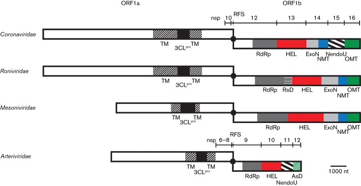
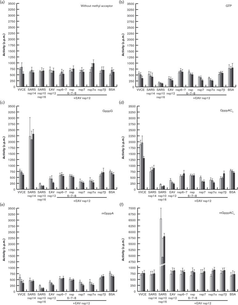
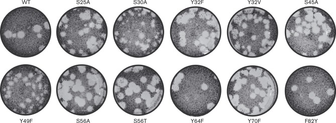
The 3'-terminal domain of the most conserved ORF1b in three of the four families of the order Nidovirales (except for the family Arteriviridae) encodes a (putative) 2'-O-methyltransferase (2'-O-MTase), known as non structural protein (nsp) 16 in the family Coronaviridae and implicated in methylation of the 5' cap structure of nidoviral mRNAs. As with coronavirus transcripts, arterivirus mRNAs are assumed to possess a 5' cap although no candidate MTases have been identified thus far. To address this knowledge gap, we analysed the uncharacterized nsp12 of arteriviruses, which occupies the ORF1b position equivalent to that of the nidovirus 2'-O-MTase (coronavirus nsp16). In our in-depth bioinformatics analysis of nsp12, the protein was confirmed to be family specific whilst having diverged much further than other nidovirus ORF1b-encoded proteins, including those of the family Coronaviridae. Only one invariant and several partially conserved, predominantly aromatic residues were identified in nsp12, which may adopt a structure with alternating α-helices and β-strands, an organization also found in known MTases. However, no statistically significant similarity was found between nsp12 and the twofold larger coronavirus nsp16, nor could we detect MTase activity in biochemical assays using recombinant equine arteritis virus (EAV) nsp12. Our further analysis established that this subunit is essential for replication of this prototypic arterivirus. Using reverse genetics, we assessed the impact of 25 substitutions at 14 positions, yielding virus phenotypes ranging from WT-like to non-viable. Notably, replacement of the invariant phenylalanine 109 with tyrosine was lethal. We concluded that nsp12 plays an essential role during EAV replication, possibly by acting as a co-factor for another enzyme.


Figures

Fig. 1.
Fig. 1.
Organization of key replicase domains encoded by nidovirus ORFs 1a and 1b. Proteolytic cleavage products described in the text for the coronaviruses and arteriviruses are indicated. Matching colours/patterns indicate domain conservation between families. Domains (putatively) involved in capping (HEL, NMT, OMT and AsD) are depicted in bright colours. TM, transmembrane domain; 3CLpro, 3C-like protease; black dot and RFS, ribosomal frameshift site; RdRp, RNA-dependent RNA polymerase; HEL, ZBD, helicase core (HEL1) and variable additional domains; ExoN, exoribonuclease; NMT, N7-MTase; NendoU, endoribonuclease; OMT, 2′-O-MTase; RsD, ronivirus-specific domain; AsD, arterivirus-specific domain (nsp12). Genomic organizations are shown for Beluga whale coronavirus SW1 (family Coronaviridae), Gill-associated virus (family Roniviridae), Nam Dinh virus (family Mesoniviridae) and PRRSV, North American genotype (family Arteriviridae). Depicted is a simplified domain organization as most enzymes are multidomain proteins. Note that viruses of the family Coronaviridae that do not belong to the subfamily Coronavirinae encode a truncated version of NMT. Adapted from Lauber et al. (2013).
Fig. 2.
Fig. 2.
Similarity density plots of the C-terminal region of pp1ab of different nidovirus (sub)families. Values above and below average similarities are indicated in black and grey, respectively. HEL1, helicase core domain; ExoN, exoribonuclease; NMT, N 7-MTase (t, truncated); NendoU, endoribonuclease; OMT, 2′-O-MTase. For the sake of simplicity, we have applied the nsp nomenclature of the subfamily Coronavirinae also to the orthologous torovirus domains for which the processing of pp1a/pp1ab is yet to be fully described.
Fig. 3.
Fig. 3.
Multiple sequence alignment (MSA) and secondary structure predictions of representative arterivirus nsp12 sequences. Partially and fully conserved amino acids are highlighted in coloured boxes. Colours represent residues with similar biophysical properties; yellow, aromatic; black, hydrophobic; blue, (putative) zinc binding; green, other. Secondary structures (barrel, α-helix; arrow, β-strand) were predicted with JPred (Cole et al., 2008) (grey) or psipred (Buchan et al., 2013) (red) based on the MSA. Residue numbers are indicated for nsp12 of the EAV-Bucyrus isolate (pEAV030) (van Dinten et al., 1997), the parental strain of pEAV211 used for the reverse genetics experiments. Replaced residues are indicated below the alignment; black stars, positions where stop codons were introduced; empty circles, control residues; filled circles, conserved residues. A putative zinc finger in simian arterivirus nsp12 sequences is indicated by a dashed line. EAV, GenBank accession number AY349167; SHFV, GenBank accession numbers AF180391, JX473847, JX473848, HQ845737 and HQ845738; PRRSV, GenBank accession numbers JX138233 and JF802085; LDV, GenBank accession numbers L13298 and U15146; WPDV, GenBank accession number JN116253.
Fig. 4.
Fig. 4.
SDS-PAGE analysis of purified EAV nsps. (a) The progression of metal-ion chromatography of EAV nsp12-containing (13 kDa) E. coli lysates was monitored by Coomassie brilliant blue staining. Insoluble and soluble: proteins retained in pellet or supernatant, respectively, after cell lysis and ultracentrifugation; after binding: proteins in supernatant after removal of Talon beads. (b) Elution fractions of EAV ORF1a proteins and intermediates (nsp6–7, 29 kDa; nsp6–7–8, 34 kDa; nsp7, 26 kDa; nsp7α, 15 kDa; nsp7β, 13 kDa). Products marked with an asterisk are the remaining ubiquitin–nsp fusion proteins. Lane M, size markers (kDa).
Fig. 5.
Fig. 5.
MTase activity assays using recombinant EAV nsp12 in the presence and absence of possible co-factors. Recombinant EAV nsp12 (1 μM) and equimolar amounts of the indicated possible co-factors were incubated for 30 (light grey), 60 (dark grey) or 180 min (black) with S-[methyl-3H]adenosylmethionine and the indicated methyl acceptor. Proteins with known MTase activity served as positive controls. VVCE, Vaccinia virus capping enzyme (0.1 U μl− 1, N7-MTase); SARS nsp14 (75 nM, N 7-MTase); SARS nsp10/nsp16 (2 μM complex, 2′-O-MTase); SARS, SARS coronavirus; BSA served as negative control. Reslults are presented as mean ± sd of two independent experiments. The background variation evident for several of the protein combinations using GTP, GpppG, GpppAC4 or mGpppA most likely represents an artefact originating from a position effect, which was observed repeatedly in the employed 96-well format.
Fig. 6.
Fig. 6.
Plaque phenotypes of viable EAV nsp12 mutants. Virus-containing supernatants obtained 48 h p.t. were serially diluted and used to infect BHK-21 cells. After 72 h the cells were fixed with 4 % formaldehyde and stained with crystal violet.

References

    1. Bailey A.L., Lauck M., Sibley S.D., Pecotte J., Rice K., Weny G., Tumukunde A., Hyeroba D., Greene J., other authors Two novel simian arteriviruses in captive and wild baboons (Papio spp.) J Virol. 2014;88:13231–13239. doi: 10.1128/JVI.02203-14. - DOI - PMC - PubMed
    1. Benson D.A., Cavanaugh M., Clark K., Karsch-Mizrachi I., Lipman D.J., Ostell J., Sayers E.W. GenBank. Nucleic Acids Res. 2013;41((D1)):D36–D42. doi: 10.1093/nar/gks1195. - DOI - PMC - PubMed
    1. Bouvet M., Debarnot C., Imbert I., Selisko B., Snijder E.J., Canard B., Decroly E. In vitro reconstitution of SARS-coronavirus mRNA cap methylation. PLoS Pathog. 2010;6:e1000863. doi: 10.1371/journal.ppat.1000863. - DOI - PMC - PubMed
    1. Buchan D.W., Minneci F., Nugent T.C., Bryson K., Jones D.T. Scalable web services for the psipred Protein Analysis Workbench. Nucleic Acids Res. 2013;41((W1)):W349–W357. doi: 10.1093/nar/gkt381. - DOI - PMC - PubMed
    1. Chen Z., Faaberg K.S., Plagemann P.G. Determination of the 5′ end of the lactate dehydrogenase-elevating virus genome by two independent approaches. J Gen Virol. 1994;75:925–930. doi: 10.1099/0022-1317-75-4-925. - DOI - PubMed

Publication types

MeSH terms